. 2006 Aug;79(2):197-213.
doi: 10.1086/504411. Epub 2006 Jun 12.

The Alexander disease-causing glial fibrillary acidic protein mutant, R416W, accumulates into Rosenthal fibers by a pathway that involves filament aggregation and the association of alpha B-crystallin and HSP27

Affiliations

The Alexander disease-causing glial fibrillary acidic protein mutant, R416W, accumulates into Rosenthal fibers by a pathway that involves filament aggregation and the association of alpha B-crystallin and HSP27

Ming Der Perng et al. Am J Hum Genet. 2006 Aug.

Abstract

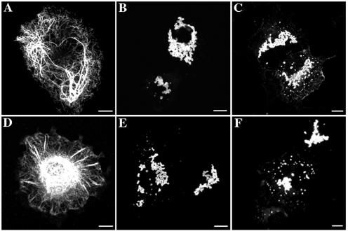
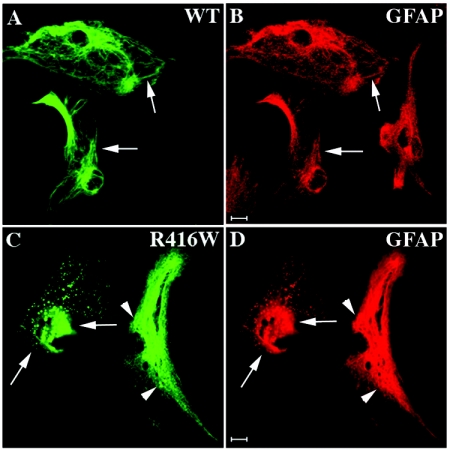
Here, we describe the early events in the disease pathogenesis of Alexander disease. This is a rare and usually fatal neurodegenerative disorder whose pathological hallmark is the abundance of protein aggregates in astrocytes. These aggregates, termed "Rosenthal fibers," contain the protein chaperones alpha B-crystallin and HSP27 as well as glial fibrillary acidic protein (GFAP), an intermediate filament (IF) protein found almost exclusively in astrocytes. Heterozygous, missense GFAP mutations that usually arise spontaneously during spermatogenesis have recently been found in the majority of patients with Alexander disease. In this study, we show that one of the more frequently observed mutations, R416W, significantly perturbs in vitro filament assembly. The filamentous structures formed resemble assembly intermediates but aggregate more strongly. Consistent with the heterozygosity of the mutation, this effect is dominant over wild-type GFAP in coassembly experiments. Transient transfection studies demonstrate that R416W GFAP induces the formation of GFAP-containing cytoplasmic aggregates in a wide range of different cell types, including astrocytes. The aggregates have several important features in common with Rosenthal fibers, including the association of alpha B-crystallin and HSP27. This association occurs simultaneously with the formation of protein aggregates containing R416W GFAP and is also specific, since HSP70 does not partition with them. Monoclonal antibodies specific for R416W GFAP reveal, for the first time for any IF-based disease, the presence of the mutant protein in the characteristic histopathological feature of the disease, namely Rosenthal fibers. Collectively, these data confirm that the effects of the R416W GFAP are dominant, changing the assembly process in a way that encourages aberrant filament-filament interactions that then lead to protein aggregation and chaperone sequestration as early events in Alexander disease.


Figures

Figure  1.
Figure 1.
The dominant effect of R416W GFAP, revealed by in vitro assembly studies. Purified GFAP at a concentration of 0.3 mg/ml was assembled in vitro by stepwise dialysis into assembly buffer, as described in the “Material and Methods” section. Assembled filaments were negatively stained and were visualized by transmission electron microscopy. Under these assembly conditions, wild-type GFAP assembled into typical 10-nm filaments with length of several microns (A), whereas R416W GFAP alone and in different proportions with wild-type protein formed short filamentous intermediates that had a strong tendency to aggregate (B–E). It is difficult to see the structural detail of the aggregates formed by R416W GFAP (D) and mixtures thereof (B and C) when negatively stained with uranyl acetate. Sometimes, less aggregated material can be found, and then, at higher magnification (E), the filamentous structures that comprise the aggregates are clearly seen. Mixing wild-type GFAP in either 75:25 (B) or 50:50 (C) proportions with R416W GFAP failed to rescue intermediate filament formation, and similar aggregates were formed (B and C). A low-speed sedimentation assay was used to assess the extent of this aggregation. F, Wild-type (WT) (lanes 1 and 2) and R416W GFAP (lanes 7 and 8) were assembled, either individually or in mixtures of 75:25 (lanes 3 and 4) or 50:50 (lanes 5 and 6) WT:R416W GFAP. After assembly, the samples were subjected to low-speed centrifugation, and the resulting supernatant (S) and pellet (P) fractions were analyzed by SDS-PAGE and were visualized by Coomassie blue staining. Whereas only one-third of assembled wild-type GFAP was sedimented (lane 1), almost all the R416W mutant was found in the pellet fraction (lane 8). Mixing wild-type GFAP with the R416W mutant in different proportions did not dramatically increase the GFAP signal in either the supernatant fraction of the 50:50 mixture (lane 5) or the 75:25 mixture (lane 3). These data show that the effects of R416W GFAP on in vitro filament assembly is dominant over the wild-type protein. Bars = 1 μm, except in panel E, where it is 0.1μm.
Figure  2.
Figure 2.
Effect of R416W and R239C mutations on the de novo GFAP IF network formation in IF-free cells. SW13/cl.2 (A–C) and primary astrocytes derived from GFAP/vimentin-null mice (D-F) were transiently transfected with either wild-type (A and D), R416W (B and E), or R239C (C and F) GFAP. At 48 h after transfection, the distribution of GFAP was assessed by confocal immunofluorescence microscopy with use of the rabbit polyclonal anti-GFAP antibody. When expressed in SW13/cl.2 cells, wild-type GFAP assembled into bundled filaments that extended throughout the cytoplasm (A). In contrast, cells transfected with either R416W (B) or R239C (C) GFAP-expression plasmids exhibited only GFAP-positive aggregates. In the IF-free mouse astrocytes, wild-type GFAP assembled into extended filaments at the cell periphery with some perinuclear accumulations (D), whereas R416W mutant GFAP formed punctuate aggregates scattered throughout the cytoplasm without any detectable filaments (E). Expression of R239C GFAP also induced numerous GFAP aggregates in the cytoplasm (F). For both R416W and R239C GFAP, all the transfected cells had aggregates. Bars = 10 μm.
Figure  3.
Figure 3.
The network-forming abilities of the wild-type and R416W GFAP in SW13/cl.1 cells. SW13/cl.1 cells transiently transfected with either wild-type (A and B) or R416W (C and D) GFAP were fixed at 48 h after transfection and were processed for double-label confocal immunofluorescence microscopy. GFAP immunofluoresence is shown in the green channel (A and C), whereas the counterstaining for vimentin is in the red channel (B and D). Notice that wild-type GFAP (A) incorporated into endogenous vimentin (Vim) (B) networks, whereas this is not the case for R416W GFAP (C). Whereas some transfected cells exhibited one large inclusion with small aggregates at the cell periphery (arrowheads in C), other transfected cells displayed bundled filaments (arrows in C) that coaligned with the endogenous vimentin (arrows in D). Bars = 10 μm.
Figure  4.
Figure 4.
Expression of R416W mutant in MCF7 cells resulted in the formation of GFAP aggregates. MCF7 cells transfected with either wild-type (A and B) or R416W GFAP (C and D) were processed at 48 h after transfection for confocal double-label immunofluorescence microscopy with use of antibodies against GFAP (A and C) and keratin (B and D). When expressed in these cells, wild-type GFAP formed extended filaments as well as perinuclear filament bundles (A) that partially colocalized with keratin IF networks (arrows in A and B). In contrast, transfected cells expressing R416W GFAP exhibited large aggregates (C) that also cocollapsed the endogenous keratin IF networks (arrows in C and D). Bars = 10 μm.
Figure  5.
Figure 5.
Analysis of wild-type and R416W GFAP expression in transfected MCF7 cells by immunoblotting. MCF7 cells were transfected with either wild-type (lanes 3 and 4) or R416W GFAP (lanes 5 and 6). Untransfected cells were used as a control (lanes 1 and 2). At 48 h after transfection, cells were collected, lysed with MEB, and centrifuged at 18,000 g for 15 min at 4°C. The resulting supernatant (S) and pellet (P) fractions were analyzed by SDS-PAGE, followed by immunoblotting using anti-GFAP (A) and anti-actin (B) antibodies. The blots were developed by the ECL system. Notice that, after transfection into MCF7 cells, both wild-type and mutant GFAP expressed at comparable levels, although proteolyzed GFAP fragments with slightly higher electrophoretic mobilities were also detected. Most of the wild-type GFAP was detected in the pellet fraction (A, lane 4) with a small proportion that remained soluble (A, lane 3), whereas the R416W GFAP was found exclusively in the pellet fraction (A, lane 6). Equal loading of each supernatant and pellet fractions was confirmed by probing with anti-actin antibody (B).
Figure  6.
Figure 6.
Ultrastructural analysis of wild-type and R416W GFAP in MCF7 cells by immunoelectron microscopy. MCF7 cells transfected with either wild-type (A and B) or R416W (C and D) GFAP were processed at 48 h after transfection for immunogold labeling, as described in the “Material and Methods” section. Immunogold labeling of ultrathin sections was stained and visualized by a transmission electron microscope. Wild-type GFAP assembled into filaments that were organized into parallel bundles (A). In contrast, cells expressing the R416W mutant formed membrane-free irregular-shaped structures composed of electron-dense aggregates at the perinuclear region (C), often in association with IFs (asterisks [*] in C). Panels B and D are higher magnification views of the boxed areas of panels A and C, respectively, showing that both the filaments (B) and aggregates (D) were decorated with 5-nm gold particles (arrows in B and D), confirming the identity of GFAP.
Figure  7.
Figure 7.
Transient expression of wild-type or R416W GFAP in primary mouse astrocytes. Primary mouse astrocytes were transfected with either human wild-type (A and B) or R416W (C and D) GFAP. At 48 h after transfection, cells were processed for double-label immunofluoresence microscopy with use of anti-human GFAP monoclonal antibody (SMI-21) and anti-panGFAP polyclonal antibodies (3270). When expressed in mouse primary astrocytes, wild-type GFAP formed filaments (arrows in A) that colocalized with the endogenous mouse GFAP (arrows in B). The expression of human R416W GFAP resulted in both filamentous (arrowheads in C) and aggregate staining patterns (arrows in C), which also costained with the endogenous mouse GFAP (arrows and arrowheads, respectively, in D). Bars = 10 μm.
Figure  8.
Figure 8.
Characterization of R416W GFAP-specific antibodies and demonstration of its presence in Rosenthal fibers. A, Immunoblots, performed as described in the “Material and Methods” section, with use of purified, recombinant human wild-type (hGF-WT) and R416W (hGF-R416W) GFAP and lysates from brain samples taken from either control human (WT) or patients with Alexander disease that harbor either an R239C mutation (R239C) or an R416W mutation (R416W) in GFAP. The general anti-human GFAP monoclonal antibody (SMI-21) reacts with all samples, whereas the anti-R416W monoclonal antibodies (1A3 and 19.2) produce signals from R416W-containing samples only, with the pattern for the R416W patient lysate identical to that of SMI-21. The identity of the immunopositive bands above and below the prominent GFAP-positive band are as yet unknown. The lower bands most likely correspond to degradation products, since these are normally seen in control brain samples. The upper bands are common to both the R239C and R416W samples, suggesting these are a common feature of Alexander disease pathology, but, as yet, the reason for their slower electrophoretic mobility is unknown. Panels B and C are striatum in a control (normal) brain stained with standard polyclonal GFAP antibody (B) and by R416W monoclonal antibody 19.2 (C). Note that the R416W antibody does not crossreact with normal human brain tissue. Panels D–F are brain sections from a patient with the R416W GFAP mutation that are stained with the monoclonal 19.2 antibody and then are visualized by either peroxidase- (D) or rhodamine-tagged secondary antibodies (F) or are stained with the rabbit polyclonal GFAP antibody (Dako) and then are detected with FITC-tagged secondary antibodies (E). Nuclei are counterstained with Hoechst 33258 (E and F [see the “Material and Methods” section for procedure details]) to assist comparison of the panels E and F. Numerous Rosenthal fibers are stained around their periphery (arrows in E and F), a feature often reported for these aggregates (e.g., the work of Tomokane et al.6). Normal-looking GFAP filaments are also stained by the R416W-specific mAb (arrowheads in F) and can be detected by the diffuse staining in other parts of the section.
Figure  9.
Figure 9.
The similarity between Rosenthal fibers and GFAP aggregates formed in transfected human astrocytoma cells. U343MG cells were transiently transfected with R416W GFAP and were routinely stained with rabbit polyclonal antibodies (3270) to GFAP (A, C, E, and G) and then were double stained with mouse monoclonal antibodies specific to R416W GFAP (B), αB-crystallin (αB-cry) (D), or HSP27 (F). Notice that the GFAP containing aggregates are also positive for both αB-crystallin and HSP27 (arrows in C–F). To demonstrate the presence of ubiquitin in the GFAP aggregates, cells were cotransfected with His6-myc ubiquitin as well as R416W GFAP and then were stained with rabbit polyclonal antibodies to GFAP (G) and the mouse monoclonal antibodies that recognize the myc epitope (H), showing that the GFAP aggregates contain ubiquitin. Bars = 10 μm. I, Wild-type and R416W GFAP (R416W) were transiently expressed in the human astrocyte cell line U343MG, and supernatant (S) and pellet (P) fractions were prepared from these culture and were compared with mock transfected cells. Cell fractionation used HEB, which almost completely solubilized wild-type GFAP. R416W GFAP, on the other hand, remained in the pellet fraction. Immunoblots of the cell fractions were probed with antibodies to GFAP, αB-crystallin, HSP27, HSP70, and finally actin, which was used as a loading control. Notice that, when cells were transfected with R416W GFAP, a significant proportion of the HSP27 and αB-crystallin but not HSP70 remained in the pellet fraction along with the R416W GFAP. Both the sHSPs and R416W GFAP were more resistant to extraction compared with these proteins in the wild-type GFAP transfected cells.
Figure  10.
Figure 10.
GFAP aggregation caused by the R416W mutation induces sHSP association and the association of ubiquitin as early events in the etiology of Alexander disease. The presence of the R416W GFAP mutation decreases the solubility of the GFAP filaments, probably by altering the filament-filament interactions in a manner that encourages aggregation. This is accompanied by the sequestration of the sHSP protein chaperones—αB-crystallin and HSP27 (shaded circles)—and GFAP into aggregates. Both proteins also localize to Rosenthal fibers, which also contain ubiquitin (Ub). The filament aggregates undergo a maturing process, with the additional posttranslational modification of integral components, such as the phosphorylation and ubiquitination of αB-crystallin to form the Rosenthal fibers. The model is not exclusive to R416W GFAP, since Rosenthal fibers are a characteristic diagnostic feature of Alexander disease. Other GFAP mutations differ in the details of the mechanism by which they produce aberrant filament-filament interactions leading to the formation of stabilized aggregates, but, once formed, they then follow a common pathway to Rosenthal fiber formation. Increased GFAP filament stability and the specific association of sHSPs are predicted to be the earliest events in the development of Alexander disease.

Similar articles

Cited by

References

Web Resources

    1. Alexander Disease Web site, http://www.waisman.wisc.edu/alexander/home.htmlx
    1. GenBank, http://www.ncbi.nlm.nih.gov/Genbank/ (for GFAP [accession number J04569])
    1. Intermediate Filament Disease Mutation Database, http://www.interfil.org/
    1. Online Mendelian Inheritance in Man (OMIM), http://www.ncbi.nlm.nih.gov/Omim/ (for Alexander disease)

References

    1. Alexander WS (1949) Progressive fibrinoid degeneration of fibrillary astrocytes associated with mental retardation in a hydrocephalic infant. Brain 72:373–381 - PubMed
    1. Russo LS Jr, Aron A, Anderson PJ (1976) Alexander’s disease: a report and reappraisal. Neurology 26:607–614 - PubMed
    1. Neal JW, Cave EM, Singhrao SK, Cole G, Wallace SJ (1992) Alexander’s disease in infancy and childhood: a report of two cases. Acta Neuropathol (Berl) 84:322–327 - PubMed
    1. Deprez M, D’Hooghe M, Misson JP, de Leval L, Ceuterick C, Reznik M, Martin JJ, D’Hooge M (1999) Infantile and juvenile presentations of Alexander’s disease: a report of two cases. Acta Neurol Scand 99:158–165 - PubMed
    1. Rodriguez D, Gauthier F, Bertini E, Bugiani M, Brenner M, N’Guyen S, Goizet C, Gelot A, Surtees R, Pedespan J-M, Hernandorena X, Troncoso M, Uziel G, Messing A, Ponsot G, Pham-Dinh D, Dautigny A, Boespflug-Tanguy O (2001) Infantile Alexander disease: spectrum of GFAP mutations and genotype-phenotype correlation. Am J Hum Genet 69:1134–1140 - PMC - PubMed

Publication types

MeSH terms