

Oral tolerance induction by mucosal administration of cholera toxin B-coupled antigen involves T-cell proliferation in vivo and is not affected by depletion of CD25+ T cells
- PMID: 16827892
- PMCID: PMC1782302
- DOI: 10.1111/j.1365-2567.2006.02368.x
Oral tolerance induction by mucosal administration of cholera toxin B-coupled antigen involves T-cell proliferation in vivo and is not affected by depletion of CD25+ T cells
Abstract
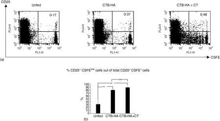
Oral administration of antigens coupled to the B subunit of the cholera toxin (CTB) can dramatically reduce the amount of antigen needed for tolerance induction and has been used in several animal models to suppress conditions where the immune system overreacts to foreign and self-antigens. In this study, the cellular events following oral administration of CTB-coupled antigen was investigated. As a model system, limited numbers of CSFE-labelled cells from influenza haemagglutinin peptide (HApep) T-cell transgenic mice were transferred to wild type mice and the mice were then given CTB-coupled HApep orally. The inductive events of CTB-induced tolerance was characterized by extensive proliferation of HApep-specific T cells in the mesenteric lymph nodes (MLNs) and in the spleen. The proliferating cells up-regulated the gut homing molecule alpha4beta7 and down-regulated the high endothelial venule binding molecule L-selectin. Addition of the whole cholera toxin (CT) to CTB-HApep showed a similar pattern as CTB-HApep feeding, with antigen-specific proliferation in the MLN and spleen and expression of alpha4beta7 on the proliferating cells. However, addition of CT to CTB-HApep, produced a stronger and faster proliferative response and abrogated CTB-HA mediated oral tolerance. Feeding of CTB-HApep expanded CD25+ cells in the MLNs. CTB-induced oral tolerance could, however, not be explained by CD25+ dependent regulatory activity, as oral administration of CTB-HApep to mice depleted of CD25+ cells still gave rise to systemic tolerance. Thus, several mechanisms might co-orchestrate the systemic tolerance seen in response to feeding with CTB-coupled antigen.
Figures


Similar articles
-
Sublingual tolerance induction with antigen conjugated to cholera toxin B subunit induces Foxp3+CD25+CD4+ regulatory T cells and suppresses delayed-type hypersensitivity reactions.Scand J Immunol. 2006 Sep;64(3):251-9. doi: 10.1111/j.1365-3083.2006.01823.x. Scand J Immunol. 2006. PMID: 16918694
-
Induction of antigen-specific regulatory T cells in the liver-draining celiac lymph node following oral antigen administration.Immunology. 2005 Nov;116(3):362-72. doi: 10.1111/j.1365-2567.2005.02236.x. Immunology. 2005. PMID: 16236126 Free PMC article.
-
Regional oral tolerance in transgenic 2C mice.Surgery. 2005 Aug;138(2):141-9. doi: 10.1016/j.surg.2005.05.010. Surgery. 2005. PMID: 16153420
-
ADP-ribosylating bacterial enzymes for the targeted control of mucosal tolerance and immunity.Ann N Y Acad Sci. 2004 Dec;1029:193-208. doi: 10.1196/annals.1309.036. Ann N Y Acad Sci. 2004. PMID: 15681758 Review.
-
Mucosally induced immunological tolerance, regulatory T cells and the adjuvant effect by cholera toxin B subunit.Scand J Immunol. 2010 Jan;71(1):1-11. doi: 10.1111/j.1365-3083.2009.02321.x. Scand J Immunol. 2010. PMID: 20017804 Review.
Cited by
-
Cholera-like enterotoxins and Regulatory T cells.Toxins (Basel). 2010 Jul;2(7):1774-95. doi: 10.3390/toxins2071774. Epub 2010 Jul 6. Toxins (Basel). 2010. PMID: 22069660 Free PMC article. Review.
-
Nasal cardiac myosin peptide treatment and OX40 blockade protect mice from acute and chronic virally-induced myocarditis.J Autoimmun. 2011 May;36(3-4):210-20. doi: 10.1016/j.jaut.2011.01.006. Epub 2011 Feb 17. J Autoimmun. 2011. PMID: 21333491 Free PMC article.
-
Polymeric Nanoparticles as Oral and Intranasal Peptide Vaccine Delivery Systems: The Role of Shape and Conjugation.Vaccines (Basel). 2024 Feb 15;12(2):198. doi: 10.3390/vaccines12020198. Vaccines (Basel). 2024. PMID: 38400181 Free PMC article.
-
Stable production of peptide antigens in transgenic tobacco chloroplasts by fusion to the p53 tetramerisation domain.Transgenic Res. 2010 Aug;19(4):703-9. doi: 10.1007/s11248-009-9348-y. Epub 2009 Dec 2. Transgenic Res. 2010. PMID: 19953346
-
LPS enhances CTB-INSULIN induction of IDO1 and IL-10 synthesis in human dendritic cells.Cell Immunol. 2019 Apr;338:32-42. doi: 10.1016/j.cellimm.2019.03.003. Epub 2019 Mar 19. Cell Immunol. 2019. PMID: 30910218 Free PMC article.
References
-
- Tarkowski A, Sun JB, Holmdahl R, Holmgren J, Czerkinsky C. Treatment of experimental autoimmune arthritis by nasal administration of a type II collagen-cholera toxoid conjugate vaccine. Arthritis Rheum. 1999;42(8):1628–34. - PubMed
-
- Wiedermann U, Jahn-Schmid B, Lindblad M, Rask C, Holmgren J, Kraft D, Ebner C. Suppressive versus stimulatory effects of allergen/cholera toxoid (CTB) conjugates depending on the nature of the allergen in a murine model of type I allergy. Int Immunol. 1999;11(10):1717–24. - PubMed
-
- Rask C, Holmgren J, Fredriksson M, Lindblad M, Nordstrom I, Sun JB, Czerkinsky C. Prolonged oral treatment with low doses of allergen conjugated to cholera toxin B subunit suppresses immunoglobulin E antibody responses in sensitized mice. Clin Exp Allergy. 2000;30(7):1024–32. - PubMed
Publication types
MeSH terms
Substances
LinkOut - more resources
Full Text Sources
Research Materials
