. 2006 Jul;118(3):361-71.
doi: 10.1111/j.1365-2567.2006.02381.x.

The fate of effector CD8 T cells in vivo is controlled by the duration of antigen stimulation

Affiliations

The fate of effector CD8 T cells in vivo is controlled by the duration of antigen stimulation

Xiaopei Huang et al. Immunology. 2006 Jul.

Abstract

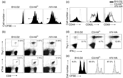
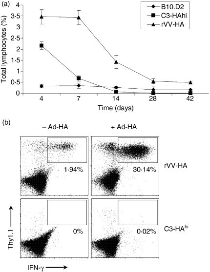
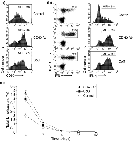
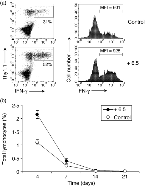
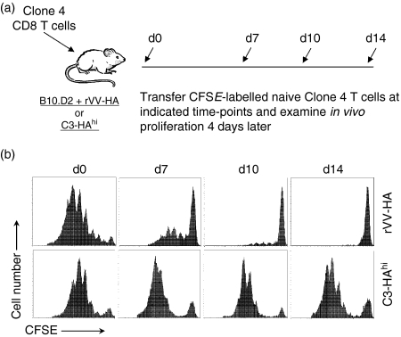
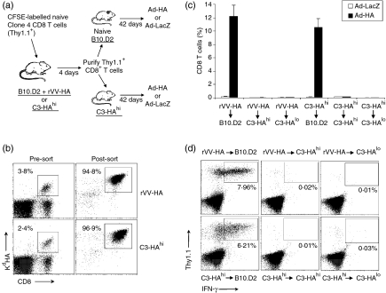
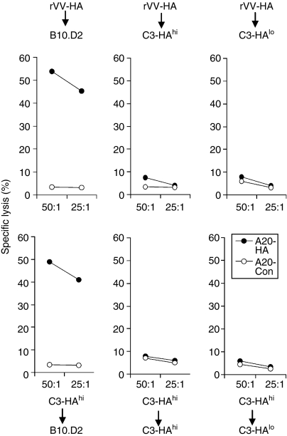
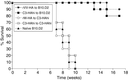
What controls the fate of the T-cell response remains incompletely defined. Gain of effector function facilitated by costimulation has been thought to be a crucial factor in determining the outcome of the T-cell response, i.e. long-term memory in the presence of costimulation versus tolerance induction in the absence of costimulation. In this study, we show that while costimulation or cognate CD4 helps to promote the acquisition of effector function during the initial phase of the CD8 T-cell response, the fate of effector CD8 T cells is controlled by the duration of subsequent antigenic stimulation. Effector CD8 T cells differentiate into memory cells only after clearance of antigen, whereas in the presence of persistent antigen, effector CD8 T cells are tolerized. Furthermore, protective immunity against tumour cannot develop in the persisting antigen environment. These results suggest that removal of persisting antigen by other means might be a prerequisite for effective immunotherapy in cancer.


Figures

Figure 1
Figure 1
Effector CD8 T cells to viral antigen versus self-antigen. Naive HA-specific CD8 T cells were adoptively transferred into non-transgenic B10.D2, C3-HAhi or B10.D2 mice infected with rVV-HA (rVV-HA). After 4 days, lymphocytes from peripheral lymph nodes were harvested for subsequent analyses. (a) In vivo divisions of CFSE-labelled HA-specific T cells. Events were gated on CD8+ Thy-1.1+ T cells. Arrows indicate cells with at least three divisions which were gated for sorting described in Fig. 6. (b) Expansion of HA-specific T cells. Cells were stained with either Thy-1.1 or KdHA tetramer (KdHA518-526) in combination with CD8. Percentage of double-positive cells is indicated. (c) Phenotypic characterization of HA-specific T cells. Cells were stained with activation marker CD44, CD62L or CD69. Events were gated on Thy-1.1+CD8+ T cells. (d) Functional analysis of HA-specific T cells. After in vitro re-stimulation with KdHA peptide (2 µg/ml) for 6 hr, cells were stained with CD8, Thy-1.1 and IFN-γ intracellularly. The percentages of IFN-γ-producing clonotypic CD8 T cells are indicated. Events were gated on CD8+ T cells. (e) In vivo CTL assay. Mice were injected intravenously with an equal number mixture of KdHA peptide-pulsed CFSEhi-labelled (right peak) and control peptide-pulsed CFSElo-labelled (left peak) syngeneic targets. Six hours later, cells from peripheral lymph nodes were examined to detect and quantify CFSE-labelled cells. Percentage of specific lysis is indicated. Representative data of three independent experiments are shown.
Figure 2
Figure 2
Different fates of effector CD8 T cells. Naive HA-specific CD8 T cells were adoptively transferred into non-transgenic B10.D2, C3-HAhi or B10.D2 mice infected with rVV-HA (rVV-HA). (a) The fate of clonotypic T cells. Cells were harvested 4, 7, 14, 28 or 42 days later for the percentage of HA-specific CD8 T cells (CD8+Thy-1.1+ cells ± SD) in total lymphocytes for all mice in each group is indicated. (b) Recall response: 42 days later, mice were challenged with (+ Ad-HA) or without (– Ad-HA) Ad-HA. Seven days after the challenge, cells were harvested for the percentages of IFN-γ-producing clonotypic CD8 T cells by IFN-γ intracellular staining. Events were gated on CD8+ T cells. (c) Representative data of three independent experiments are shown.
Figure 3
Figure 3
Co-stimulation does not dictate the fate of effector CD8 T cells. Naive HA-specific CD8 T cells were adoptively transferred into C3-HAhi mice treated with 10 µg of anti-CD40 antibody (CD40 Ab) or 10 nmol CpG ODN (CpG), or the control rat IgG (Control). After 24 hr, cells were harvested from the peripheral lymph nodes and stained for CD11c and CD80. Data represent the expression of CD80 in CD11c+ DCs with mean fluorescence intensity (MFI) indicated (a). Four days later, lymphocytes from peripheral lymph nodes were harvested for the percentage and MFI of IFN-γ-secreting cells among clonotypic HA-specific CD8 T cells (b). Four, 7, 14, 28 or 42 days later, the percentages of clonotypic HA-specific CD8 T cells in total lymphocytes were analysed (c). Representative data of two independent experiments are shown.
Figure 4
Figure 4
CD4 help does not result in the formation of memory CD8 T cells. Naive HA-specific CD8 T cells were adoptively transferred into C3-HAhi mice alone (Control) or with HA-specific CD4 T cells derived from 6.5 TCR-HA transgenic mice (+ 6.5). Four days later, lymphocytes from peripheral lymph nodes were harvested for the percentage and MFI of IFN-γ-secreting cells among clonotypic HA-specific CD8 T cells (a). Four, 7, 14, or 21 days later, the percentages of clonotypic HA-specific CD8 T cells in total lymphocytes were analysed (b).
Figure 5
Figure 5
Antigen duration in CD8 T-cell memory versus tolerance. (a) A schematic view of the experiments outlined below. (b) Naive HA-specific CD8 T cells were adoptively transferred into B10.D2 mice infected with rVV-HA (rVV-HA) or C3-HAhi mice on day 0. At days 0 (d0), 7 (d7), 10 (d10) or 14 (d14), CFSE-labelled naive HA-specific T cells were transferred into these mice and in vivo divisions of CFSE-labelled cells harvested from the spleen were analysed 4 days later. Events were gated on CD8+ Thy-1.1+ T cells. Representative data of three independent experiments are shown.
Figure 6
Figure 6
Effector CD8 T cells fail to differentiate into memory cells in persisting antigen environment. (a) Purification and transfer of effector CD8 T cells into antigen-free versus antigen-persistent hosts. A schematic view of the adoptive transfer experiments. (b) CFSE-labelled naive HA-specific CD8 T cells were adoptively transferred into B10.D2 mice infected with rVV-HA (rVV-HA) or C3-HAhi mice. Four days later, cells were harvested from peripheral lymph nodes and spleen, and CD8+ Thy-1.1+ T cells were enriched by negative selection using magnetic beads before sorting gated on CD8, Thy-1.1 and at least three divisions by CFSE as shown in Fig. 1(a). Cells were stained with CD8 and KdHA tetramer. The percentage of clonotypic T cells before enrichment and sorting (Pre-sort) and the purity of clonotypic T cells after cell sorting (Post-sort) are indicated. (c) HA-specific effector CD8 T cells purified from rVV-HA infected B10.D2 mice (rVV-HA) were transferred into naive B10.D2 (rVV-HA (r) B10.D2), C3-HAhi (rVV-HA (r) C3-HAhi), or C3-HAlo (rVV-HA (r) C3-HAlo), and those effector cells from C3-HAhi mice were transferred into naive B10.D2 (C3-HAhi (r) B10.D2), C3-HAhi (C3-HAhi (r) C3-HAhi), or C3-HAlo (C3-HAhi (r) C3-HAlo). After 42 days, mice were challenged with Ad-HA or Ad-LacZ; 7 days after the challenge, cells were analysed for the expansion of clonotypic T cells. The percentages of clonotypic T cells (CD8+ Thy-1.1+ cells ± SD) in total CD8 T cells for all mice in each group are indicated. (d) Function of clonotypic CD8 T cells after challenge with Ad-HA were analysed by IFN-γ intracellular staining and the percentages of IFN-γ-producing clonotypic CD8 T cells in total CD8 T cells are indicated. Representative data of three independent experiments are shown.
Figure 7
Figure 7
Ex vivo cytolytic assay. The same clonotypic CD8 T cells harvested from the experiments outlined in Fig. 6 were assayed for their cytolytic activity on A20 lymphoma targets (H-2d) pulsed with Kd-HA peptide (A20-HA) or an irrelevant peptide (A20-Con) by a 6-hr JAM test. Percentage of specific lysis as a function of effector to target ratio is indicated. Representative data of three independent experiments are shown.
Figure 8
Figure 8
Protective immunity against tumour cannot be generated in the persisting antigen environment. Purified HA-specific effector CD8 T cells from rVV-HA infected B10.D2-BALB/c F1 mice were transferred into B10.D2-BALB/c F1 (rVV-HA (r) B10.D2, n = 10) or C3-HAhi-BALB/c F1 (rVV-HA (r) C3-HAhi, n = 10), and those effector cells from C3-HAhi-BALB/c F1 mice were transferred into B10.D2-BALB/c F1 (C3-HAhi (r) B10.D2, n = 10) or C3-HAhi-BALB/c F1 (C3-HAhi (r) C3-HAhi, n = 10). After 42 days, these mice along with naive B10.D2-BALB/c F1 mice (Naive B10.D2, n = 10) were challenged with A20-HA lymphoma intravenously. The graph indicates the percentage of tumour-free survival over time from tumour inoculation. Representative results of two independent experiments are shown.

References

    1. Lafferty KJ, Cunningham AJ. A new analysis of allogeneic interactions. Aust J Exp Biol Med Sci. 1975;53:27–42. - PubMed
    1. Schwartz RH. Models of T cell anergy: is there a common molecular mechanism? J Exp Med. 1996;184:1–8. - PMC - PubMed
    1. Mellman I, Steinman RM. Dendritic cells: specialized and regulated antigen processing machines. Cell. 2001;106:255–8. - PubMed
    1. Matzinger P. The danger model: a renewed sense of self. Science. 2002;296:301–5. - PubMed
    1. Steinman RM, Turley S, Mellman I, Inaba K. The induction of tolerance by dendritic cells that have captured apoptotic cells. J Exp Med. 2000;191:411–16. - PMC - PubMed

Publication types

MeSH terms