. 2006 Jul 7:4:20.
doi: 10.1186/1741-7007-4-20.

Genetic context determines susceptibility to intraocular pressure elevation in a mouse pigmentary glaucoma

Affiliations

Genetic context determines susceptibility to intraocular pressure elevation in a mouse pigmentary glaucoma

Michael G Anderson et al. BMC Biol. .

Abstract

Background: DBA/2J (D2) mice develop an age-related form of glaucoma. Their eyes progressively develop iris pigment dispersion and iris atrophy followed by increased intraocular pressure (IOP) and glaucomatous optic nerve damage. Mutant alleles of the Gpnmb and Tyrp1 genes are necessary for the iris disease, but it is unknown whether alleles of other D2 gene(s) are necessary for the distinct later stages of disease. We initiated a study of congenic strains to further define the genetic requirements and disease mechanisms of the D2 glaucoma.

Results: To further understand D2 glaucoma, we created congenic strains of mice on the C57BL/6J (B6) genetic background. B6 double-congenic mice carrying D2-derived Gpnmb and Tyrp1 mutations develop a D2-like iris disease. B6 single-congenics with only the Gpnmb and Tyrp1 mutations develop milder forms of iris disease. Genetic epistasis experiments introducing a B6 tyrosinase mutation into the congenic strains demonstrated that both the single and double-congenic iris diseases are rescued by interruption of melanin synthesis. Importantly, our experiments analyzing mice at ages up to 27 months indicate that the B6 double-congenic mice are much less prone to IOP elevation and glaucoma than are D2 mice.

Conclusion: As demonstrated here, the Gpnmb and Tyrp1 iris phenotypes are both individually dependent on tyrosinase function. These results support involvement of abnormal melanosomal events in the diseases caused by each gene. In the context of the inbred D2 mouse strain, the glaucoma phenotype is clearly influenced by more genes than just Gpnmb and Tyrp1. Despite the outward similarity of pigment-dispersing iris disease between D2 and the B6 double-congenic mice, the congenic mice are much less susceptible to developing high IOP and glaucoma. These new congenic strains provide a valuable new resource for further studying the genetic and mechanistic complexity of this form of glaucoma.


Figures

Figure 1
Figure 1
The B6 genetic background is permissive to iris phenotypes individually caused by Tyrp1b and GpnmbR150X mutations. (A) The Tyrp1b and (B) the GpnmbR150X congenic intervals on chromosome 4 and chromosome 6. After 10 generations of backcrossing, the selected D2-derived intervals (filled red box), as well as a limited amount of flanking DNA (empty red box), were present in an essentially B6-derived background (thin blue line). The chromosome 4 interval is delimited proximally by D4Mit214-D4Mit151 and distally by D4Mit185-D4Mit146; the chromosome 6 interval is delimited proximally by D6Mit268-D6Mit207 and distally by D6Mit277-D6Mit16. B6.Tyrp1b irides at (C) 4 months, (D) 9 months, and (E) 20 months. From 1–6 months, B6.Tyrp1b irides were indistinguishable from wild-type B6. Thereafter, B6.Tyrp1b eyes were characterized by a gradual atrophy of iris stromal morphology. Beginning at 6 months, a population of small phagocytic clump cells began to be discernible across the iris surface. With age, the underlying vasculature became obscured, and irides appeared increasingly coarse and atrophic, particularly at the pupil margin where a narrow white band of underlying tissue was exposed. With advanced age, full-thickness iris holes occurred, but rarely before 2 years of age (n = 16 eyes at 1–6 months, 20 eyes at 7–11 months, and 42 eyes at 12+ months). B6.GpnmbR150X irides at (F) 4 months, (G) 9 months, and (H) 20 months. From 1–5 months, B6.GpnmbR150X irides were indistinguishable from wild-type B6. Near 6 months, B6.GpnmbR150X eyes developed a pronounced peripupillary swelling accompanied by pronounced accumulation of clump cells on the iris surface. These swellings remained prominent to the oldest ages examined. The iris surface maintained an overall normal morphology during the first year, after which time it became increasingly atrophic, and accumulations of dispersed pigment were visible on the lens and cornea. No sex-specific differences were evident in the phenotypes of either strain. (n = 16 eyes at 1–6 months, 20 eyes at 7–11 months, and 46 eyes at 12+ months).
Figure 2
Figure 2
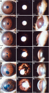
Severe iris disease in double-congenic B6. Tyrp1b GpnmbR150X mice. Cohorts of B6.Tyrp1isa GpnmbR150X mice were aged and analyzed by slit-lamp examination; representative eyes of indicated ages are shown. Each row contains three images of the same eye. The left column shows broad-beam illumination. The middle column shows transillumination defects. The right column shows the relative dimensions of the anterior chamber. The degree of pigment dispersion and iris atrophy is remarkably similar in both timing and severity to that of D2 mice (see reference [19] for comparable image of D2 eyes). (A to C) Until 5 months, B6.Tyrp1bGpnmbR150X eyes were indistinguishable from wild-type, with a complex iris morphology, no transillumination, and anterior chambers of normal dimension with a closely juxtaposed cornea and iris. (D to F) By 6 months, all B6.Tyrp1b GpnmbR150X eyes exhibit a clear phenotype characterized by slight swelling of peripupillary tissue. This timing and phenotype closely resembles the initial stages of the D2 iris disease. (G to I) In 9-month-old eyes, the peripupillary region becomes notably atrophic, transillumination is obvious, and dispersed pigment is present on both the lens and cornea. Beyond this age, a steadily worsening course ensues, which at (J to L) 12 months, (M to O) 14 months, and (P to R) 18 months is characterized by increasing degrees of iris atrophy that include full-thickness iris holes, profound transillumination, pigment dispersion and frequent pigment accumulation on the lens and cornea, and changes to the dimensions of the anterior chamber. No sex-specific differences were evident in these phenotypes. This synopsis of disease progression involved >146 eyes aged 2–20+ months, with each of the cohorts described above involving groups of at least 14 eyes.
Figure 3
Figure 3
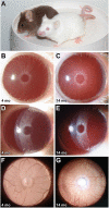
Tyrosinase deficiency prevents iris disease in B6. Tyrp1b GpnmbR150X mice. Intercrosses generated mice homozygous for various genotypic combinations of the Tyrp1b and GpnmbR150X mutations with the albino-inducing Tyrc-2J mutation. These mice were aged and analyzed by slit-lamp examination; representative B6.Tyrp1b GpnmbR150X eyes are shown. (A) The normally brown pigmented coat of a mouse on the left compared with a triple homozygous Tyrc-2J Tyrp1b GpnmbR150X mouse on the right. (B, D, F) Different views emphasize the clinical morphology of the albino iris. Eyes of B6 mice that are homozygous for the Tyrc-2J mutation only appear pink as they lack melanin, but otherwise the iris and its vasculature have normal morphology. (C, E, G) Homozygosity for Tyrc-2J completely prevents iris disease in B6.Tyrp1b GpnmbR150X Tyrc-2J mice. The iris morphology of the triple mutant is indistinguishable from that of B6 mice homozygous for Tyrc-2J only (compare 3B to 3C), lacks peripupillary abnormalities (compare 3D to 3E), and has a healthy uninterrupted vasculature (3F to 3G). n = 14 eyes, all 12+ months.
Figure 4
Figure 4
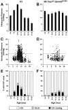
B6.Tyrp1b GpnmbR150X mice are relatively resistant to IOP elevation. Graphs of IOP and age for D2 mice (left column) versus B6.Tyrp1b GpnmbR150X mice (right column), with the same data represented as: (A and B) bar graphs of mean IOP +/- SEM (>30 mice per bin), (C and D) scatter plots, and (E and F) plots showing percent of mice with IOP of indicated values. IOP was recorded from a total of 336 B6.Tyrp1b GpnmbR150X mice and 1437 D2 mice. All groups contained >30 mice and included both male and female mice.
Figure 5
Figure 5
B6. Tyrp1b GpnmbR150X mice have optic nerve phenotypes similar to standard B6 mice. Optic nerve cross-sections were stained by the PPD method, which darkly stains the myelin sheath of all axons and the axoplasm only of diseased or dying axons. The majority of optic nerves had only the mild degree of damage that is typical for aged mice, as shown here in a comparison of optic nerves from: (A) typical 22-month-old B6, versus (B) typical 22-month-old B6.Tyrp1b GpnmbR150X. This was true even in mice with the highest IOPs recorded in the study, as shown here with (C) 14-month-old B6.Tyrp1b GpnmbR150X and (D) 22-month-old B6.Tyrp1b GpnmbR150X. Scale bar = 10 μm. (E and F) The overall distribution of nerve damage was almost identical in both standard B6 and B6.Tyrp1b GpnmbR150X mice. Although some mice of each genotype developed a "moderate" degree of damage with age, the degree of damage in these mice only just met inclusion criteria for the "moderate" level and never involved significant axon loss. It was, therefore, much milder in all of these "moderate" mice than is typical of "moderate" D2 mice [42, 43]. A single B6.Tyrp1b GpnmbR150X nerve had severe damage. The similar damage distributions of both standard B6 and B6.Tyrp1b GpnmbR150X mice indicates that the damage reflects age-related B6 changes that are not related to Gpnmb and Tyrp1 glaucoma. For each group, the number of optic nerves graded for B6 and B6.Tyrp1bGpnmbR150X were: (4–6 months) 10 and 8, (11–12 months) 19 and 22, (13–15 months) 29 and 38, (16–19 months) 28 and 28, (20–26 months) 24 and 17. (E) Quantitative axon counts (mean ± SEM) show no detectable axon loss in B6.Tyrp1b GpnmbR150X compared with standard B6 mice. The similar damage distributions and lack of axon loss in both strains indicates that the detected damage reflects age-related B6 changes that are not related to Gpnmb and Tyrp1 glaucoma. For each group, the number of randomly selected nerves utilized in quantitative axon counting for B6 and B6.Tyrp1bGpnmbR150X were: (4- months) 6 and 6, (22–27 months) 8 and 10.

Similar articles

Cited by

References

    1. Allingham RR, Damji KF, Freedman S, Moroi SE, Shafranov G, Shields MB. Shields' Textbook of Glaucoma. 5. Philadelphia: Lippincott Williams and Wilkins;; 2004.
    1. Nickells RW, Jampel HD, Zack DJ. Glaucoma. In: Rimoin DL, Conner MJ, Pyeritz RE, Korf BR, editor. Emery & Rimoins Principles and Practices of Medical Genetics. 4. Vol. 3. Churchill Livingstone; 2002. pp. 3491–3512.
    1. Ritch R, Shields MB, Krupin T. The Glaucomas. St Louis Mosby; 1996.
    1. Palmberg PF, Wiggs JL. Mechanisms of glaucoma. In: Yanoff M, Duker JS, editor. Ophthalmology. 2. St Louis: Mosby; 2004.
    1. Weinreb RN, Khaw PT. Primary open-angle glaucoma. Lancet. 2004;363:1711–1720. doi: 10.1016/S0140-6736(04)16257-0. - DOI - PubMed

Publication types