

Source memory retrieval is affected by aging and prefrontal lesions: behavioral and ERP evidence
- PMID: 16828722
- PMCID: PMC2365725
- DOI: 10.1016/j.brainres.2006.06.013
Source memory retrieval is affected by aging and prefrontal lesions: behavioral and ERP evidence
Abstract
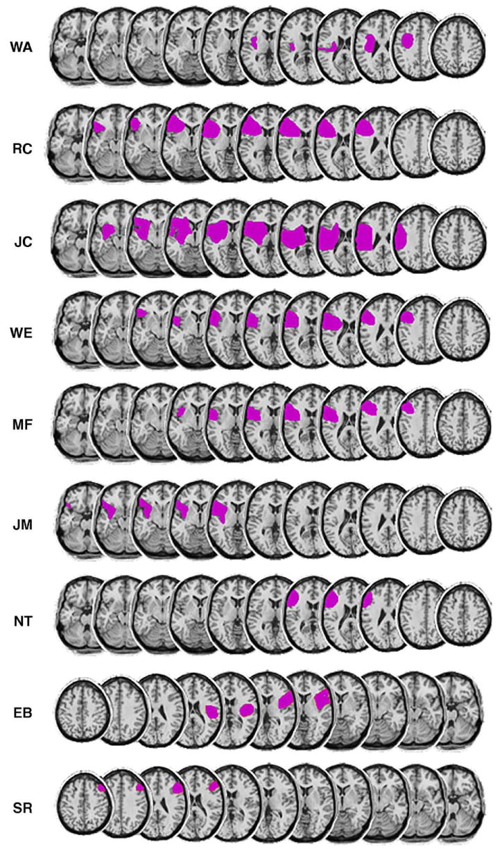
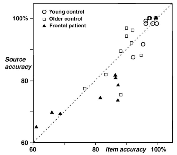
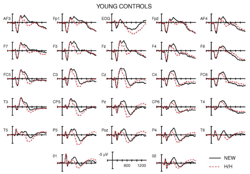
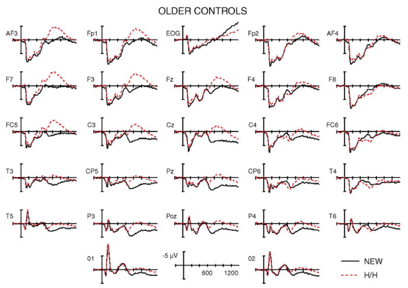
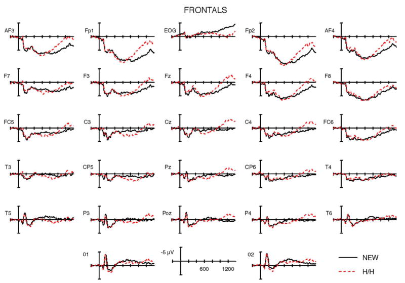
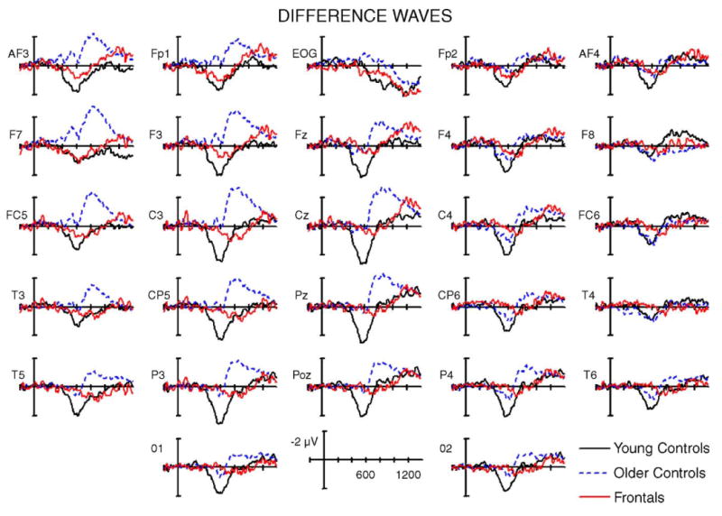
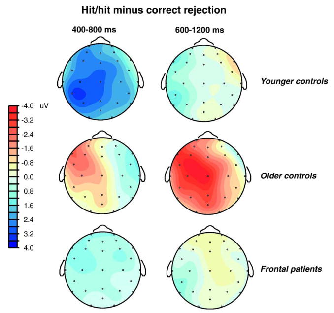
Age-related deficits in source memory have been attributed to alterations in prefrontal cortex (PFC) function, but little is known about the neural basis of such changes. The present study examined the time course of item and source memory retrieval by recording event-related potentials (ERPs) in patients with focal lesions in lateral PFC and in healthy older and young controls. Both normal aging and PFC lesions were associated with decrements in item and source memory. However, older controls showed a decrease in item hit rate with no change in false alarms, whereas patients showed the opposite pattern. Furthermore, ERPs revealed notable differences between the groups. The early positive-going old/new effect was prominent in the young but reduced in patients and older adults, who did not differ from each other. In contrast, older adults displayed a prominent left frontal negativity (600-1200 ms) not observed in the young. This left frontal effect was substantially smaller and delayed in the patients. The current results provide novel insights into the effects of aging on source memory and the role of the lateral PFC in these processes. Older controls appeared to adopt alternate memory strategies and to recruit compensatory mechanisms in left PFC to support task performance. In contrast, the lateral frontal patients were unable to use these mechanisms, thus exhibiting difficulties with strategic memory and monitoring processes.
Figures
Similar articles
-
Individual differences in executive functioning modulate age effects on the ERP correlates of retrieval success.Neuropsychologia. 2010 Oct;48(12):3540-53. doi: 10.1016/j.neuropsychologia.2010.08.003. Epub 2010 Aug 13. Neuropsychologia. 2010. PMID: 20709089
-
Neural correlates of source memory retrieval in young, middle-aged and elderly adults.Biol Psychol. 2012 Apr;90(1):33-49. doi: 10.1016/j.biopsycho.2012.02.004. Epub 2012 Feb 17. Biol Psychol. 2012. PMID: 22366225
-
Age-related deficits in selective attention during encoding increase demands on episodic reconstruction during context retrieval: An ERP study.Neuropsychologia. 2016 Jun;86:66-79. doi: 10.1016/j.neuropsychologia.2016.04.009. Epub 2016 Apr 16. Neuropsychologia. 2016. PMID: 27094851 Free PMC article.
-
Age differences in the neural correlates of novelty processing: The effects of item-relatedness.Brain Res. 2015 Jul 1;1612:2-15. doi: 10.1016/j.brainres.2014.08.006. Epub 2014 Aug 19. Brain Res. 2015. PMID: 25149192 Review.
-
Association between prefrontal activity and volume change in prefrontal and medial temporal lobes in aging and dementia: a review.Ageing Res Rev. 2013 Mar;12(2):479-89. doi: 10.1016/j.arr.2012.11.001. Epub 2012 Nov 24. Ageing Res Rev. 2013. PMID: 23183352 Review.
Cited by
-
Do young and older adults rely on different processes in source memory tasks? A neuropsychological study.J Exp Psychol Learn Mem Cogn. 2008 Jul;34(4):809-22. doi: 10.1037/0278-7393.34.4.809. J Exp Psychol Learn Mem Cogn. 2008. PMID: 18605870 Free PMC article.
-
Compensatory effects of pointing and predictive cueing on age-related declines in visuospatial working memory.Mem Cognit. 2016 Aug;44(6):950-65. doi: 10.3758/s13421-016-0611-1. Mem Cognit. 2016. PMID: 27126873 Free PMC article.
-
The cognitive aging of episodic memory: a view based on the event-related brain potential.Front Behav Neurosci. 2013 Aug 26;7:111. doi: 10.3389/fnbeh.2013.00111. eCollection 2013. Front Behav Neurosci. 2013. PMID: 23986668 Free PMC article.
-
Effects of age on the neural correlates of retrieval cue processing are modulated by task demands.J Cogn Neurosci. 2009 Jan;21(1):1-17. doi: 10.1162/jocn.2009.21001. J Cogn Neurosci. 2009. PMID: 18476757 Free PMC article.
-
Orbito-frontal cortex is necessary for temporal context memory.J Cogn Neurosci. 2010 Aug;22(8):1819-31. doi: 10.1162/jocn.2009.21316. J Cogn Neurosci. 2010. PMID: 19642880 Free PMC article.
References
-
- Alexander MP, Stuss DT, Fansabedian N. California Verbal Learning Test: performance by patients with focal frontal and non-frontal lesions. Brain. 2003;126:1493–1503. - PubMed
-
- Buckner RL, Tulving E. Neuroimaging studies of memory: theory and recent PET results. In: Grafman J, editor. Handbook of Neuropsychology. Vol. 10. Elsevier; Amsterdam: 1995. pp. 439–466.
-
- Butters MA, Kaszniak AW, Glisky EL, Eslinger PJ, Schacter DL. Recency discrimination deficits in frontal lobe patients. Neuropsychology. 1994;8:343–353.
-
- Baldo JV, Delis D, Kramer J, Shimamura AP. Memory performance on the California Verbal Learning Test—II: findings from patients with focal frontal lesions. J Int Neuropsychol Soc. 2002;8:539–546. - PubMed
-
- Cabeza R, Anderson ND, Locantore JK, McIntosh AR. Aging gracefully: compensatory brain activity in high-performing older adults. NeuroImage. 2002;17:1394–1402. - PubMed
Publication types
MeSH terms
Grants and funding
LinkOut - more resources
Full Text Sources
Medical
Miscellaneous
