

Current staging of hepatocellular carcinoma: imaging implications
- PMID: 16829469
- PMCID: PMC1693764
- DOI: 10.1102/1470-7330.2006.0014
Current staging of hepatocellular carcinoma: imaging implications
Abstract
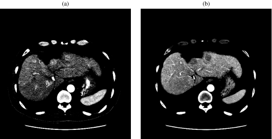
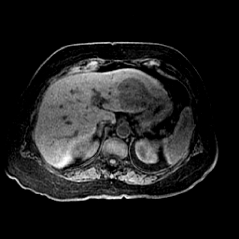
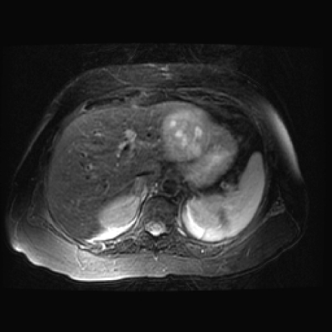
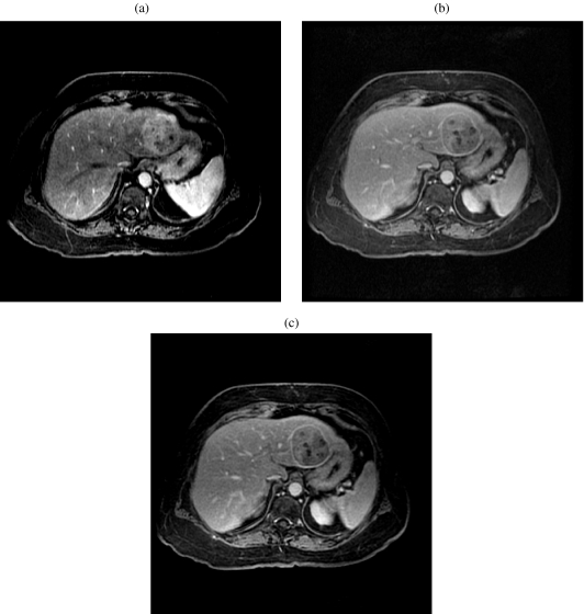
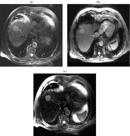
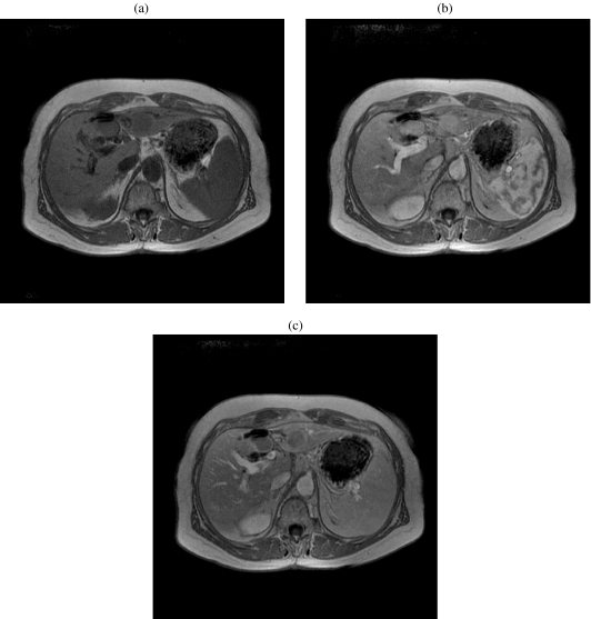
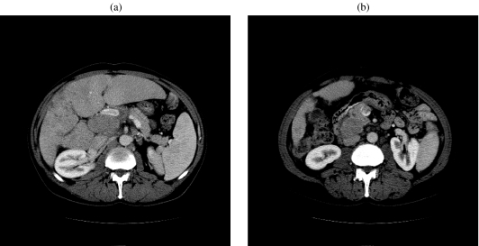
The incidence of hepatocellular carcinoma has been rising in the USA in the past two decades. Hepatocellular carcinoma primarily affects older people and reaches its highest prevalence among those aged between 50 and 70 years. Chronic infection by the hepatitis B virus is the most common cause of this disease. Since hepatocellular carcinoma is an indolent tumor, it has a low life expectancy. In patients with suspected hepatocellular carcinoma, CT, MRI, and ultrasound techniques are useful for formulating the diagnosis based on vascularity and specific enhancement features. In this paper we will discuss the multimodal approach for diagnosis and surveillance of hepatocellular carcinoma. We will also furnish the latest staging and treatment, epidemiology, clinical presentation, pathology and laboratory findings in hepatocellular carcinoma.
Figures
Similar articles
-
Imaging in the diagnosis, staging, treatment, and surveillance of hepatocellular carcinoma.AJR Am J Roentgenol. 2003 Feb;180(2):441-54. doi: 10.2214/ajr.180.2.1800441. AJR Am J Roentgenol. 2003. PMID: 12540450 No abstract available.
-
Imaging diagnosis and staging of hepatocellular carcinoma.Liver Transpl. 2011 Oct;17 Suppl 2:S34-43. doi: 10.1002/lt.22369. Liver Transpl. 2011. PMID: 21739567 Review. No abstract available.
-
[Imaging diagnosis of hepatocellular carcinomas].Gan To Kagaku Ryoho. 1989 Jan;16(1):25-33. Gan To Kagaku Ryoho. 1989. PMID: 2536268 Japanese.
-
CT and MRI of hepatocellular carcinoma: an update.Expert Rev Anticancer Ther. 2010 Apr;10(4):507-19. doi: 10.1586/era.10.24. Expert Rev Anticancer Ther. 2010. PMID: 20397916 Review.
-
[Medical imaging investigation and project of hepatocellular carcinoma].Zhonghua Gan Zang Bing Za Zhi. 2003 Sep;11(9):517-8. Zhonghua Gan Zang Bing Za Zhi. 2003. PMID: 14552706 Chinese. No abstract available.
Cited by
-
Feasibility analysis of arterial CT radiomics model to predict the risk of local and metastatic recurrence after radical cystectomy for bladder cancer.Discov Oncol. 2024 Feb 19;15(1):40. doi: 10.1007/s12672-024-00880-x. Discov Oncol. 2024. PMID: 38369583 Free PMC article.
-
Combined treatment of vitamin K2 and angiotensin-converting enzyme inhibitor ameliorates hepatic dysplastic nodule in a patient with liver cirrhosis.World J Gastroenterol. 2007 Jun 21;13(23):3259-61. doi: 10.3748/wjg.v13.i23.3259. World J Gastroenterol. 2007. PMID: 17589909 Free PMC article.
-
Diagnostic and Prognostic Role of Serum Interleukin-6 in Malignant Transformation of Liver Cirrhosis.Euroasian J Hepatogastroenterol. 2018 Jan-Jun;8(1):23-30. doi: 10.5005/jp-journals-10018-1253. Epub 2018 May 1. Euroasian J Hepatogastroenterol. 2018. PMID: 29963457 Free PMC article.
-
Guide for diagnosis and treatment of hepatocellular carcinoma.World J Hepatol. 2015 Jun 28;7(12):1632-51. doi: 10.4254/wjh.v7.i12.1632. World J Hepatol. 2015. PMID: 26140083 Free PMC article. Review.
References
-
- Clark HP, Carson WF, Kavanagh PV, Ho CP, Shen P, Zagoria RJ. Staging and current treatment of hepatocellular carcinoma. Radiographics. 2005;25(1):S3–S23. - PubMed
-
- Merle P. Epidemiology, natural history and pathogenesis of hepatocellular carcinoma. Cancer Radiother. 2005;9:452–7. - PubMed
-
- Lai EC, Lau WY. The continuing challenge of hepatic cancer in Asia. Surgeon. 2005;3:210–15. - PubMed
-
- Oral contraceptives and liver cancer: results of the Multicentre International Liver Tumor Study (MILTS) Contraception. 1997;56:275–84. - PubMed
-
- Pawlik TM, Delman KA, Vauthey JN, Nagorney DM, Ng IO, Ikai I, et al. Tumor size predicts vascular invasion and histologic grade: Implications for selection of surgical treatment for hepatocellular carcinoma. Liver Transpl. 2005;11:1086–92. - PubMed
Publication types
MeSH terms
LinkOut - more resources
Full Text Sources
Medical
Miscellaneous