. 2006 Sep 8;281(36):26370-81.
doi: 10.1074/jbc.M603093200. Epub 2006 Jul 7.

The C-terminal zinc finger of UvrA does not bind DNA directly but regulates damage-specific DNA binding

Affiliations

The C-terminal zinc finger of UvrA does not bind DNA directly but regulates damage-specific DNA binding

Deborah L Croteau et al. J Biol Chem. .

Abstract

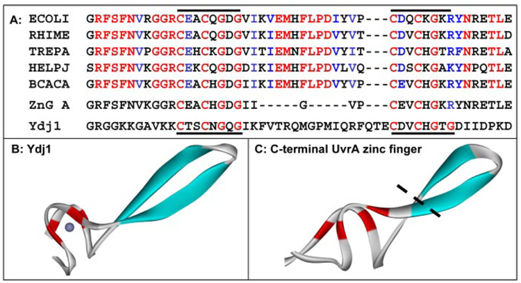
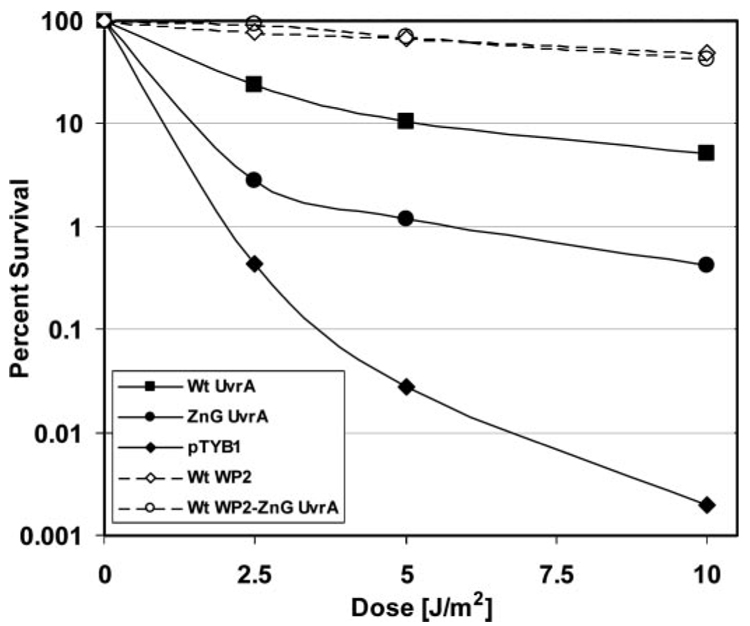
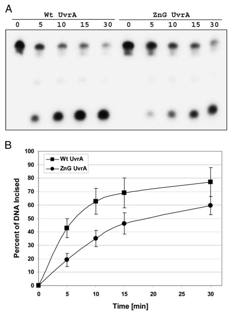
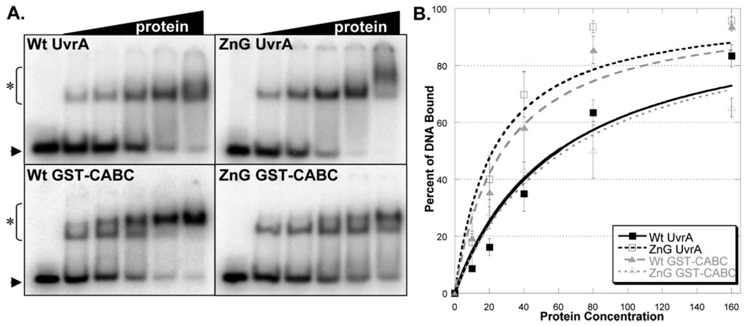
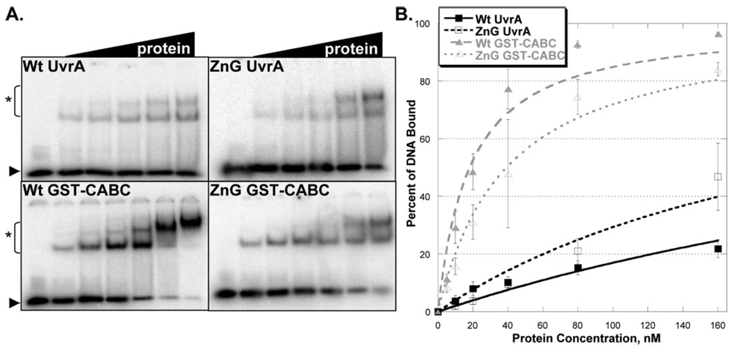
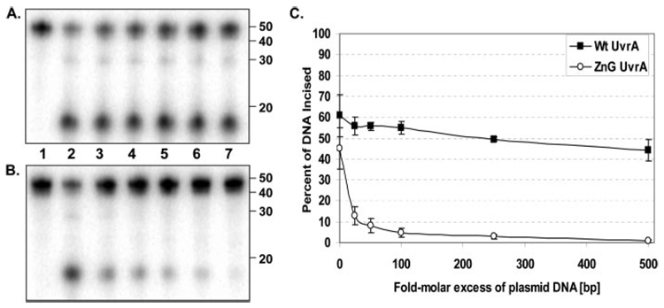
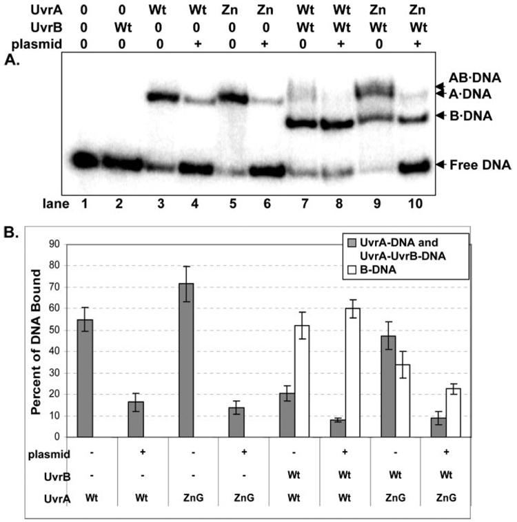
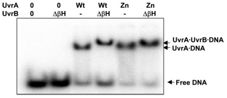
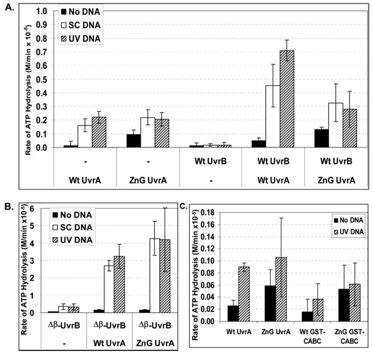
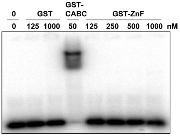
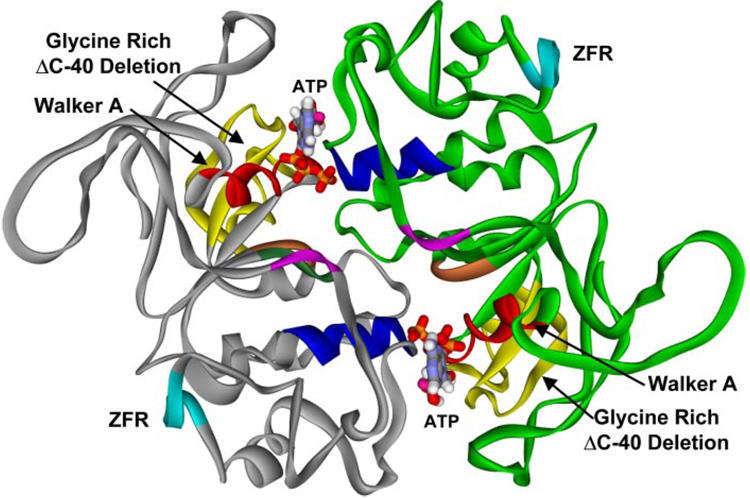
In prokaryotic nucleotide excision repair, UvrA recognizes DNA perturbations and recruits UvrB for the recognition and processing steps in the reaction. One of the most remarkable aspects of UvrA is that it can recognize a wide range of DNA lesions that differ in chemistry and structure. However, how UvrA interacts with DNA is unknown. To examine the role that the UvrA C-terminal zinc finger domain plays in DNA binding, an eleven amino acid deletion was constructed (ZnG UvrA). Biochemical characterization of the ZnG UvrA protein was carried out using UvrABC DNA incision, DNA binding and ATPase assays. Although ZnG UvrA was able to bind dsDNA slightly better than wild-type UvrA, the ZnG UvrA mutant only supported 50-75% of wild type incision. Surprisingly, the ZnG UvrA mutant, while retaining its ability to bind dsDNA, did not support damage-specific binding. Furthermore, this mutant protein only provided 10% of wild-type Bca UvrA complementation for UV survival of an uvrA deletion strain. In addition, ZnG UvrA failed to stimulate the UvrB DNA damage-associated ATPase activity. Electrophoretic mobility shift analysis was used to monitor UvrB loading onto damaged DNA with wild-type UvrA or ZnG UvrA. The ZnG UvrA protein showed a 30-60% reduction in UvrB loading as compared with the amount of UvrB loaded by wild-type UvrA. These data demonstrate that the C-terminal zinc finger of UvrA is required for regulation of damage-specific DNA binding.


Figures

FIGURE 1
FIGURE 1. Sequence alignment and homology modeling of the C-terminal zinc finger of UvrA
A, sequence alignment of the C-terminal zinc finger found in five UvrA homologues. Color coding of the alignment is based on the homology among 22 UvrA proteins in the seed alignment for UvrA within the HAMAP project (record MF_00205, Ref. 45). Red indicates at least 95% conservation; blue denotes that the amino acid is functionally conserved (K/R, D/E, F/Y, M/V/I/L) in 95% of 22 homologues. The red letters within ZnG UvrA and Ydj1 and the black bars denote the position of the conserved motif. The Entrez protein accession numbers are as follows: E. coli UvrA, (ECOLI) P0A698; Rhizobium meliloti UvrA, (RHIME) P56899; Treponema pallidum UvrA, (TREPA) O83527; Helicobacter pylori J99 UvrA, (HELPJ) Q9ZLD6, and B. caldotenax UvrA,(BCACA) AAK29748. B, crystal structure of the ZnII domain of Ydj1, the yeast dnaJ homologue is shown (PDB accession code 1NLT). C, model of the C-terminal zinc finger of Bca UvrA containing 35 residues. The black dotted line represents where the zinc finger would be truncated in the ZnG UvrA mutant. The protein images were generated with DS ViewerPro (Accelrys).
FIGURE 2
FIGURE 2. UV survival of E. coli WP2 strains
WP2 (trp, uvrA) cells were transformed with pT7pol26, a plasmid encoding an IPTG-inducible T7 polymerase, and pTYB1 or pTYB1-Wt UvrA or pTYB1-ZnG UvrA. In addition, WP2 (trp) cells with endogenous UvrA were transformed with pT7pol26 and pTYB1 or pTYB1-ZnG UvrA. After individual colonies were selected and grown to an A600 of ~1.0, the cell culture was diluted 2-fold, and the proteins were induced by addition of IPTG (0.1 mm) for 1 h at 37 °C. Serial dilutions of each sample of culture (100 µl) was then spread onto a nutrient rich-media and UV-irradiated with a 254-nm germicidal light source. The numbers of colonies visible after 20 h of growth at 37 °C was recorded, and the fraction of cells surviving after each dose of UV was calculated based on the plating efficiency of the unirradiated controls. The mean of two or three independent experiments is reported. Solid lines indicate the WP2 (trp, uvrA) strain and dashed lines indicate the wild-type WP2 (trp) strain. Transformants contained pT7pol26 and pTYB1 (diamonds), or pTYB1-Wt UvrA (squares), or pTYB1-ZnG UvrA (circles).
FIGURE 3
FIGURE 3. ZnG UvrA supports reduced incision activity
A, incision of the 5′-end-labeled substrate (F2650/NDB) was monitored over time. The fluorescein adducted 50-bp duplex (F2650/NDB) was incubated with UvrB (100 nm), UvrC (50 nm) and 20 nm of the indicated UvrA protein for varying times at 55 °C in reaction buffer. The reactions were terminated with stop buffer, and the incision products were analyzed on a 10% denaturing polyacrylamide gel. B, graphic representation of the incision activity at various times using the indicated UvrA proteins. Data are reported as the mean ± S.D. (n = 4).
FIGURE 4
FIGURE 4. Damaged DNA binding profiles in the presence of ATP and magnesium
A, EMSA was used to monitor the DNA binding properties of the proteins. Increasing amounts of protein (10–160 nm) were incubated with 2 nm NDB/F2650 duplex DNA in reaction buffer containing ATP (1 mm) and MgCl2 (10 mm) for 15 min at 37 °C. The reaction mixtures were separated on 3.5% polyacrylamide native gels in the presence of 1 mm ATP and 10 mm MgCl2. Asterisk (*) denotes protein-DNA complexes, and arrow denotes migration of free DNA. The data are reported as the mean ± S.D. (n = 3). B, quantitation of EMSAs in A. Binding isotherms were fitted by nonlinear regression analysis using Kaleidegraph® and the method of Schofield (17).
FIGURE 5
FIGURE 5. Damaged DNA binding profiles in the absence of ATP and magnesium
A, EMSAs contained an increasing amount of protein (5–160 nm) and 2 nm NDB/F2650 duplex DNA in reaction buffer without ATP and MgCl2. Proteins were allowed to incubate with DNA for 15 min at 37 °C then were loaded onto a 3.5% native polyacrylamide gel. Asterisk (*) denotes protein-DNA complexes and arrow denotes migration of free DNA. The data are reported as the mean ± S.D. (n = 3). B, quantitation of EMSAs in A. Binding isotherms were fitted by nonlinear regression analysis using Kaleidegraph® and the method of Schofield (17).
FIGURE 6
FIGURE 6. Excess plasmid DNA inhibits incision assays initiated by ZnG UvrA
A, Wt UvrA (20 nm) or B, ZnG UvrA (20 nm) was incubated with increasing concentrations of pUC19 DNA (25–500 molar excess in bp DNA, relative to oligonucleotide concentration) for 10 min at room temperature prior to addition of 2 nm 5′ end-labeled "damaged" oligonucleotide duplex (F2650/NDB), UvrB (100 nm), and UvrC (50 nm) in reaction buffer. Proteins were incubated for 30 min at 55 °C then the reactions were terminated with stop buffer, and the incision products were analyzed on a 10% denaturing polyacrylamide gel. C, graphic representation of the incision activity with various concentrations of excess plasmid DNA. Data are reported as the mean ± S.D. (n = 3).
FIGURE 7
FIGURE 7. Reduced loading of UvrB onto sites of DNA damage by ZnG UvrA
A, Wt UvrA (Wt, 20 nm) or ZnG UvrA (Zn, 20 nm) were incubated alone or with Wt UvrB protein (100 nm) for 30 min at 55 °C in the presence of 2 nm F2650/NDB duplex DNA. The protein-DNA complexes were separated on 4% native polyacrylamide gels containing ATP (1 mm) and MgCl2 (10 mm). B, quantitation of EMSAs in A, reporting the percent of DNA bound to the various protein-DNA complexes. The data are reported as the mean ± S.D. (n = 3). Gray bars indicate the percentage of DNA bound to UvrA either as the A2·DNA or AB·DNA complex, while white bars represent the B·DNA complexes.
FIGURE 8
FIGURE 8. ZnG UvrA forms normal protein-protein interactions with Δβ-Hairpin UvrB
Wt UvrA (20 nm) or ZnG UvrA (20 nm) were incubated alone or with Δβ-Hairpin UvrB (ΔβH, 100 nm) for 15 min at 55 °C in the presence of 2 nm NDB/F2650 duplex DNA in reaction buffer. The protein-DNA complexes were separated on 4% native polyacrylamide gels containing ATP (1 mm) and MgCl2 (10 mm). Representative gel (n = 3).
FIGURE 9
FIGURE 9. ZnG UvrA fails to unlock UvrB's crytpic ATPase
A, conversion of ATP to ADP by Wt UvrA (50 nm), ZnG UvrA (50 nm), and Wt UvrB (100 nm) at 55 °C was monitored using a coupled enzyme assay system consisting of pyruvate kinase and lactic dehydrogenase, which links the hydrolysis of ATP to the oxidation of NADH (see "Experimental Procedures"). The data are reported as the mean ± S.D. (n = 3 or 4). B, ATPase activity of Wt UvrA or ZnG UvrA (50 nm) in the presence of Δβ-Hairpin UvrB (100 nm) was assayed at 55 °C. The data are reported as the mean ± S.D. (n = 3) C, ATPase activity of Wt UvrA, ZnG UvrA, Wt GST-CABC, or ZnG GST-CABC (50 nm) was examined at 37 °C. Data are reported as the mean and range, n = 2. Black bar indicates ATPase assayed in the absence of DNA (No DNA), white bar indicates addition of supercoiled plasmid DNA (SC DNA, 10 ng/µl) and hatched bar indicated addition of 10 ng/µl UV-irradiated plasmid DNA.
FIGURE 10
FIGURE 10. No apparent DNA binding by GST-ZnF
GST (125 nm or 1 µm), GST-CABC (50 nm) or increasing concentrations of GST-ZnF (125 nm to 1 µm) were incubated with F2650/NDB in reaction buffer for 15 min at 37 °C. The samples were then loaded onto a 4% native polyacrylamide gel, and electrophoresis was carried out for 1 h at 100 V.
FIGURE 11
FIGURE 11. Structural model of the B. caldotenax UvrA dimer
A model of the UvrA dimer was created based on the structural similarity between the ABC ATPase domains of UvrA and Rad50 (PDB codes: 1F2T, 1F2U). The dimer consists of two monomers, one in gray and the other in green. The ABC ATPase motifs: the Walker A (red), signature sequence (blue), Q-loop (purple), Walker B (dark green), and His loop (orange) are shown. The zinc finger region, containing 88 amino acids, has been deleted in the model, and six alanines were substituted (pale blue, ZFR). The position of the glycine-rich ΔC-40 deletion is depicted in yellow.

Similar articles

Cited by

References

    1. Van Houten B, Croteau DL, DellaVecchia MJ, Wang H, Kisker C. Mutat. Res. 2005;577:92–117. - PubMed
    1. Truglio JJ, Croteau DL, Van Houten B, Kisker C. Chem. Rev. 2006;106:233–252. - PubMed
    1. Croteau DL, Dellavecchia MJ, Skorvaga M, Van Houten B. In: DNA Damage Recognition. Wolfram S, Kow YW, Doetsch PW, editors. Boca Raton, FL: Taylor & Francis; 2005. pp. 111–138.
    1. Reardon JT, Sancar A. Cell Cycle. 2004;3:141–144. - PubMed
    1. Doolittle RF, Johnson MS, Husain I, Van Houten B, Thomas DC, Sancar A. Nature. 1986;323:451–453. - PubMed

Publication types

MeSH terms

LinkOut - more resources