The paperclip triplex: understanding the role of apex residues in tight turns
- PMID: 16829568
- PMCID: PMC1562401
- DOI: 10.1529/biophysj.106.084137
The paperclip triplex: understanding the role of apex residues in tight turns
Abstract
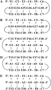
In this study, we investigate the role of the apex nucleotides of the two turns found in the intramolecular "paperclip" type triplex DNA formed by 5'-TCTCTCCTCTCTAGAGAG-3'. Our previously published structure calculations show that residues C7-A18 form a hairpin turn via Watson-Crick basepairing and residues T1-C6 bind into the major groove of the hairpin via Hoogsteen basepairing resulting in a broad turn of the T1-T12 5'-pyrimidine section of the DNA. We find that only the C6C7/G18 apex triad (and not the T12A13/T1 apex triad) is required for intramolecular triplex formation, is base independent, and occurs whether the purine section is located at the 5' or 3' end of the sequence. NMR spectroscopy and molecular dynamics simulations are used to investigate a bimolecular complex (which retains only the C6C7/G18 apex) in which a pyrimidine strand 5'- TCTCTCCTCTCT-3' makes a broad fold stabilized by the purine strand 5'-AGAGAG-3' via Watson Crick pairing to the T8-T12 and Hoogsteen basepairing to T1-T5 of the pyrimidine strand. Interestingly, this investigation shows that this 5'-AGAGAG-3' oligo acts as a new kind of triplex forming oligonucleotide, and adds to the growing number of triplex forming oligonucleotides that may prove useful as therapeutic agents.
Figures


Similar articles
-
Proton NMR studies of 5'-d-(TC)(3) (CT)(3) (AG)(3)-3'--a paperclip triplex: the structural relevance of turns.Biophys J. 2002 Jun;82(6):3170-80. doi: 10.1016/S0006-3495(02)75659-2. Biophys J. 2002. PMID: 12023241 Free PMC article.
-
Nuclear magnetic resonance structural studies of intramolecular purine.purine.pyrimidine DNA triplexes in solution. Base triple pairing alignments and strand direction.J Mol Biol. 1991 Oct 20;221(4):1403-18. J Mol Biol. 1991. PMID: 1942059
-
Thermodynamic, kinetic, and conformational properties of a parallel intermolecular DNA triplex containing 5' and 3' junctions.Biochemistry. 1998 Oct 27;37(43):15188-98. doi: 10.1021/bi980057m. Biochemistry. 1998. PMID: 9790683
-
Evidence for a DNA triplex in a recombination-like motif: I. Recognition of Watson-Crick base pairs by natural bases in a high-stability triplex.J Mol Recognit. 2001 Mar-Apr;14(2):122-39. doi: 10.1002/jmr.528. J Mol Recognit. 2001. PMID: 11301482
-
Triplex DNA structures.Annu Rev Biochem. 1995;64:65-95. doi: 10.1146/annurev.bi.64.070195.000433. Annu Rev Biochem. 1995. PMID: 7574496 Review.
Cited by
-
Molecular Assembly of Triplex of Duplexes from Homothyminyl-Homocytosinyl Cγ(S/R)-Bimodal Peptide Nucleic Acids with dA8/dG6 and the Cell Permeability of Bimodal Peptide Nucleic Acids.ACS Omega. 2021 Jul 19;6(30):19757-19770. doi: 10.1021/acsomega.1c02451. eCollection 2021 Aug 3. ACS Omega. 2021. PMID: 34368563 Free PMC article.
-
The triple helix: 50 years later, the outcome.Nucleic Acids Res. 2008 Sep;36(16):5123-38. doi: 10.1093/nar/gkn493. Epub 2008 Aug 1. Nucleic Acids Res. 2008. PMID: 18676453 Free PMC article. Review.
References
-
- Gilbert, D. E., and J. Feigon. 1999. Multistranded DNA structures. Curr. Opin. Struct. Biol. 9:305–314. - PubMed
-
- Felsenfeld, G., D. R. Davies, and A. Rich. 1957. Formation of a three-stranded polynucleotide molecule. J. Am. Chem. Soc. 79:2023–2024.
-
- Frank-Kamenetskii, M. D., and S. M. Mirkin. 1995. Triplex DNA structures. Annu. Rev. Biochem. 64:65–95. - PubMed
-
- Callahan, D. E., T. L. Trapane, P. S. Miller, P. O. P. Ts'o, and L.-S. Kan. 1991. Comparative circular dichroism and fluorescence studies of oligodeoxyribonucleotide and oligodeoxyribonucleoside methylphosphonate pyrimidine strands in duplex and triplex formation. Biochemistry. 30:1650–1655. - PubMed
-
- Kan, L.-S., D. E. Callahan, T. L. Trapane, P. S. Miller, P. O. P. Ts'o, and D. H. Huang. 1991. Proton NMR and optical spectroscopy studies on the DNA triplex formed by d-A(G-A)7-A and d-C-(T-C)7-T. J. Biomol. Struct. Dyn. 8:911–933. - PubMed
Publication types
MeSH terms
Substances
LinkOut - more resources
Full Text Sources
Miscellaneous

