. 2006 Jul 18;103(29):10973-8.
doi: 10.1073/pnas.0600923103. Epub 2006 Jul 7.

Whirlin complexes with p55 at the stereocilia tip during hair cell development

Affiliations

Whirlin complexes with p55 at the stereocilia tip during hair cell development

Philomena Mburu et al. Proc Natl Acad Sci U S A. .

Abstract

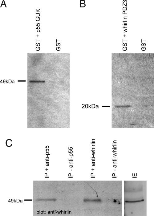
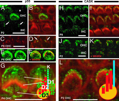
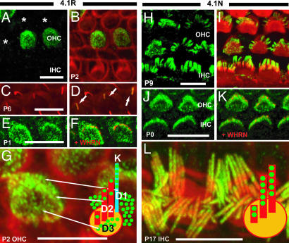
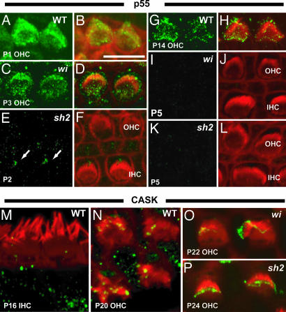
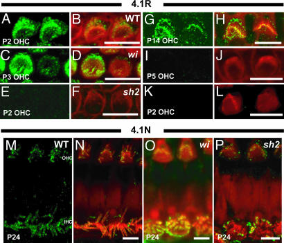
Hearing in mammals depends upon the proper development of actin-filled stereocilia at the hair cell surface in the inner ear. Whirlin, a PDZ domain-containing protein, is expressed at stereocilia tips and, by virtue of mutations in the whirlin gene, is known to play a key role in stereocilia development. We show that whirlin interacts with the membrane-associated guanylate kinase (MAGUK) protein, erythrocyte protein p55 (p55). p55 is expressed in outer hair cells in long stereocilia that make up the stereocilia bundle as well as surrounding shorter stereocilia structures. p55 interacts with protein 4.1R in erythrocytes, and we find that 4.1R is also expressed in stereocilia structures with an identical pattern to p55. Mutations in the whirlin gene (whirler) and in the myosin XVa gene (shaker2) affect stereocilia development and lead to early ablation of p55 and 4.1R labeling of stereocilia. The related MAGUK protein Ca2+-calmodulin serine kinase (CASK) is also expressed in stereocilia in both outer and inner hair cells, where it is confined to the stereocilia bundle. CASK interacts with protein 4.1N in neuronal tissue, and we find that 4.1N is expressed in stereocilia with an identical pattern to CASK. Unlike p55, CASK labeling shows little diminution of labeling in the whirler mutant and is unaffected in the shaker2 mutant. Similarly, expression of 4.1N in stereocilia is unaltered in whirler and shaker2 mutants. p55 and protein 4.1R form complexes critical for actin cytoskeletal assembly in erythrocytes, and the interaction of whirlin with p55 indicates it plays a similar role in hair cell stereocilia.


Conflict of interest statement

Conflict of interest statement: No conflicts declared.

Figures

Fig. 1.
Fig. 1.
In vitro (A and B) and in vivo (C) pull-down assays between whirlin and p55. (A and B) GST-tagged GUK domain of p55 or GST-tagged whirlin PDZ3 domain were expressed in E. coli K12 ER2508 cells using pGEX-4T1. Immobilized proteins were incubated with in vitro translated 35S-labeled short whirlin isoform (A) and the C-terminal region (Lys-323 through Tyr-466), including the GUK domain of p55 (B). GST protein alone was used as a negative control. (C) Cochlear proteins were incubated with p55 or the short whirlin isoform-specific antibodies. Immunoprecipitates from each were then subjected to immunoblot analysis with the short isoform of whirlin. IE, cochlear protein extract.
Fig. 2.
Fig. 2.
Localization of p55 and CASK at the apical surface of mouse hair cells. (A and B) Cochlear whole mounts were stained with antibody to p55 (green) and phalloidin (red). (A) p55 labeling of P2 hair cells. (B) Merge of p55 and phalloidin. Some OHCs are strongly labeled, others weakly, and some are not labeled (asterisk). In postnatal stages, no significant levels of p55 staining were detected in stereocilia of IHCs (data not shown). (C) β-Tubulin (red) staining highlights microtubules in P0 OHCs. Arrows indicate kinocilia. (D) Merged image with p55. The merged image shows that p55 and β-tubulin colocalize in the kinocilia. (E and F) p55 (green) colocalizes with whirlin (red) at the stereocilia tips in hair cells. (E) p55 localization in P6 OHC. (F) Merged image with whirlin. (G) More highly magnified image of p55 labeling in P4 OHCs; p55 is detected in stereocilia (domain, D2) as well as strial (D1), neural (D3) domains of the OHCs, and kinocilia (k). (H–L) Cochlear whole mounts were stained with antibody to CASK (green) and phalloidin (red). (H) CASK labeling of P9 OHCs and IHCs. (I) Merge of CASK and phalloidin. CASK was localized at stereocilia tips of both OHCs and IHCs. (J and K) CASK (green) colocalizes with whirlin (red) at the stereocilia tips in hair cells. (K) Merged image with whirlin localization. (L) More highly magnified image of CASK labeling in P2 OHCs. CASK labeling is detected at stereocilia tips. (Scale bar, 5 μm.)
Fig. 3.
Fig. 3.
Localization of 4.1R (A–G) and 4.1N (H–L) at the apical surface of mouse hair cells. (A and B) Cochlear whole mounts were stained with antibody to 4.1R (green) and phalloidin (red). (A) 4.1R labeling of P2 hair cells. (B) Merge of 4.1R and phalloidin. Some cells are strongly labeled, others weakly, and some are not labeled (asterisks). In postnatal stages, no significant 4.1R staining was detected in IHCs (data not shown). (C and D) 4.1R labeling was also observed in the kinocilia. (C) β-Tubulin (red) staining highlights microtubules in P6 OHCs. Arrows indicate kinocilia. (D) Merged image with 4.1R labeling. The merged image shows that the 4.1R and β-tubulin colocalize in the kinocilia. (E and F) 4.1R (green) colocalizes with whirlin (red) at the stereocilia tips in hair cells. (E) 4.1R localization in P1 OHC. (F) Merged image with whirlin. (G) More highly magnified image of 4.1R labeling in P2 OHCs. 4.1R is detected in stereocilia (domain, D2) as well as neural (D3), strial (D1) domains of the OHCs, and kinocilia (k). (H–L) Cochlear whole mounts were stained with antibody to 4.1N (green) and phalloidin (red). (H) 4.1N labeling of P9 OHCs and IHCs. (I) Merge of 4.1N and phalloidin. 4.1N was localized at stereocilia bundles of both OHCs and IHCs. (J and K) 4.1N (green) colocalizes with whirlin (red) at the stereocilia tips in hair cells. (K) Merged image with whirlin. (L) 4.1N localization in P17 hair cells. (L) More highly magnified image of 4.1N labeling in P17 IHCs. 4.1N labeling was not detected along the whole length of stereocilia but was concentrated at the apical regions of stereocilia bundles, including the tips. (Scale bar, 5 μm.)
Fig. 4.
Fig. 4.
Localization of p55 and CASK at the apical surface of hair cells in WT, whirler (wi), and shaker2 (sh2) mutant mice during cochlear development. All images were taken from the apical turn of the cochlea. (A–L) Localization of p55 in WT and mutant mice during cochlea development. In WT mice, p55 labeling declines significantly around P14 (G and H) and fades out at P17 (data not shown). In the wi mutant, expression of p55 is normal until the P4 stage (P3 stage illustrated in C and D). However, labeling of p55 fades out from P5 in the wi mutant (I and J). Expression of p55 in stereocilia is completely ablated in the sh2 mutant (E, F, K, and L), although labeling of kinocilia (arrows) is still detected (E and F). (M–O) Localization of CASK in WT and mutant mice in mature hair cells. In WT mice, CASK signals were detected in stereocilia tips into adulthood in OHCs (N), but IHC labeling was not seen after P16 (M). CASK labeling is unaffected in both mutants in OHCs and IHCs (O and P and data not shown).
Fig. 5.
Fig. 5.
Localization of 4.1R and 4.1N at the apical surface of hair cells in WT, whirler (wi), and shaker2 (sh2) mutant mice during cochlear development. All images were taken from the apical turn of the cochlea. (A–L) Localization of 4.1R in WT and mutant mice during cochlea development. 4.1R shows a nearly identical pattern of labeling to that of p55 in the WT. Labeling of 4.1R fades out from P5 in the whirler mutant (C, D, I, and J). Expression of 4.1R in stereocilia is completely ablated in the shaker2 mutant (E, F, K, and L), although labeling of kinocilia is still detected (data not shown). (M–P) Localization of 4.1N in WT and mutant mice in mature hair cells. In WT mice, 4.1N signals were detected in stereocilia into adulthood as with CASK (M and N). 4.1N labeling is unaffected in both mutants (O and P).

References

    1. Tilney L. G., Tilney M. S., DeRosier D. J. Annu. Rev. Cell Biol. 1992;8:257–274. - PubMed
    1. Forge A., Souter M., Denman-Johnson K. Semin. Cell Dev. Biol. 1997;8:225–237. - PubMed
    1. Denman-Johnson K., Forge A. J. Neurocytol. 1999;28:821–835. - PubMed
    1. Anniko M. Anat. Embryol. (Berl.) 1983;166:355–368. - PubMed
    1. Furness D., Richardson G. P., Russell I. J. Hear. Res. 1989;38:95–110. - PubMed

Publication types

MeSH terms