

Quantification of photoreceptor layer thickness in normal eyes using optical coherence tomography
- PMID: 16829808
- PMCID: PMC1933486
- DOI: 10.1097/01.iae.0000236468.33325.74
Quantification of photoreceptor layer thickness in normal eyes using optical coherence tomography
Abstract
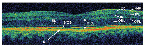
Objective: To demonstrate the ability to segment and analyze individual intraretinal layers, including the outer retinal complex (ORC; outer nuclear layer and inner and outer segments of the photoreceptor cells), in healthy eyes using images acquired from the latest commercially available optical coherence tomography (OCT) system (StratusOCT; Carl Zeiss Meditec, Inc., Dublin, CA) and from the ultrahigh resolution OCT (UHR-OCT) prototype.
Methods: Thirty-seven eyes from 37 healthy subjects underwent complete ophthalmologic examination using StratusOCT and UHR-OCT. ORC was identified and measured using a segmentation algorithm.
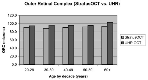
Results: For StratusOCT, mean weighted ORC thickness +/- SD was 91.1 +/- 7.9 microm, and mean weighted total retinal thickness +/- SD was determined to be 258.9 +/- 10.1 microm. For UHR-OCT, mean weighted ORC thickness +/- SD was 96.4 +/- 6.3 microm, and mean weighted total retinal thickness +/- SD was determined to be 263.4 +/- 9.2 mum. There was a higher rate of algorithm failure with UHR-OCT images.
Conclusions: Photoreceptor layer thickness can be calculated by measuring ORC on OCT images using a macular segmentation algorithm. ORC values may serve as a useful objective parameter in determining the efficacy of various therapeutic modalities that target the photoreceptor layer in various diseases.
Figures
References
-
- Swanson EA, Izatt JA, Hee MR, et al. In vivo retinal imaging by optical coherence tomography. Opt Lett. 1993;18:1864–1866. - PubMed
-
- Hee MR, Izatt JA, Swanson JA, et al. Optical coherence tomography of the human retina. Arch Ophthalmol. 1995;113:325–332. - PubMed
-
- Puliafito CA, Hee MR, Lin CP, et al. Imaging of macular diseases with optical coherence tomography. Ophthalmology. 1995;102:217–229. - PubMed
-
- Hee MR, Puliafito CA, Wong C, et al. Quantitative assessment of macular edema with optical coherence tomography. Arch Ophthalmol. 1995;113:1019–1029. - PubMed
Publication types
MeSH terms
Grants and funding
LinkOut - more resources
Full Text Sources
Other Literature Sources
