. 1991 Nov 1;254(5032):726-9.
doi: 10.1126/science.1683005.

Pre-Bötzinger complex: a brainstem region that may generate respiratory rhythm in mammals

Affiliations

Pre-Bötzinger complex: a brainstem region that may generate respiratory rhythm in mammals

J C Smith et al. Science. .

Abstract

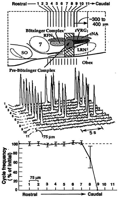
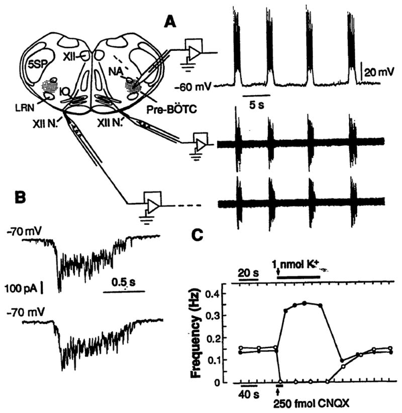
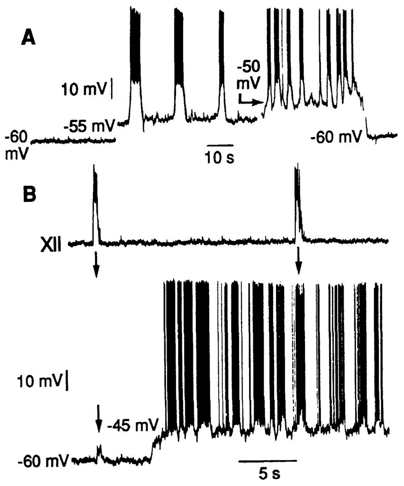
The location of neurons generating the rhythm of breathing in mammals is unknown. By microsection of the neonatal rat brainstem in vitro, a limited region of the ventral medulla (the pre-Bötzinger Complex) that contains neurons essential for rhythmogenesis was identified. Rhythm generation was eliminated by removal of only this region. Medullary slices containing the pre-Bötzinger Complex generated respiratory-related oscillations similar to those generated by the whole brainstem in vitro, and neurons with voltage-dependent pacemaker-like properties were identified in this region. Thus, the respiratory rhythm in the mammalian neonatal nervous system may result from a population of conditional bursting pacemaker neurons in the pre-Bötzinger Complex.


Figures

Fig. 1
Fig. 1
Perturbations of respiratory motor pattern with serial microsections of neonatal rat medulla in vitro. (Top) Sagittal view of medulla showing pre-Bötzinger Complex and neighboring regions. Hatched rectangular area indicates critical region for rhythmogenesis. Pre-Bötzinger Complex (shaded area) within the critical region extends from caudal end of retrofacial nucleus ~200 μm toward obex. SO, superior olive; 7, facial nucleus; LRN, lateral reticular nucleus; RFN, retrofacial nucleus; rVRG, rostral ventral respiratory group; cNA, caudal (semicompact) division of nucleus ambiguus. (Middle) Traces show integrated phrenic motoneuron population discharge on C4 spinal ventral roots after 75-μm sectioning in the rostral to caudal direction. The steady-state discharge shown is after sections made from the level of caudal facial nucleus through the caudal end of pre-Bötzinger Complex (1 through 11). Sections at the level of pre-Bötzinger Complex (sections 8 to 10) eliminated rhythmic motor output of all spinal and cranial (IX, X, XII) (not shown) respiratory motoneuron populations. (Bottom) A single 75-μm section through the rostral boundary of the pre-Bötzinger Complex caused a reduction in cycle frequency and instabilities of the rhythm (that is, an increase in cycle-to-cycle variation of period). The mean cycle period in the experiment shown was 7.5 ± 0.4 s (n = 15 cycles) after sectioning just rostral to pre-Bötzinger Complex and 11.1 ± 2.9 s after a single transection within this region.
Fig. 2
Fig. 2
(A) Rhythmic activity of a medullary slice (500 μm thick) containing pre-Bötzinger Complex (pre-BÖTC, shaded regions). NA, nucleus ambiguus; 5SP, spinal trigeminal nucleus; XII, hypoglossal nucleus; XII N., hypoglossal nerve; and IO, inferior olive. Traces at right show respiratory motor discharge recorded bilaterally from hypoglossal nerve roots (lower) (18) and whole-cell recording from a rhythmically active neuron in pre-Bötzinger Complex (upper). (B) (Upper) Synaptic drive currents of pre-Bötzinger Complex neuron under voltage-clamp (−70 mV) consisted of rapidly peaking, slowly decrementing envelope. (Lower) Identical pattern of synaptic currents of respiratory neuron in en bloc medulla. (C) Unilateral microinjection of solution with elevated K+ concentration (upper arrow and bar) (40 nl, 25 mM; 1 nmol) in the pre-Bötzinger Complex in slice reversibly elevated motor burst frequency bilaterally (solid dots). Microinjection of 250 fmol CNQX (10 nl, 25 μM) (lower arrow and bar) in pre-Bötzinger Complex reduced the burst frequency (open points) and transiently blocked rhythmogenesis, which gradually returned with local washout of antagonist. Data are average values computed from continuous recordings of motor output from slice.
Fig. 3
Fig. 3
Voltage-dependent oscillatory properties of neurons in the pre-Bötzinger Complex. (A) One population of neurons generated large-amplitude oscillatory membrane potentials in response to membrane depolarization. Recording was obtained in a slice 300 μm thick without rhythmic motor output. (B) In slices generating hypoglossal (XII) motor output (top trace), neurons exhibiting intrinsic bursting properties (bottom trace) received synaptic inputs that synchronize neuron depolarization and bursting (19); synchronizing inputs are evident as small-amplitude (~5 mV) depolarizing potentials (arrow at left in bottom trace), which cause the neuron to generate a burst of action potentials coincident with the rest of the network at more depolarized baseline potentials (arrow at right). Voltage-dependent bursting properties become evident with depolarization to −45 mV, which results in membrane potential oscillations and bursting at a higher frequency than that of slice motor output.
Fig. 4
Fig. 4
Fluorescence photomicrographs of horizontally sliced sections in the plane of the caudal (A) and contiguous rostral (B) portions of the lateral tegmental field containing the ventral respiratory group (14) and pre-Bötzinger Complex. Note the nonuniform distribution of three classes of retrogradely labeled neurons: (i) bulbospinal premotoneurons labeled with Fluoro-Gold (Fluorochrome, Inc., Englewood, Colorado) (yellow); (ii) propriobulbar interneurons labeled with rhodamine-impregnated latex beads (red); and (iii) vagal motoneurons labeled with fast blue. Bulbospinal neurons and vagal motoneurons are concentrated at caudal levels (A), whereas in the pre-Bötzinger Complex (B) there is a high density of propriobulbar neurons and few bulbospinal neurons and vagal motoneurons. The caudal boundary of pre-Bötzinger Complex is indicated by the arrow at left in (B); rostral boundary is at the right margin. The level of obex is indicated by the arrow in (A); the bottom is toward the midline. Length bar, 200 μm.

References

    1. Fluorens P. C R Acad Sci (Paris) 1851;33:437.
    1. Cohen M. Physiol Rev. 1979;59:1105. - PubMed
    2. Feldman JL. Handbook of Physiology. In: Bloom FE, editor. The Nervous System IV. American Physiological Society; Washington, D.C: 1986. pp. 463–524.
    3. Ezure K. Prog Neurobiol (NY) 1990;35:429. - PubMed
    1. Suzue T. J Physiol (London) 1984;354:173. - PMC - PubMed
    2. Smith JC, Feldman JL. J Neurosci Methods. 1987;21:321. - PubMed
    1. Smith JC, Greer JJ, Liu G, Feldman JL. J Neurophysiol. 1990;64:1149. - PubMed
    1. Harada Y, Kuno M, Wang YZ. J Physiol (London) 1986;368:679. - PMC - PubMed
    2. McCrimmon DR, Smith JC, Feldman JL. J Neurosci. 1989;9:1910. - PMC - PubMed
    3. Hilaire G, Monteau R, Errchidi S. Brain Res. 1989;485:325. - PubMed
    4. Liu G, Feldman JL, Smith JC. J Neurophysiol. 1990;64:423. - PubMed

Publication types

Substances

LinkOut - more resources