Clinical Trial
. 2007 Mar;56(3):381-9.
doi: 10.1007/s00262-006-0197-8. Epub 2006 Jul 8.

Successful induction of clinically competent dendritic cells from granulocyte colony-stimulating factor-mobilized monocytes for cancer vaccine therapy

Affiliations
Clinical Trial

Successful induction of clinically competent dendritic cells from granulocyte colony-stimulating factor-mobilized monocytes for cancer vaccine therapy

Yuji Ueda et al. Cancer Immunol Immunother. 2007 Mar.

Abstract

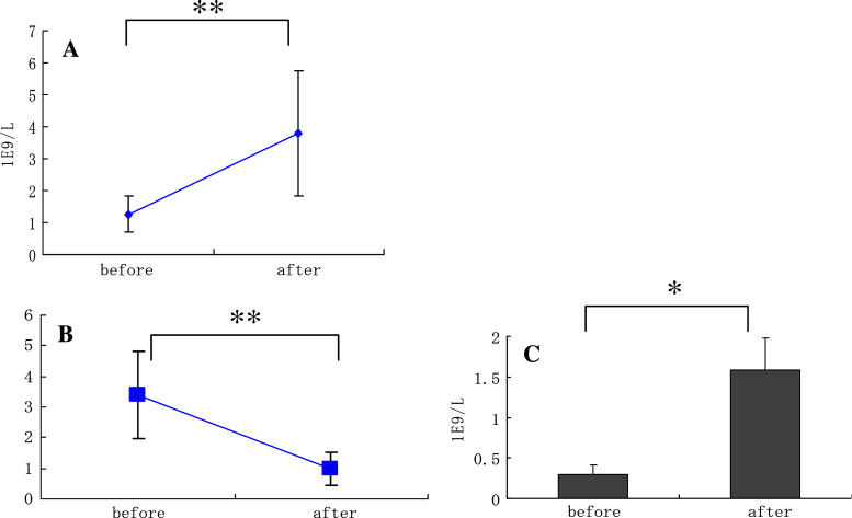
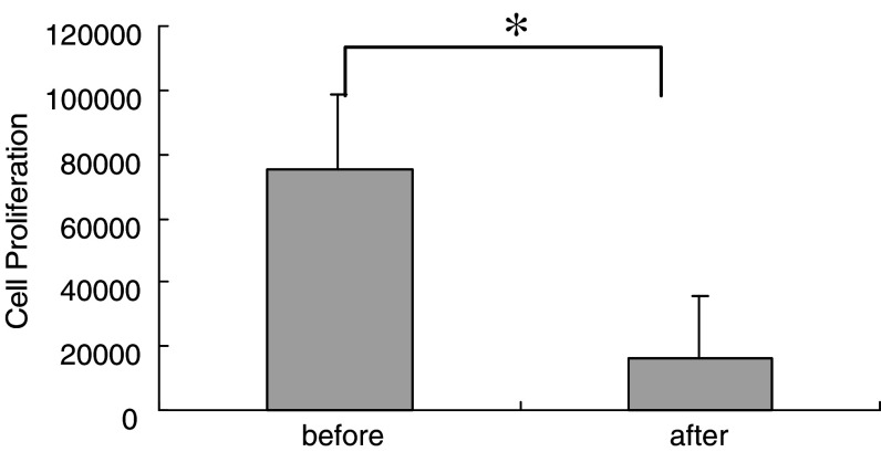
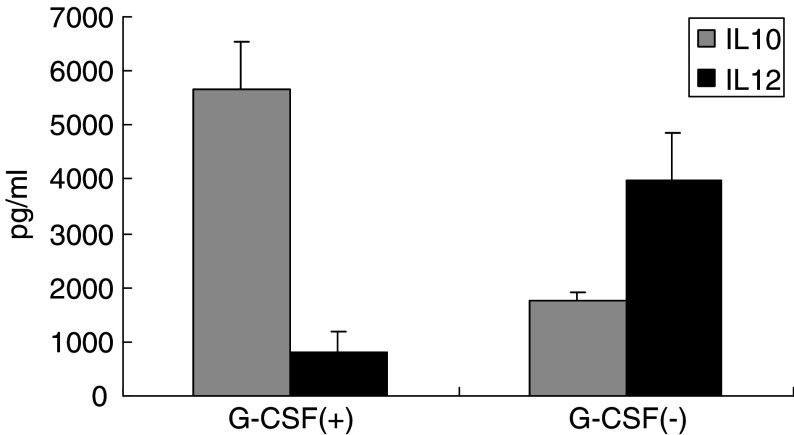
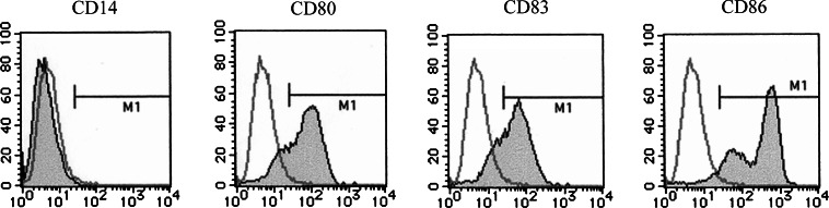
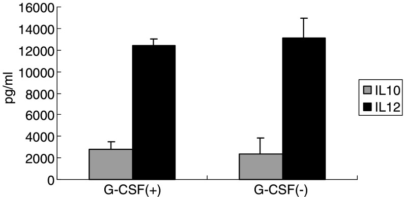
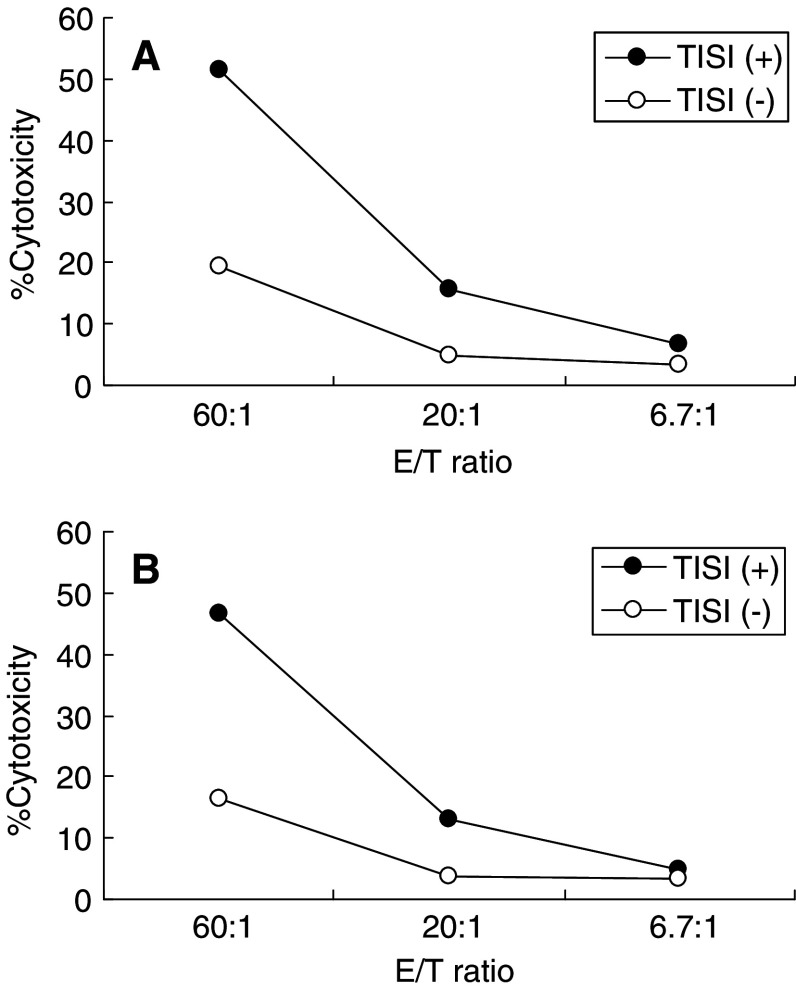
Recent studies have suggested that dendritic cell (DC)-based immunotherapy is one promising approach for the treatment of cancer. We previously studied the clinical toxicity, feasibility, and efficacy of cancer vaccine therapy with peptide-pulsed DCs. In that study, we used granulocyte colony-stimulating factor (G-CSF)-mobilized peripheral blood monocytes as a cell source of DCs. However, previous investigations have suggested that G-CSF-mobilized peripheral blood monocytes produce reduced levels of proinflammatory cytokines such as interleukin (IL)-12 and tumor necrosis factor (TNF)-alpha. These T helper (Th)-1-type cytokines are thought to promote antitumor immune response. In this study, we assessed the functional abilities of DCs generated from G-CSF-mobilized monocytes obtained from 13 patients with CEA-positive advanced solid cancers. Peripheral blood mononuclear cells were obtained from leukapheresis products collected before and after systemic administration of G-CSF (subcutaneous administration of high-dose [5-10 microg/kg] human recombinant G-CSF for five consecutive days). In vitro cytokine production profiles after stimulation with lipopolysaccharide (LPS) were compared between monocytes with and without G-CSF mobilization. DCs generated from monocytes were also examined with respect to cytokine production and the capacity to induce peptide-specific T cell responses. Administration of G-CSF was found to efficiently mobilize peripheral blood monocytes. Although G-CSF-mobilized monocytes (G/Mo) less effectively produced Th-1-type cytokines than control monocytes (C/Mo), DCs generated from G/Mo restored the same level of IL-12 production as that seen in DCs generated from C/Mo. T cell induction assay using recall antigen peptide and phenotypic analyses also demonstrated that DCs generated from G/Mo retained characteristics identical to those generated from C/Mo. Our results suggest that G-CSF mobilization can be used to collect monocytes as a cell source for the generation of DCs for cancer immunotherapy. DCs generated in this fashion were pulsed with HLA-A24-restricted CEA epitope peptide and administered to patients safely; immunological responses were induced in some patients.


Figures

Fig. 1
Fig. 1
Monocytes were efficiently mobilized into peripheral blood by G-CSF. Peripheral blood mononuclear cells (PBMCs) were harvested by leukapheresis before and after administration of G-CSF in 13 patients. Lymphocytes (CD3+) and monocytes (CD14+) were then measured by FACS analysis. The numbers of PBMCs were counted (a). The ratios of the numbers of lymphocytes to monocytes (b) and absolute numbers of monocytes harvested (c) were calculated from the results of FACS analyses. Data represent the means ± SD of 13 cases. *< 0.01, **P < 0.03
Fig. 2
Fig. 2
Decreased T cell proliferation response against phytohemagglutinin (PHA) stimulation after G-CSF mobilization. T cells were cultured in the presence of PHA and then pulsed with 3H-thymidine for 18 h. Cell proliferation activity was measured by counting 3H incorporation with a scintillation counter. Data represent the means ± SD of 13 cases. *< 0.02
Fig. 3
Fig. 3
Th-2-type cytokine production profiles observed in monocytes mobilized with G-CSF. PBMCs were obtained by leukapheresis before and after G-CSF mobilization. Monocytes were then purified with cell sorter using anti-CD14 mAb and subjected to cytokine production assay. Isolated cells (2.5 × 105/ml) were stimulated with 10 μg/ml LPS. After 48 h of incubation at 37°C, released cytokines were measured in the cell-free supernatants. The concentrations of IL-10 and IL-12 were measured by ELISA. Data represent the means ± SD, derived from two independent experiments using PBMCs obtained from patient nos. 1 to 3 and 5
Fig. 4
Fig. 4
Surface phenotypic analysis of DCs generated from G-CSF-mobilized monocytes. G-CSF-mobilized monocytes were cultured in the presence of IL-4 and GM-CSF for 7 days and then subjected to phenotypic analysis. The expression of CD14, CD80, CD83, and CD86 was assessed with the use of mAbs (closed histogram). Isotype immunoglobulins were used for controls (open histogram). Results represent the data from patient no. 11
Fig. 5
Fig. 5
Restored production of IL-12 in monocytes obtained from PBMCs mobilized with G-CSF by culturing with IL-4 and GM-CSF. To assess the ability to produce Th-1-type cytokines from DCs generated from monocytes obtained after G-CSF mobilization, DCs were stimulated with 10 μg/ml LPS. DCs were purified with the use of Human Dendritic Cell Enrichment Cocktail (Stem Cell Technologies), which is a depletion cocktail containing several mAbs (anti-CD3, anti-CD14, anti-CD16, anti-CD19, anti-CD56, anti-CD66b, and Glycophorin A), according to the manufacturer’s instruction. Cells were then cultured at the concentration of 1 × 105/ml for 48 h. The concentrations of IL-10 and IL-12 were measured by ELISA using the harvested cell-free supernatants. Data represent the means ± SD, derived from two independent experiments using PBMCs obtained from patient nos. 1 to 3 and 5. There were no significant differences with regard to the production levels of IL-10 and IL-12 between PBMCs obtained before and after G-CSF mobilization
Fig. 6
Fig. 6
Dendritic cells generated from G-CSF-mobilized monocytes induced efficient cytotoxic T cell response against peptide antigen in vitro. To assess the functional ability of DCs, T cells were stimulated by DCs generated from monocytes before (a) and after (b) G-CSF mobilization, pulsed with HLA-A24-restricted CTL epitope peptide derived from influenza nucleoprotein, and cultured for 7 days. Cytotoxic activities of effector T cells were measured by standard 51Cr-releasing assay using TISI cells (HLA-A24 positive) pulsed with or without cognate peptide at three different effector-to-target ratios. Data represent one of the two independent experiments using PBMCs obtained from patient nos. 1 to 3

Similar articles

Cited by

References

    1. Timmerman JM, Levy R. Dendritic cell vaccines for cancer immunotherapy. Annu Rev Med. 1999;50:507. doi: 10.1146/annurev.med.50.1.507. - DOI - PubMed
    1. Nestle FO, Alijagic S, Gilliet M, Sun Y, Grabbe S, Dummer R, et al. Vaccination of melanoma patients with peptide- or tumor lysates-pulsed dendritic cells. Nat Med. 1998;4:328. doi: 10.1038/nm0398-328. - DOI - PubMed
    1. Schuler TB, Schultz ES, Berger TG, Weinlich G, Ebner S, Woerl P, et al. Rapid induction of tumor-specific type 1 T helper cells in metastatic melanoma patients by vaccination with mature, cryopreserved, peptide-loaded monocyte-derived dendritic cells. J Exp Med. 2002;195:1279. doi: 10.1084/jem.20012100. - DOI - PMC - PubMed
    1. Paneli MC, Wunderlich J, Jeffries J, Wang E, Mixon A, Rosenberg SA, et al. Phase 1 study in melanoma-associated antigens MART-1 and gp100. J Immunother. 2000;23:487. doi: 10.1097/00002371-200007000-00013. - DOI - PubMed
    1. Lau R, Wang F, Jeffery G, Marty V, Kuniyoshi J, Made E, et al. Phase I trial of intravenous peptide-pulsed dendritic cells in patients with metastatic melanoma. J Immunother. 2001;24:66. doi: 10.1097/00002371-200101000-00008. - DOI - PubMed

Publication types

MeSH terms