

Osteopontin knockdown suppresses tumorigenicity of human metastatic breast carcinoma, MDA-MB-435
- PMID: 16830223
- PMCID: PMC1574364
- DOI: 10.1007/s10585-006-9013-2
Osteopontin knockdown suppresses tumorigenicity of human metastatic breast carcinoma, MDA-MB-435
Abstract
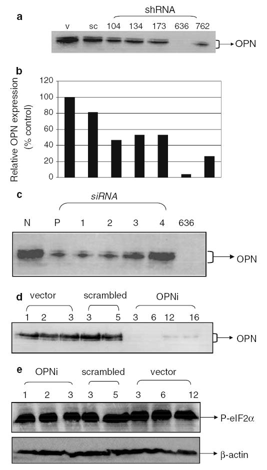
Elevated expression of osteopontin (OPN), a secreted phosphoglycoprotein, is frequently associated with many transformed cell lines. Various studies suggest that OPN may contribute to tumor progression as well as metastasis in multiple tumor types. High levels of OPN have been reported in patients with metastatic cancers, including breast. We found that the expression of OPN corroborates with the aggressive phenotype of the breast cancer cells i.e. the expression of OPN is acquired as the breast cancer cells become more aggressive. To assess the role(s) of OPN in breast carcinoma, expression of endogenous OPN was knocked down in metastatic MDA-MB-435 human breast carcinoma cells using RNA interference. We targeted multiple regions of the OPN transcript for RNA interference, along with 'scrambled' and 'non-targeting siRNA pool' controls to distinguish between target-specific and potential off-target effects including interferon-response gene (PeIF2-alpha) induction. The OPN knockdown by shRNA suppressed tumor take in immunocompromised mice. The 'silenced' cells also showed significantly lower invasion and migration in modified Boyden chamber assays and reduced ability to grow in soft agar. Thus, in addition to the widely reported roles of OPN in late stages of tumor progression, these results provide functional evidence that OPN contributes to breast tumor growth as well.
Figures


Similar articles
-
The LCC15-MB human breast cancer cell line expresses osteopontin and exhibits an invasive and metastatic phenotype.Exp Cell Res. 1998 Jun 15;241(2):273-84. doi: 10.1006/excr.1998.4029. Exp Cell Res. 1998. PMID: 9637769
-
The thrombin inhibitor Argatroban reduces breast cancer malignancy and metastasis via osteopontin-dependent and osteopontin-independent mechanisms.Breast Cancer Res Treat. 2008 Nov;112(2):243-54. doi: 10.1007/s10549-007-9865-4. Epub 2007 Dec 21. Breast Cancer Res Treat. 2008. PMID: 18097747
-
Targeting osteopontin suppresses glioblastoma stem-like cell character and tumorigenicity in vivo.Int J Cancer. 2015 Sep 1;137(5):1047-57. doi: 10.1002/ijc.29454. Epub 2015 May 21. Int J Cancer. 2015. PMID: 25620078
-
Role of osteopontin in cellular signaling and metastatic phenotype.Front Biosci. 2008 May 1;13:4276-84. doi: 10.2741/3004. Front Biosci. 2008. PMID: 18508510 Review.
-
Osteopontin: an effector and an effect of tumor metastasis.Curr Mol Med. 2010 Feb;10(1):71-81. doi: 10.2174/156652410791065381. Curr Mol Med. 2010. PMID: 20205680 Free PMC article. Review.
Cited by
-
Overcoming Irinotecan Resistance by Targeting Its Downstream Signaling Pathways in Colon Cancer.Cancers (Basel). 2024 Oct 15;16(20):3491. doi: 10.3390/cancers16203491. Cancers (Basel). 2024. PMID: 39456585 Free PMC article.
-
Nonclassical activation of Hedgehog signaling enhances multidrug resistance and makes cancer cells refractory to Smoothened-targeting Hedgehog inhibition.J Biol Chem. 2013 Apr 26;288(17):11824-33. doi: 10.1074/jbc.M112.432302. Epub 2013 Mar 18. J Biol Chem. 2013. PMID: 23508962 Free PMC article.
-
Osteopontin gene expression determines spontaneous metastatic performance of orthotopic human breast cancer xenografts.Am J Pathol. 2007 Aug;171(2):682-92. doi: 10.2353/ajpath.2007.070232. Epub 2007 Jul 9. Am J Pathol. 2007. PMID: 17620367 Free PMC article.
-
Enhanced down-regulation of ALCAM/CD166 in African-American Breast Cancer.BMC Cancer. 2014 Sep 25;14:715. doi: 10.1186/1471-2407-14-715. BMC Cancer. 2014. PMID: 25255861 Free PMC article.
-
Extracellular and intracellular mechanisms that mediate the metastatic activity of exogenous osteopontin.Cancer. 2009 Apr 15;115(8):1753-64. doi: 10.1002/cncr.24170. Cancer. 2009. PMID: 19224553 Free PMC article.
References
-
- Singhal H, Bautista DS, Tonkin KS, et al. Elevated plasma osteopontin in metastatic breast cancer associated with increased tumor burden and decreased survival. Clin Cancer Res. 1997;3(4):605–611. - PubMed
-
- Tuck AB, O’Malley FP, Singhal H, et al. Osteopontin expression in a group of lymph node negative breast cancer patients. Int J Cancer. 1998;79(5):502–508. - PubMed
-
- Agrawal D, Chen T, Irby R, et al. Osteopontin identified as lead marker of colon cancer progression using pooled sample expression profiling. J Natl Cancer Inst. 2002;94(7):513–521. - PubMed
-
- Rudland PS, Platt-Higgins A, El-Tanani M, et al. Prognostic significance of the metastasis-associated protein osteopontin in human breast cancer. Cancer Res. 2002;62:3417–3427. - PubMed
-
- Coppola D, Szabo M, Boulware D, et al. Correlation of osteopontin protein expression and pathological stage across a wide variety of tumor histologies. Clin Cancer Res. 2004;10:184–190. - PubMed
Publication types
MeSH terms
Substances
Grants and funding
LinkOut - more resources
Full Text Sources
Other Literature Sources
Medical
Research Materials
Miscellaneous
