

doi: 10.1016/j.jneuroim.2006.05.027.
Epub 2006 Jul 10.
CCR2 mediates increases in glial activation caused by exposure to HIV-1 Tat and opiates
Affiliations
- PMID: 16831471
- PMCID: PMC4310703
- DOI: 10.1016/j.jneuroim.2006.05.027
Item in Clipboard
CCR2 mediates increases in glial activation caused by exposure to HIV-1 Tat and opiates
J Neuroimmunol.
2006 Sep.
Abstract
To assess the role of CCL2/MCP-1 in opiate drug abuse and HIV-1 comorbidity, the effects of systemic morphine and intrastriatal HIV-1 Tat on macrophage/microglial and astroglial activation were assessed in wild type and CCR2 null mice. Tat and/or morphine additively increased the proportion of CCL2 immunoreactive astroglia. The effects of morphine were prevented by naltrexone. Glial activation was significantly reduced in CCR2-/- versus wild-type mice following Tat or morphine plus Tat exposure. Thus, CCR2 contributes to local glial activation caused by Tat alone or in the presence of opiates, implicating CCR2 signaling in HIV-1 neuropathogenesis in drug abusers and non-abusers.
Figures

CCL2, μ-opioid receptor (MOR), and CCR2 immunocytochemical co-localization with glial fibrillary acidic protein (GFAP) in subpopulations of striatal astrocytes in wild type (A,B,D–F) and CCR2(−/−) mice (C,G–I). A small subpopulation of astrocytes normally express MOR (arrows in A), CCL2 (arrow in B), or CCR immunoreactivity (arrows in F), although the proportion possessing MOR (A) (El-Hage et al., 2006), or CCL2 (B), immunoreactivity increases following morphine and/or Tat exposure (see Fig. 2A) (scale bar A–B, = 25 μm; C = 15 μm). MOR could be localized in a subset of astrocytes in CCR2−/− mice (arrowheads in C delineate astrocytes with highly polarized MOR and GFAP cytoplasmic immunofluorescence; Hoechst counterstained nuclei). CCR2 immunoreactivity was present in wild type (D–F; arrows in F), but not in CCR2 (−/−) null (G–I) mice (D–I; scale bar = 20 μm).
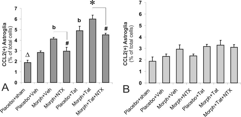
Effects of subcutaneous opiates and/or intrastriatal Tat on glial fibrillary acidic protein (GFAP) and CCL2 immunoreactivity at loci near (300 ± 100 μm) (A) and distant (600 ± 100 μm) (B) from the site of Tat injection in wild type mice. The proportion of CCL2 immunoreactive astrocytes increased significantly following morphine or Tat exposure (bP < 0.05 vs. vehicle-injected controls or morphine plus Tat treatment) (A), and showed additive increases with combined exposure (*P < 0.05 vs. other groups). The effects of morphine (alone or in combination with Tat) were antagonized by naltrexone (#P < 0.05 vs. morphine treatment) (A). Increases in CCL2+ astrocytes were seen following intrastriatal injection of vehicle compared to un-injected sham mice receiving placebo implants (ΔP < 0.05 vs. vehicle-injected, placebo controls) (A). The proportion of CCL2+ astrocytes was unchanged by morphine and/or Tat-treatment at locations distant from the site of injection (600 ± 100 μm) (B); n= 5–6 mice per group.

CCR2 deletion (−/−) significantly reduced HIV-1 Tat or morphine plus Tat-induced increases in GFAP+ astroglia (A) and F4/80+ macrophages/microglia (C) compared to wild type (+/+) mice at sites near (300 ± 100 μm) the site of Tat injection. By contrast, at sites more distant from Tat injection (600 ± 100 μm), neither Tat nor morphine plus Tat significantly increased the proportion of astroglia (B) or macrophages/microglia (D); *P < 0.05, wild type versus CCR2(−/−) mice; n= 5 mice per group.

Intrastriatal HIV-1 Tat caused significant increases in the proportion of GFAP+ astroglia (A) and F4/80+ macrophages/microglia (B) (*P < 0.05 vs. vehicle controls) compared to vehicle- or mutant TatΔ31–61- injected CCR2(+/+) mice (TatΔ31–61), or Tat-injected CCR2(−/−)(Tat [CCR2(−/−)]) mice (#P < 0.05 vs. Tat-treatment) (A-B). Moreover, in wild type-mice injected with Tat ipsilaterally and saline in the contralateral striatum (Contralateral), there no additional increases in macrophage/microglial or astroglial numbers in the contralateral striatum compared to mice only receiving saline.
References
-
- Ambati J, Anand A, Fernandez S, Sakurai E, Lynn BC, Kuziel WA, Rollins BJ, Ambati BK. An animal model of age-related macular degeneration in senescent Ccl-2- or Ccr-2-deficient mice. Nat Med. 2003;9:1390–1397. - PubMed
-
- Andjelkovic AV, Kerkovich D, Shanley J, Pulliam L, Pachter JS. Expression of binding sites for beta chemokines on human astrocytes. Glia. 1999;28:225–235. - PubMed
-
- Andjelkovic AV, Song L, Dzenko KA, Cong H, Pachter JS. Functional expression of CCR2 by human fetal astrocytes. J Neurosci Res. 2002;70:219–231. - PubMed
-
- Avison MJ, Nath A, Greene-Avison R, Schmitt FA, Bales RA, Ethisham A, Greenberg RN, Berger JR. Inflammatory changes and breakdown of microvascular integrity in early human immunodeficiency virus dementia. J Neurovirol. 2004;10:223–232. - PubMed
-
- Banisadr G, Queraud-Lesaux F, Boutterin MC, Pelaprat D, Zalc B, Rostene W, Haour F, Parsadaniantz SM. Distribution, cellular localization and functional role of CCR2 chemokine receptors in adult rat brain. J Neurochem. 2002;81:257–269. - PubMed
Publication types
MeSH terms
Substances
Grants and funding
LinkOut - more resources
Full Text Sources
Miscellaneous
