. 2006 Jul 17;174(2):231-43.
doi: 10.1083/jcb.200512085. Epub 2006 Jul 10.

Complete repair of dystrophic skeletal muscle by mesoangioblasts with enhanced migration ability

Affiliations

Complete repair of dystrophic skeletal muscle by mesoangioblasts with enhanced migration ability

Beatriz G Galvez et al. J Cell Biol. .

Erratum in

  • J Cell Biol. 2006 Aug 14;174(4):605
  • J Cell Biol. 2006 Oct 23;175(2):361. Costantin, Gabriela [corrected to Constantin, Gabriela]
  • J Cell Biol. 2013 Aug 19;202(4):715-6

Abstract

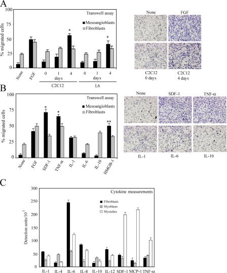
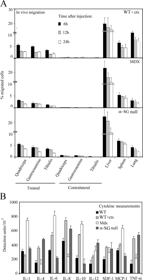
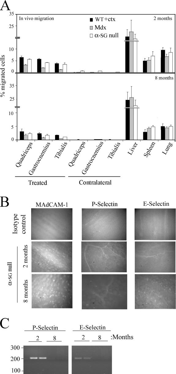
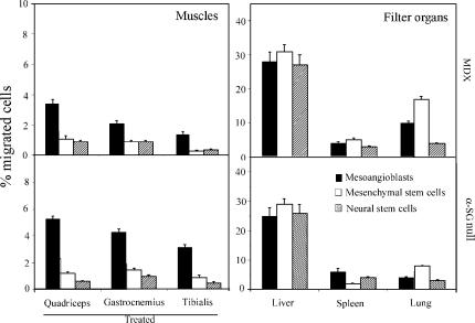
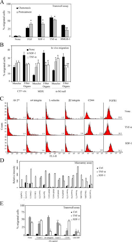
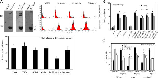
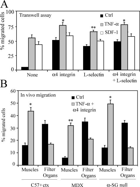
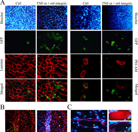
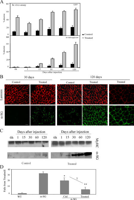
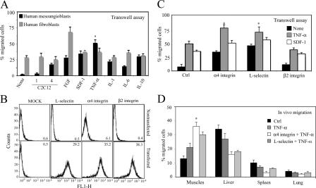
Efficient delivery of cells to target tissues is a major problem in cell therapy. We report that enhancing delivery of mesoangioblasts leads to a complete reconstitution of downstream skeletal muscles in a mouse model of severe muscular dystrophy (alpha-sarcoglycan ko). Mesoangioblasts, vessel-associated stem cells, were exposed to several cytokines, among which stromal- derived factor (SDF) 1 or tumor necrosis factor (TNF) alpha were the most potent in enhancing transmigration in vitro and migration into dystrophic muscle in vivo. Transient expression of alpha4 integrins or L-selectin also increased several fold migration both in vitro and in vivo. Therefore, combined pretreatment with SDF-1 or TNF-alpha and expression of alpha4 integrin leads to massive colonization (>50%) followed by reconstitution of >80% of alpha-sarcoglycan-expressing fibers, with a fivefold increase in efficiency in comparison with control cells. This study defines the requirements for efficient engraftment of mesoangioblasts and offers a new potent tool to optimize future cell therapy protocols for muscular dystrophies.


Figures

Figure 1.
Figure 1.
Induction of mesoangioblast transmigration in vitro by cytokines. (A) Mouse mesoangioblasts or fibroblasts were plated on endothelium-coated transwell filters and induced to migrate for 6 h in the presence of C2C12 or L6 myoblasts or differentiated myotubes. FGF was used as positive migration control. A representative out of five independent experiments run in duplicate is shown (left). Transmigrated mesoangioblasts were fixed, stained, and counted, and a representative image of different conditions is shown (right). *, α < 0.01. (B) Mesoangioblasts or fibroblasts were plated on endothelium-coated transwell filters and induced to migrate for 6 h the in presence of different cytokines. One representative out of five independent experiments run in duplicate is shown (left). A representative image of the transmigrated mesoangioblasts under the different conditions is also shown (right). *, α < 0.005; **, α < 0.01. (C) Supernatants from fibroblast, myoblast, or myotube cultures were collected after 4 d, and different cytokines were detected using the mouse multicytokine-detection system. One out of two experiments is shown.
Figure 2.
Figure 2.
In vivo mesoangioblast migration. (A) Mesoangioblasts were injected into the right femoral artery (treated muscles) of 2-mo-old WT (preinjected with ctx), mdx, or α-SG–null mice. After 6, 12, or 24 h, different organs were collected and the number of migrated cells was calculated by real-time PCR for GFP. A mean of three independent experiments run in triplicate is shown. (B) Muscles from 2-mo-old WT (either control or preinjected with ctx), mdx, or α-SG–null mice were homogenized and analyzed for cytokine expression with the mouse multicytokine-detection system. One out of two experiments is shown.
Figure 3.
Figure 3.
Mesoangioblast migration depends on age. (A) GFP mesoangioblasts were injected into the right femoral artery (treated muscles) of 2- or 8-mo-old WT (preinjected with ctx), mdx, or α-SG–null mice, and after 6 h, organs were collected and the percentage of migrated cells was calculated by real-time PCR for GFP. A mean of three independent experiments run in triplicate is shown. (B) Intravital microscopy of Alexa 488 E- and P-selectin and MAdCAM-1 on the vascular endothelium from striated muscles were performed on α-SG–null mice of 2 and 8 mo. Isotype antibodies controls are also shown. A representative image out of three independent experiments is shown. (C) mRNA expression of P- and E-selectin in muscles taking from two α-SG–null mice of 2 or 8 mo analyzed by PCR.
Figure 4.
Figure 4.
Migration to dystrophic muscle of different types of stem cells. Mesoangioblasts, mesenchymal, or neural stem cells derived from homo-EGFP mice were injected into the femoral artery of α-SG–null mice, and after 6 h, treated muscles and filter organs were collected. Samples were subjected to real-time PCR for the presence of GFP. A mean of three independent experiments run in triplicate is shown.
Figure 5.
Figure 5.
SDF-1 and TNF-α favor mesoangioblast migration. (A) GFP mesoangioblasts pretreated with different cytokines (without pretreatment for chemotaxis assays) were plated on endothelium-coated filters and induced to migrate for 6 h (in the presence of different cytokines for chemotaxis assays). FGF was used as positive control. One representative out of five independent experiments run in duplicate is shown. * α < 0.005; **, α < 0.01. (B) GFP mesoangioblasts pretreated with 50 ng/ml SDF-1 or TNF-α were injected into the femoral artery of 2-mo-old WT (preinjected with ctx), mdx, or α-SG–null mice, and after 6 h, muscles and filter organs were analyzed by real-time PCR for the presence of migrated cells. *, α < 0.01; **, α < 0.05. (C) SDF-1– or TNF-α–pretreated mice mesoangioblasts were analyzed by flow cytometry for the presence of different surface molecules. One representative out of two independent experiments is shown. Mean fluorescence intensity is shown at the bottom right of each plot. (D) A gene array containing oligos corresponding to different mouse adhesion molecules was hybridized with cDNAs probes retrotranscribed from the RNA of mesoangioblasts pretreated with TNF-α or SDF-1. Relative RNA levels of selected mRNAs normalized to β-actin expression are shown. Data presented are the mean of two independent experiments. (E) SDF-1– or TNF-α–pretreated mesoangioblasts were incubated with MMP inhibitor GM1489 or with antibodies against different surface molecules and induced to migrate for 6 h through endothelium-coated filters. A representative out of three independent experiments run in duplicate is shown. *, α < 0.005; **, α < 0.01.
Figure 6.
Figure 6.
α4 integrin increases mesoangioblast migration. (A) Mesoangioblasts were transfected with expression vectors encoding different surface molecules and EGFP and lysed with Laemmli buffer. Protein and GFP expression was checked by Western blot (left). Expression at the cell surface was also analyzed by flow cytometry (right). Mean fluorescence intensity is also shown in each plot. Transfected or pretreated mesoangioblasts were subjected to skeletal muscle differentiation assays. The mean of three different experiments is shown (bottom). (B) Mesoangioblasts transfected with one, two, or all three vectors expressing the different surface molecules and EGFP were plated on endothelium-coated filters and induced to migrate for 6 h. A representative out of five independent experiments run in duplicate is shown. *, α < 0.005. (C) Mesoangioblasts transfected with vectors expressing α4 integrin or L-selectin and GFP were injected into the right femoral artery of 2-mo-old α-SG–null mice, and after 6 h, muscles and filter organs were collected. Percentage of migrated cells was calculated after performing real-time PCR for GFP. A mean of three independent experiments run in triplicate is shown. * α < 0.005; **, α < 0.01.
Figure 7.
Figure 7.
Combination of TNF-α and α4 integrin increases mesoangioblast homing. (A) TNF-α– or SDF-1–pretreated mesoangioblasts transfected with vectors expressing α4- and/or L-selectin and GFP were induced to migrate for 6 h through endothelium-coated filters. A mean of three independent experiments run in duplicate is shown. *, α < 0.005; **, α < 0.05. (B) TNF-α–pretreated mesoangioblasts transfected with vectors expressing α4 integrin–EGFP were injected through the femoral artery of WT (preinjected with ctx), mdx, or α-SG–null mice, and after 6 h, muscles and filter organs were collected and analyzed by real-time PCR for the presence of GFP. A mean of three independent experiments run in triplicate is shown. *, α < 0.01; **, α < 0.05.
Figure 8.
Figure 8.
Treated mesoangioblast cross-muscles vessels. (A) Muscles from α-SG–null mice killed 6 h after injection with TNF-α–pretreated mesoangioblasts transfected with α4 integrin–EGFP vectors or with control mesoangioblasts were frozen. Sections were immunostained with anti-GFP, anti-PECAM, and/or anti-laminin antibodies as indicated. DAPI was used for nuclear staining. Bar, 100 μm. (B) Muscles from α-SG–null mice killed 48 h after injection with treated mesoangioblasts were stained with anti-GFP (green), anti-laminin (red), and anti-PECAM (magenta) antibodies. Hoechst was used as nuclear staining. Bar, 100 μm. (C) Muscles from α-SG–null mice killed 2 wk after injection with treated mesoangioblasts were stained with anti-GFP (green), anti-myf5 (red; left), anti-myosin (red; right), and/or anti-laminin (magenta) antibodies. Hoechst was used as nuclear staining. Arrows indicate GFP-positive cells located underneath the muscle fiber basal lamina and expressing the satellite cell marker Myf5. Bars, 5 μm.
Figure 9.
Figure 9.
Treated mesoangioblasts improve α-SG expression. (A) Muscles from α-SG–null mice injected with mesoangioblasts pretreated with TNF-α and transfected with the α4 integrin–EGFP vector or with control mesoangioblasts were collected after different days and subjected to real-time PCR for the presence of α-SG or GFP. A mean of two independent experiments run in triplicate is shown. (B) Immunostaining of tibialis anterior from α-SG–null mice collected 30 or 120 d after injection with control mesoangioblasts or with mesoangioblasts pretreated with TNF-α and transfected with α4. Laminin is shown in red, and α-SG or GFP protein is shown in green. Bars: (left) 100 μm; (right) 20 μm. (C) Western blot for α-SG protein of lysates from tibialis from α-SG–null mice collected at different times after injection with control mesoangioblasts or with mesoangioblasts pretreated with TNF-α and transfected with α4 integrin. Myosin heavy chain (MyHC) is shown as protein level control. One representative out of four independent experiments is shown. (D) α-SG–null mice injected or not with mesoangioblast control or pretreated with TNF-α and transfected with α4 integrin were exercised after 1 mo on a treadmill at 7 m/min. The number of times they fell onto the grid was recorded. C57 WT mice are presented as control. Bars represent mean and standard deviation of two independent tests (n = 4 mice for each group). *, α < 0.01; **, α < 0.005 (α-SG vs. WT); γ (nontreated vs. treated mesoangioblasts) = 0.01.
Figure 10.
Figure 10.
Pretreatment of human mesoangioblasts improves migration to muscles. (A) Human mesoangioblasts or fibroblasts pretreated with different cytokines were plated on endothelium-coated filters and induced to migrate for 6 h in presence of C2C12 myoblasts or differentiated myotubes. FGF was used as positive migration control. One representative out of three independent experiments run in duplicate is shown. *, α < 0.005. (B) Human mesoangioblasts were transfected with expression vectors encoding different surface molecules and GFP, and their expression at the cell surface was analyzed by flow cytometry. Mean fluorescence intensity is shown in each plot. (C) Human mesoangioblasts transfected with vectors expressing different surface molecules and pretreated or not with different cytokines were plated on endothelium-coated filters and induced to migrate for 6 h. One representative out of three independent experiments run in duplicate is shown. *, α < 0.005. (D) Human mesoangioblasts transfected with vectors expressing α4 integrin or L-selectin and GFP and pretreated with TNF-α were injected into the right femoral artery of 2-mo-old mdx-SCID mice, and after 6 h, muscles and filter organs were collected. The percentage of migrated cells was calculated after performing real-time PCR for GFP with the different samples. A mean of three independent experiments run in triplicate is shown. *, α < 0.01.

References

    1. Beauchamp, J.R., L. Heslop, D.S.W. Yu, S. Tajbakhsh, R.G. Kelly, A. Wernig, M.E. Buckingham, T.A. Partridge, and P.S. Zammit. 2000. Expression of CD34 and Myf5 defines the majority of quiescent adult skeletal muscle satellite cells. J. Cell Biol. 151:1221–1234. - PMC - PubMed
    1. Bensidhoum, M., A. Chapel, S. Francois, C. Demarquay, C. Mazurier, L. Fouillard, S. Bouchet, J. Bertho, P. Gourmelon, J. Aigueperse, et al. 2004. Homing of in vitro expanded Stro-1-or Stro-1+ human mesenchymal stem cells into the NOD/SCID mouse and their role in supporting human CD34 cell engraftment. Blood. 103:3313–3319. - PubMed
    1. Butcher, E. 1991. Leukocyte-endothelial cell recognition: three (or more) steps to specificity and diversity. Cell. 67:1033–1036. - PubMed
    1. Corbel, S., A. Lee, L. Yi, J. Duenas, T. Brazelton, H. Blau, and F. Rossi. 2003. Contribution of hematopoietic stem cells to skeletal muscle. Nat. Med. 9:1528–1532. - PubMed
    1. De Bari, C., F. Dell'Accio, F. Vandenabeele, J. Vermeesch, J. Raymackers, and F. Luyten. 2003. Skeletal muscle repair by adult human mesenchymal stem cells from synovial membrane. J. Cell Biol. 160:909–918. - PMC - PubMed

Publication types

MeSH terms