. 2006 Jul 17;174(2):289-99.
doi: 10.1083/jcb.200601087. Epub 2006 Jul 10.

BDNF mobilizes synaptic vesicles and enhances synapse formation by disrupting cadherin-beta-catenin interactions

Affiliations

BDNF mobilizes synaptic vesicles and enhances synapse formation by disrupting cadherin-beta-catenin interactions

Shernaz X Bamji et al. J Cell Biol. .

Abstract

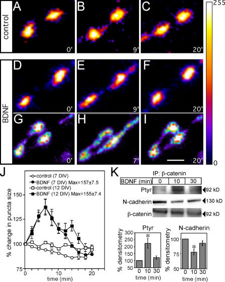
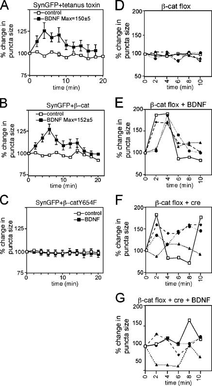
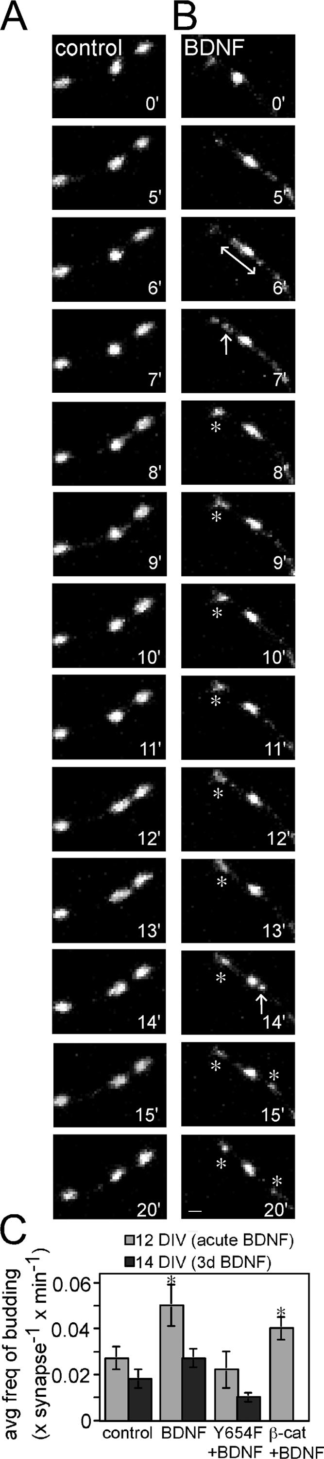
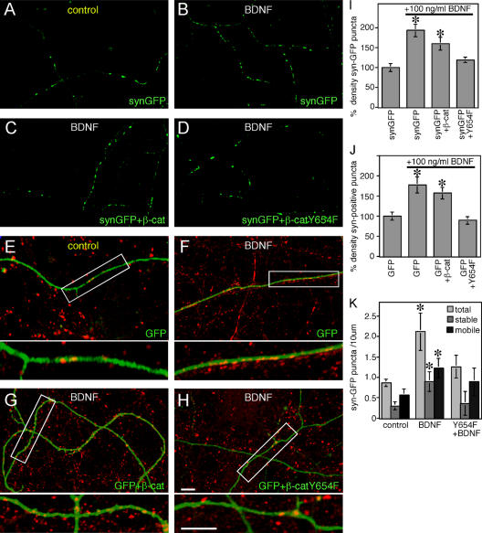
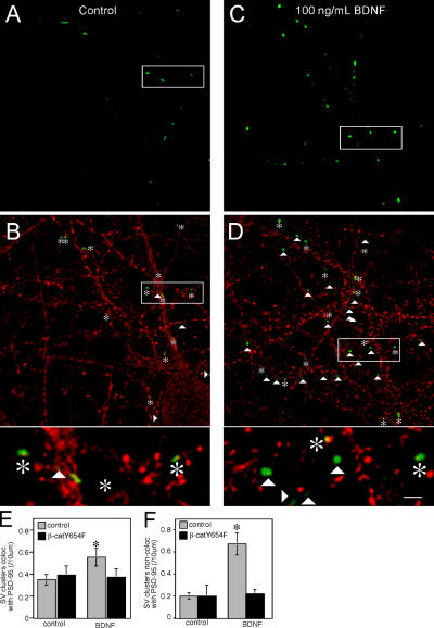
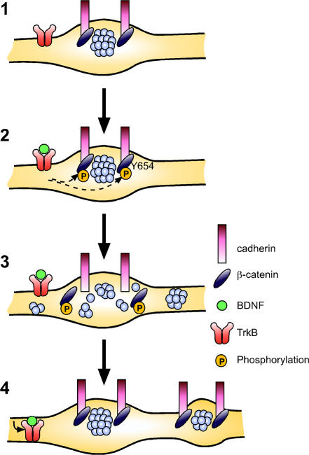
Neurons of the vertebrate central nervous system have the capacity to modify synapse number, morphology, and efficacy in response to activity. Some of these functions can be attributed to activity-induced synthesis and secretion of the neurotrophin brain-derived neurotrophic factor (BDNF); however, the molecular mechanisms by which BDNF mediates these events are still not well understood. Using time-lapse confocal analysis, we show that BDNF mobilizes synaptic vesicles at existing synapses, resulting in small clusters of synaptic vesicles "splitting" away from synaptic sites. We demonstrate that BDNF's ability to mobilize synaptic vesicle clusters depends on the dissociation of cadherin-beta-catenin adhesion complexes that occurs after tyrosine phosphorylation of beta-catenin. Artificially maintaining cadherin-beta-catenin complexes in the presence of BDNF abolishes the BDNF-mediated enhancement of synaptic vesicle mobility, as well as the longer-term BDNF-mediated increase in synapse number. Together, this data demonstrates that the disruption of cadherin-beta-catenin complexes is an important molecular event through which BDNF increases synapse density in cultured hippocampal neurons.


Figures

Figure 1.
Figure 1.
BDNF mediates a transient diffusion of SVs by increasing β-catenin phosphorylation and decreasing cadherin–β-catenin interactions. (A–I) Time-lapse confocal images of 12 DIV hippocampal neurons transfected with synaptophysin-GFP, pseudocolored for fluorescence intensity, and filtered in Photoshop with a Gaussian blur of 2 pixels to reduce the appearance of pixilation. Representative images show untreated cells (A–C) or cells treated with BDNF (D–I) at three time points. Numbers on bottom right of each image represent the time in minutes after the addition of media alone or 100 ng/ml BDNF to cultures. Maximal diffusion of synaptophysin-GFP fluorescence after BDNF treatment varied from puncta to puncta, and two examples are shown in E and H. (J) Quantification of the average percentage of change in the length of the major axis of each synaptophysin-GFP puncta over time ± SEM (n = at least 50 puncta). “Max” represents the average maximum length of the major axis for all puncta, irrespective of time, ± SEM. (K) BDNF induces tyrosine phosphorylation of β-catenin and decreases cadherin–β-catenin interactions. 100 ng/ml BDNF, or media alone, was added to hippocampal cultures for 10 or 30 min. Lysates were immunoprecipitated with anti–β-catenin and blotted for anti-phosphotyrosine and anti–N-cadherin. n = 6. Error bars denote ± SEM. Asterisks indicate P < 0.05. Bar, 2 μm.
Figure 2.
Figure 2.
Quantitative analysis of BDNF-mediated vesicle dispersal. Graphic representation of the percentage of change in the length of the major axis of synaptophysin-GFP puncta versus time. BDNF was applied to the bath, where it is indicated at a concentration of 100 ng/ml. “Max” represents the average maximum percentage of increase in length, irrespective of time, ± SEM. (A) Treatment with tetanus toxin does not affect BDNF-induced SV cluster dispersal. Cells were preincubated in 10 nM tetanus toxin for 16 h before BDNF treatment. After treatment with BDNF, there is a transient diffusion of SVs into perisynaptic regions, as indicated by an increase in the length of synaptophysin-GFP fluorescence along the axon. n = 17 puncta over three experiments. (B and C) Phosphorylation of β-catenin at Y654 is required for BDNF-promoted vesicle dispersion. In the absence of BDNF, the size of synaptic puncta in cells expressing either wild-type β-catenin (B) or β-catenin Y654F (C) were similar to that of controls; however, expression of β-catenin Y654F abolished BDNF-induced SV dispersal (C), whereas expression of wild-type β-catenin did not significantly alter the response of SVs to BDNF (B). For A–C, n = at least 50 puncta per condition from a minimum of 10 neurons over at least three separate cultures. Error bars represent the SEM. (D–G) Dynamic regulation of synaptophysin-GFP puncta by β-catenin and BDNF. Graphic representation of the percentage of change in the length of the major axis of four representative individual synaptophysin-GFP puncta over time (representatives of a total n = 25 over three experiments were examined for each condition). Hippocampal neurons were cultured from β-catenin flox (β-catenin fl/fl) mice and transfected with synaptophysin-GFP. In the presence of β-catenin and the absence of exogenous BDNF, the sizes of each of four synaptophysin-GFP clusters were relatively stable over time (D). After treatment of β-catenin–expressing neurons with BDNF, there is a transient diffusion of SVs into perisynaptic regions as indicated by an increase in the length of synaptophysin-GFP fluorescence along the axons (E). Expression of Cre recombinase to ablate β-catenin, results in increased dynamics of SVs, as indicated by dramatic fluctuations in synaptophysin-GFP puncta length along the axon (F). Addition of BDNF to cells lacking β-catenin does not further increase the dynamics of SV cluster length (G) compared with the fluctuations observed in cells not treated with BDNF, but also lacking β-catenin (F).
Figure 3.
Figure 3.
BDNF enhances the splitting of stable SV clusters. Time-lapse confocal images of synaptophysin-GFP–labeled SV clusters in 12 DIV control cultures (A) or cultures treated with 100 ng/ml BDNF (B). Numbers on the bottom right of each image represent the time in minutes after the addition of media alone or BDNF to cultures. In control cultures, SV clusters are relatively stable over time. After BDNF application, SVs diffuse along the axon transiently (double-headed arrow at 6 min), resulting in the splitting of a cluster of SVs (arrow at 7 min). This splitting results in the formation of a new stable puncta (stable over 8–20 min; denoted by asterisks). SV splitting is also seen at 14 min (arrow), resulting in the formation of a new, apparently stable punctum (right, asterisks at 15 and 20 min). (C) Graphic representation of the frequency of SV cluster splitting in cells acutely treated with BDNF, or treated with BDNF for 3 d (11–14 DIV). n = at least 25–30 puncta per condition from at least 10 neurons over at least three separate cultures. Error bars denote ± SEM. Asterisks denote P < 0.05. Bar, 1 μm.
Figure 4.
Figure 4.
Maintenance of the association between cadherin and β-catenin inhibits BDNF-dependent increases in the density of SV clusters and mature synapses. (A–H) Confocal images of hippocampal neurons transfected with the indicated constructs at 10 DIV and imaged at 14 DIV after treatment of BDNF for 3 d (11–14 DIV). (E–H) Immunolabeling with anti-synaptophysin to identify clusters of endogenous synaptophysin present within GFP-filled processes. Synaptophysin-positive clusters that do not overlap with GFP-filled processes represent presynaptic clusters in untransfected cells. (I–K) Quantification of the density of SV clusters. Changes in the density of SV clusters along the axon after BDNF treatment were determined by quantifying the density of synaptophysin-GFP clusters along the axon (I), or the density of endogenous synaptophysin-immunopositive puncta along a GFP-labeled axon (J). To examine the density of stable and mobile clusters, synaptophysin-GFP dynamics were imaged over a 20-min period, and each synaptophysin-GFP puncta was scored for mobility (K). For each condition, cultures grown in the presence of BDNF were compared with neurons grown without BDNF. Results in I and J demonstrate that 3 d of BDNF treatment results in an increased density of endogenous synaptophysin and synaptophysin-GFP clusters. These increases are prevented by expression of Y654F mutant β-catenin, but not WT β-catenin. Results in K demonstrate that 3 d of BDNF increases the density of both stable and mobile synaptic vesicle clusters and that this increase is prevented by expression of the Y654F mutant of β-catenin. n = at least 13 neurons. Error bars represent ± SEM. Asterisks denote P < 0.05. Bars, 5 μm.
Figure 5.
Figure 5.
Increased density of puncta in excitatory synapses in neurons treated with BDNF. Compared with untreated cells (A), there is an overall increase in the number of synaptophysin-GFP–positive puncta in cells treated with BDNF (C). (B and D) Immunolabeling for PSD-95. Synaptophysin-GFP puncta that were colocalized with PSD-95 (asterisks) and synaptophysin-GFP puncta not colocalized with PSD-95 (arrowheads) were identified. Bar, 10 μm. All green and yellow puncta observed at 63×, 4× zoom were counted. Higher magnification images illustrating examples of synaptophysin-GFP puncta that colocalized or did not colocalize with PSD-95 are shown at bottom of B and D. Bar, 3.3 μm. BDNF treatment increases the density of synaptophysin-GFP puncta that colocalize with PSD-95 (E) and do not colocalize with PSD-95 (F). This increase was abolished in cells expressing the β-catenin Y654F mutant. Error bars depict ± SEM. Asterisks denote P < 0.05.
Figure 6.
Figure 6.
Model for the role of cadherin–β-catenin interactions in BDNF-mediated presynaptic plasticity. (1) In the absence of TrkB-mediated signaling, β-catenin is not phosphorylated at Y654 and is associated with cadherins at the synapse, providing a scaffold through the β-catenin PDZ domain interaction motif for recruitment of scaffold proteins and synaptic vesicle to the synapse. (2) Activation of TrkB receptor tyrosine kinase by BDNF results in the phosphorylation of β-catenin on Y654. This causes the dissociation of β-catenin from cadherins, and disruption of the signals responsible for localizing SVs to the presynaptic compartment. (3) Subsequently, SVs disperse along the axon into perisynaptic regions. (4) β-catenin dephosphorylation and reassociation with cadherin may occur after the internalization of TrkB that can occur within 5 min of BDNF treatment. As a result, SVs recluster at synaptic zones; however, presynaptic compartments are altered and there is a persistent increase in the rate of small SV clusters splitting away from the SV cluster at the active zone. We hypothesize that the increased rate of SV cluster splitting may lead to an increase in the number of mobile SV clusters, and help to promote an increase in the overall density of synapses along the axon as well.

References

    1. Ahmari, S.E., J. Buchanan, and S.J. Smith. 2000. Assembly of presynaptic active zones from cytoplasmic transport packets. Nat. Neurosci. 3:445–451. - PubMed
    1. Applegate, M.D., D.S. Kerr, and P.W. Landfield. 1987. Redistribution of synaptic vesicles during long-term potentiation in the hippocampus. Brain Res. 401:401–406. - PubMed
    1. Bamji, S.X. 2005. Cadherins: actin with the cytoskeleton to form synapses. Neuron. 47:175–178. - PubMed
    1. Bamji, S.X., K. Shimazu, N. Kimes, J. Huelsken, W. Birchmeier, B. Lu, and L.F. Reichardt. 2003. Role of beta-catenin in synaptic vesicle localization and presynaptic assembly. Neuron. 40:719–731. - PMC - PubMed
    1. Benson, D.L., and H. Tanaka. 1998. N-cadherin redistribution during synaptogenesis in hippocampal neurons. J. Neurosci. 18:6892–6904. - PMC - PubMed

Publication types