

Cellular and molecular mechanisms of synovial joint and articular cartilage formation
- PMID: 16831907
- PMCID: PMC2697570
- DOI: 10.1196/annals.1346.010
Cellular and molecular mechanisms of synovial joint and articular cartilage formation
Abstract
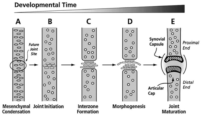
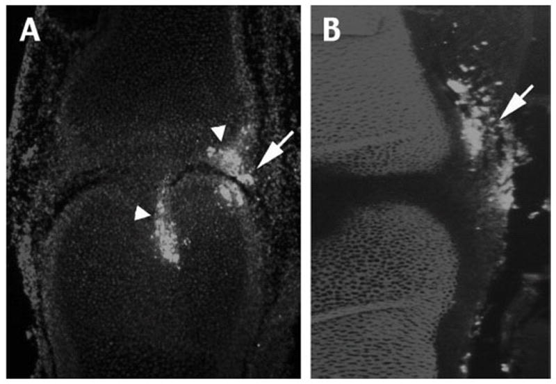
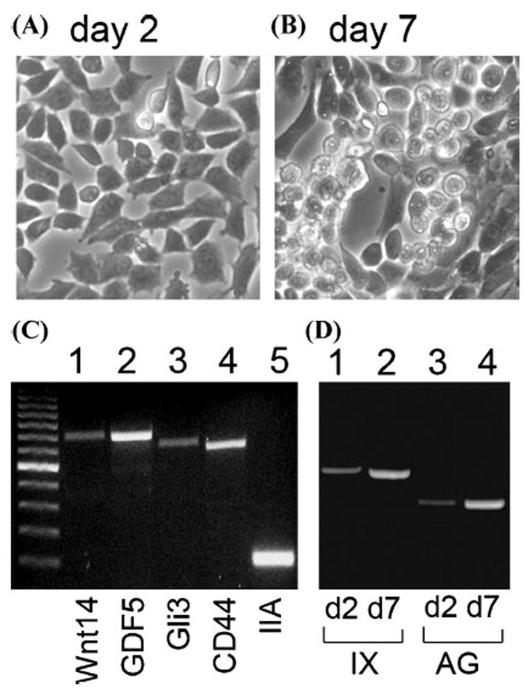
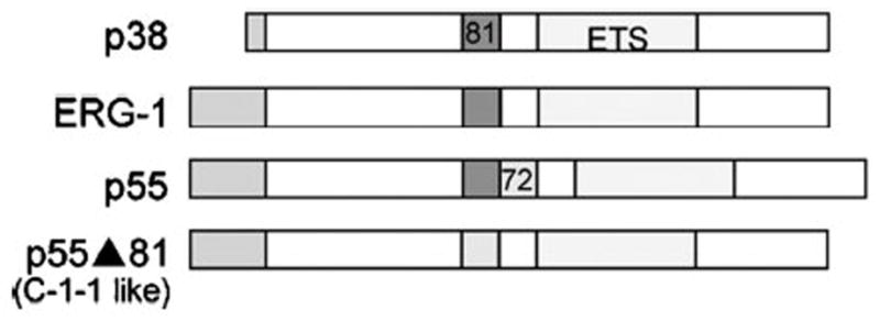
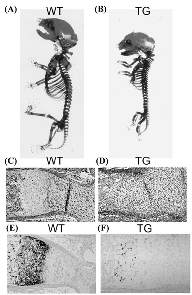
Synovial joints and articular cartilage play crucial roles in the skeletal function, but relatively little is actually known about their embryonic development. Here we first focused on the interzone, a thin mesenchymal cell layer forming at future joint sites that is widely thought to be critical for joint and articular cartilage development. To determine interzone cell origin and fate, we microinjected the vital fluorescent dye DiI at several peri-joint sites in chick limbs and monitored the behavior and fate of labeled cells over time. Peri-joint mesenchymal cells located immediately adjacent to incipient joints migrated, became part of the interzone, and were eventually found in epiphyseal articular layer and joint capsule. Interzone cells isolated and reared in vitro expressed typical phenotypic markers, including GDF-5, Wnt-14, and CD-44, and differentiated into chondrocytes over time. To determine the molecular mechanisms of articular chondrocyte formation, we carried out additional studies on the ets transcription factor family member ERG and its alternatively spliced variant C-1-1 that we previously found to be expressed in developing avian articular chondrocytes. We cloned the human counterpart of avian C-1-1 (ERGp55Delta81) and conditionally expressed it in transgenic mice under cartilage-specific Col2 gene promotor-enhancer control. The entire transgenic mouse limb chondrocyte population exhibited an immature articular-like phenotype and a virtual lack of growth plate formation and chondrocyte maturation compared to wild-type littermate. Together, our studies reveal that peri-joint mesenchymal cells take part in interzone and articular layer formation, interzone cells can differentiate into chondrocytes, and acquisition of a permanent articular chondrocyte phenotype is aided and perhaps dictated by ets transcription factor ERG.
Figures
Similar articles
-
A distinct cohort of progenitor cells participates in synovial joint and articular cartilage formation during mouse limb skeletogenesis.Dev Biol. 2008 Apr 1;316(1):62-73. doi: 10.1016/j.ydbio.2008.01.012. Epub 2008 Jan 26. Dev Biol. 2008. PMID: 18295755 Free PMC article.
-
Formation of synovial joints and articular cartilage.Folia Morphol (Warsz). 2013 Aug;72(3):181-7. doi: 10.5603/fm.2013.0031. Folia Morphol (Warsz). 2013. PMID: 24068678 Review.
-
Transcription factor ERG and joint and articular cartilage formation during mouse limb and spine skeletogenesis.Dev Biol. 2007 May 1;305(1):40-51. doi: 10.1016/j.ydbio.2007.01.037. Epub 2007 Feb 7. Dev Biol. 2007. PMID: 17336282 Free PMC article.
-
Hox11 expression characterizes developing zeugopod synovial joints and is coupled to postnatal articular cartilage morphogenesis into functional zones in mice.Dev Biol. 2021 Sep;477:49-63. doi: 10.1016/j.ydbio.2021.05.007. Epub 2021 May 16. Dev Biol. 2021. PMID: 34010606 Free PMC article.
-
Mechanisms of synovial joint and articular cartilage development.Cell Mol Life Sci. 2019 Oct;76(20):3939-3952. doi: 10.1007/s00018-019-03191-5. Epub 2019 Jun 14. Cell Mol Life Sci. 2019. PMID: 31201464 Free PMC article. Review.
Cited by
-
Joint Development Involves a Continuous Influx of Gdf5-Positive Cells.Cell Rep. 2016 Jun 21;15(12):2577-87. doi: 10.1016/j.celrep.2016.05.055. Epub 2016 Jun 9. Cell Rep. 2016. PMID: 27292641 Free PMC article.
-
TGF-beta signaling is essential for joint morphogenesis.J Cell Biol. 2007 Jun 18;177(6):1105-17. doi: 10.1083/jcb.200611031. J Cell Biol. 2007. PMID: 17576802 Free PMC article.
-
The elusive path to cartilage regeneration.Adv Mater. 2009 Sep 4;21(32-33):3419-24. doi: 10.1002/adma.200801957. Adv Mater. 2009. PMID: 20882507 Free PMC article.
-
Generation of articular chondrocytes from human pluripotent stem cells.Nat Biotechnol. 2015 Jun;33(6):638-45. doi: 10.1038/nbt.3210. Epub 2015 May 11. Nat Biotechnol. 2015. PMID: 25961409
-
Synovial joint cavitation initiates with microcavities in interzone and is coupled to skeletal flexion and elongation in developing mouse embryo limbs.Biol Open. 2022 Jun 15;11(6):bio059381. doi: 10.1242/bio.059381. Epub 2022 Jun 15. Biol Open. 2022. PMID: 35608281 Free PMC article.
References
-
- Archer CW, et al. The Biology of the Synovial Joint. London: Harwood Academics; 1999.
-
- Pacifici M, Liu M, Koyama E. Joint formation: new findings shed more light on this critical process in skeletogenesis. Curr Opin Orthop. 2002;13:339–344.
-
- Archer CW, Dowthwaite GP, Francis-West P. Development of synovial joints. Birth Defects Research. 2003;69:144–155. - PubMed
-
- Holder N. An experimental investigation into the early development of the chick elbow joint. J Embryol Exp Morphol. 1977;39:115–127. - PubMed
Publication types
MeSH terms
Substances
Grants and funding
LinkOut - more resources
Full Text Sources
Other Literature Sources
Miscellaneous
