

Case Reports
doi: 10.1001/archopht.124.7.1048.
Ocular pathologic features of Hermansky-Pudlak syndrome type 1 in an adult
Affiliations
- PMID: 16832032
- PMCID: PMC2478744
- DOI: 10.1001/archopht.124.7.1048
Item in Clipboard
Case Reports
Ocular pathologic features of Hermansky-Pudlak syndrome type 1 in an adult
Arch Ophthalmol.
2006 Jul.
No abstract available
Figures
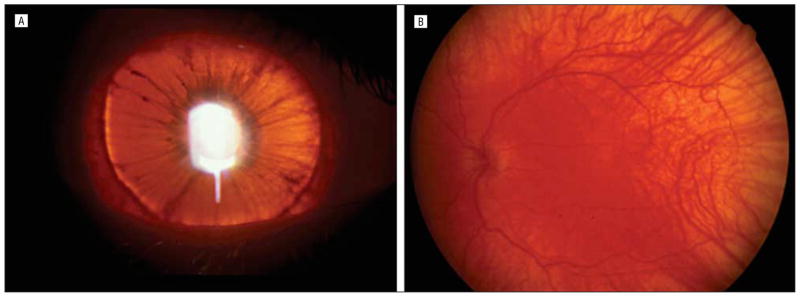
Patient with Hermansky-Pudlak syndrome type 1 described herein. A, The right iris is light brown with marked transillumination such that the edge of the crystalline lens is clearly visible. B, The fundus of the left eye shows clear choroidal vasculature with residual pigmentation in the posterior pole.
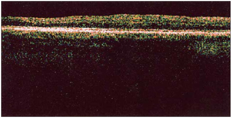
Optical coherence tomography with scan series through the macular area of the right eye retina demonstrates absence of foveal depression.
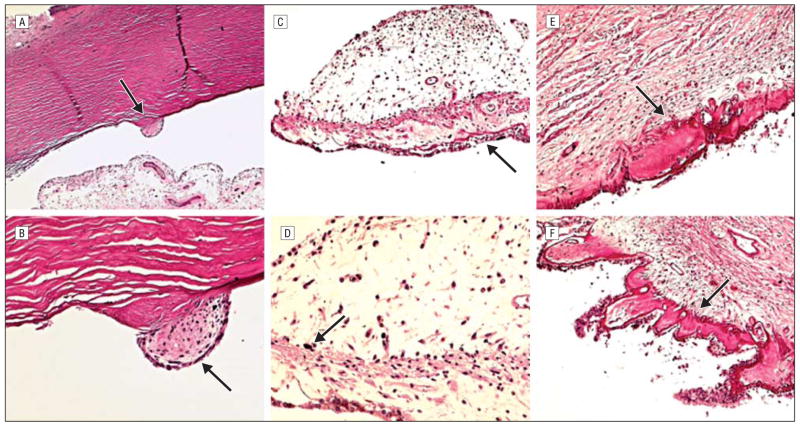
Patient with Hermansky-Pudlak syndrome type 1 described herein. A, Schwalbe ring (arrow) is prominently enlarged and displaced anteriorly (posterior embryotoxon). B, A small cluster of mesenchymal cells (arrow) is adherent to the prominent Schwalbe ring. C, Iris melanocytes are generally absent. Iris pigmented epithelial cells show loss of melanin granules resulting in a lacy appearance (arrow). D, Only small aggregates of large melanin pigment granules (arrow) were disclosed in the iris stroma at the pupillary margin. E and F, Moderate hyalinization (arrows) of the ciliary body was seen in both eyes (hematoxylineosin, original magnification ×50 [A], ×200 [B and D], and ×100 [C, E, and F]).
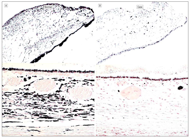
Pigmentation comparisons. A, Healthy white subject with brown irises. B, Patient with Hermansky-Pudlak syndrome type 1 described herein showing paucity of pigmentation of the iris, choroid, and retinal pigment epithelium (Fontana-Masson, original magnification ×100 [iris] and ×200 [choroid]).
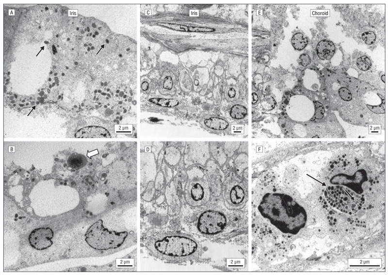
Transmission electron micrographs. A, A few melanocytes that contained sparse immature stage III melanosomes (0.2–0.6 μm in diameter) with incomplete round and oblong melanized granules (arrows). B, Rare membrane-limited lipofuscinlike material with fat globules (open arrow) is noted. C and D, The iris pigmented epithelial cells are enlarged, contain a decreased number of cytoplasmic microorganelles, and have thickened basement membranes. E and F, In the choroids, many melanocytes were swollen and degenerated. Some melanocytes contained sparse, small, immature melanosomes, and some contained an aggregation of stage IV melanosomes (arrow). U-shaped structures were occasionally seen (open arrow) (scale bar, 2 μm).
References
-
- Gahl WA, Brantly M, Kaiser-Kupfer MI, et al. Genetic defects and clinical characteristics of patients with a form of oculocutaneous albinism (Hermansky-Pudlak syndrome) N Engl J Med. 1998;338:1258–1264. - PubMed
-
- Summers CG, Knobloch WH, Witkop CJ, Jr, King RA. Hermansky-Pudlak syndrome: ophthalmic findings. Ophthalmology. 1988;95:545–554. - PubMed
-
- Tsilou ET, Rubin BI, Reed GF, et al. Milder ocular findings in Hermansky-Pudlak syndrome type 3 compared with Hermansky-Pudlak syndrome type 1. Ophthalmology. 2004;111:1599–1603. - PubMed
-
- Huizing M, Boissy RE, Gahl WA. Hermansky-Pudlak syndrome: vesicle formation from yeast to man. Pigment Cell Res. 2002;15:405–419. - PubMed
