. 2006 Jul 18;103(29):10883-8.
doi: 10.1073/pnas.0604607103. Epub 2006 Jul 10.

WNK1 and OSR1 regulate the Na+, K+, 2Cl- cotransporter in HeLa cells

Affiliations

WNK1 and OSR1 regulate the Na+, K+, 2Cl- cotransporter in HeLa cells

Anthony N Anselmo et al. Proc Natl Acad Sci U S A. .

Abstract

Oxidative stress-responsive kinase (OSR) 1 and sterile20-related, proline-, alanine-rich kinase (SPAK) are Ste20p-related protein kinases that bind to the sodium, potassium, two chloride cotransporter, NKCC. Here we present evidence that the protein kinase with no lysine [K] (WNK) 1 regulates OSR1, SPAK, and NKCC activities. OSR1 exists in a complex with WNK1 in cells, is activated by recombinant WNK1 in vitro, and is phosphorylated in a WNK1-dependent manner in cells. Depletion of WNK1 from HeLa cells by using small interfering RNA reduces OSR1 kinase activity. In addition, depletion of either WNK1 or OSR1 reduces NKCC activity, indicating that WNK1 and OSR1 are both required for NKCC function. OSR1 and SPAK are likely links between WNK1 and NKCC in a pathway that contributes to volume regulation and blood pressure homeostasis in mammals.


Conflict of interest statement

Conflict of interest statement: No conflicts declared.

Figures

Fig. 1.
Fig. 1.
Interaction of OSR1 with WNK1. (A) Two-hybrid analysis of the interaction of N-terminal and C-terminal domains of OSR1 with fragments of WNK1. The residues of fragments of WNK1 and OSR1 tested for interaction are as indicated. (B) Immunoprecipitation of OSR1 and mass spectrometric analysis of the major bands. The affinity-purified OSR1 antibody was used to immunoprecipitate OSR1 and associated proteins from HeLa lysates. Coomassie blue-stained proteins in one of three similar immunoprecipitations are shown. The protein stain of an experiment using an unrelated antibody is shown for comparison.
Fig. 2.
Fig. 2.
Phosphorylation and activation of OSR1 1–344 by WNK1 193–483. (A) Phosphorylation of OSR1 with WNK1 in vitro. Phosphorylation reactions were in 30 μl of 1× kinase buffer (20 mM Hepes, pH 7.6/50 μM ATP (5 μCi of [γ-32P]ATP)/10 mM MgCl2/10 mM β-glycerol phosphate) at 30°C for 30 min. The autoradiogram is shown. (B) Activation of OSR1 by WNK1. OSR1 was incubated with WNK1 for the indicated times in the presence of 0.5 mM ATP, followed by the addition of PAK1 1–231 for an additional 15 min. The autoradiogram of substrate phosphorylation is shown. PAK1 1–231 was phosphorylated for 40 min by WNK1 alone in the first lane and by OSR1 alone in the second lane. (C) The time course of activation of OSR1 is plotted. (D) Activation of OSR1 requires the kinase activity of WNK1. Inactive WNK1 in which the obligate autophosphorylation site S382 has been mutated to alanine was used as the enzyme for comparison to active WNK1. OSR1 D164A is a catalytically inactive mutant. OSR1 T173D is an activation loop mutation without apparent effect on activity. (D Upper) The Coomassie-blue stained PAK1 1–231 in the assays. (D Lower) The autoradiogram of phosphorylated PAK1 1–231. AC show one of five similar experiments. D shows one of two similar experiments.
Fig. 3.
Fig. 3.
Specificity of WNK1 and WNK4 toward OSR1 and SPAK. (A) WNK1 and WNK4 differentially activate OSR1. Equal amounts (lanes 1 and 2) and equal units of activity calculated by using myelin basic protein (MBP) as the substrate (lanes 3 and 4) of WNK1 and WNK4 were compared for their capacities to activate OSR1. (A Top) Kinase assays by using the model substrate MBP. (A Middle) OSR1 phosphorylation. A slight gel shift is associated with OSR1 activation. (A Bottom) The activity of OSR1 after preincubation with WNK1 or WNK4 by using PAK1 1–231 as substrate. Shown is one of three similar experiments. (B) Mutation of the essential activation loop threonine prevents activation of SPAK by WNK1. SPAK 63–390, either T243D or wild type, was incubated with WNK1, and the activity then was measured with PAK1 as indicated above. The autoradiogram shows PAK1 phosphorylation by SPAK and is one of two similar experiments.
Fig. 4.
Fig. 4.
WNK1 is required for OSR1 activation. (A) Endogenous OSR1 was immunoprecipitated from cells that previously had been transfected with siRNA directed against rat or human WNK1 or against human OSR1. Twenty-four hours after transfection with oligonucletides, cells were placed in serum-free medium; after an additional 12 h, the cells were placed in medium with or without 0.5 M sorbitol for 30 min. Cells then were lysed, and the kinase activity of endogenous OSR1 (see Materials and Methods) was assayed by using a fragment from the N terminus of NKCC2, GST-SLC12-A1 1–175, as a substrate. Shown is one of three comparable experiments. (B) Methods were as in A, except that after 12 h of starvation, cells were placed in phosphate-free medium (Invitrogen) to which 0.125 mCi/ml 32P was added. Endogeneous OSR1 was immunopurified and washed three times with 10 mM Hepes, pH 7.4/1 M NaCl/0.1% TX-100 and then once with 10 mM Hepes, pH 7.4. The autoradiogram is shown. (C) HEK 293 cells were transfected by using FuGENE with p3XFLAG-OSR1 and either pCMV5-mycGFP or pCMV5-mycWNK1(D368A) (kinase-dead) and treated as described in A, without sorbitol. The kinase activity of immunopurified 3× FLAG-OSR1 was assayed by using GST-SLC12A1(N terminus) as substrate. To detect 3× Flag-OSR1 in immunoprecipitates or lysates, anti-Flag antibody was used.
Fig. 5.
Fig. 5.
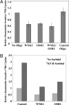
Reduced expression of WNK1 or OSR1 inhibits endogenous NKCC1 activity. HeLa cells were treated with the indicated siRNAs for 48 h. 86Rb influx assays, performed as described in Materials and Methods, were normalized to uptake under the control condition. (A) Average of six independent experiments. Error bars representing the standard error of the mean are shown. The control oligonucleotide is an siRNA that targets endogeneous RanBP2. (B) HeLa cells were treated with the indicated siRNAs for 48 h. Cells were treated for 30 min with medium minus or plus 0.5 M sorbitol. 86Rb uptake was assayed. Shown is one of three similar experiments.

Similar articles

Cited by

References

    1. Dan I., Watanabe N. M., Kusumi A. Trends Cell Biol. 2001;11:220–230. - PubMed
    1. Leberer E., Dignard D., Harcus D., Thomas D. Y., Whiteway M. EMBO J. 1992;11:4815–4824. - PMC - PubMed
    1. Ramer S. W., Davis R. W. Proc. Natl. Acad. Sci. USA. 1993;90:452–456. - PMC - PubMed
    1. Bokoch G. M. Annu. Rev. Biochem. 2003;72:743–781. - PubMed
    1. Tamari M., Daigo Y., Nakamura Y. J. Hum. Genet. 1999;44:116–120. - PubMed

Publication types

MeSH terms

Substances