. 2006 Jul 18;103(29):10979-84.
doi: 10.1073/pnas.0603625103. Epub 2006 Jul 10.

The NF-kappaB regulator Bcl-3 and the BH3-only proteins Bim and Puma control the death of activated T cells

Affiliations

The NF-kappaB regulator Bcl-3 and the BH3-only proteins Bim and Puma control the death of activated T cells

Anette Bauer et al. Proc Natl Acad Sci U S A. .

Abstract

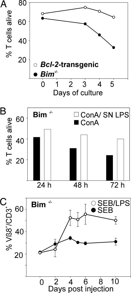
Apoptosis of activated T cells is critical for the termination of immune responses. Here we show that adjuvant-stimulated dendritic cells secrete cytokines that prime activated T cells for survival and analyze the roles of the NF-kappaB regulator Bcl-3 and the proapoptotic Bcl-2 family members Bim and Puma. Bcl-3 overexpression increased survival, and activated bcl-3-/- T cells died abnormally rapidly. Cytokines from adjuvant-stimulated dendritic cells induced Bcl-3, but survival through cytokine priming was Bcl-3-independent. Apoptosis inhibition by Bcl-3 involved blockade of Bim activation, because Bim was overactivated in Bcl-3-deficient cells, and Bcl-3 failed to increase survival of bim-/- T cells. However, adjuvants increased survival also in Bim-deficient T cells. This Bim-independent death pathway is at least in part regulated by Puma, as shown by analysis of puma-/- and noxa-/- T cells. IL-1, IL-7, and IL-15 primed T cells for survival even in the absence of Bim or Puma. Our data define interrelations and a Bim-independent pathway to activated T cell death.


Conflict of interest statement

Conflict of interest statement: No conflicts declared.

Figures

Fig. 1.
Fig. 1.
DC-derived factors promote survival of activated T cells. (A) Spleen cells from C57BL/6 mice were stimulated in vitro for 3 days with ConA with or without LPS. On day 3, T cells were purified and transferred to 96-well plates. T cell survival was measured directly after washing and after 20 h of incubation in simple medium (no growth factors or mitogens). Values are mean ± SD from four independent experiments (∗, P < 0.001). (B) T cells were purified from spleen by negative sorting and stimulated for 3 days with ConA, with or without the addition of LPS (1 μg/ml), supernatant from unstimulated BMDDC (SN C, 50% vol/vol) or supernatant from LPS-stimulated BMDDC (SN LPS, 50% vol/vol). On day 3, cells were washed and transferred into flat bottom 96-well plates, incubated for 20 h and cell death was measured by flow cytometry. Results are mean/SD of three independent experiments.
Fig. 2.
Fig. 2.
Adjuvants cause up-regulation of Bcl-3 protein but can still improve T cell survival in its absence. (A) (Left) Spleen cells from WT or bcl-3−/− mice were stimulated in vitro for 3 days with or without addition of LPS. T cells were then purified and subjected to Western blot analysis. Similar results were seen in three separate experiments. Spleen cells from bcl-3−/− mice were used as a specificity control and detection of β-actin served as a loading control. (Right) Purified splenic T cells were stimulated in vitro for 3 days in six-well cell culture plates with ConA, ConA plus LPS, or ConA plus supernatant from unstimulated (SN C) or LPS-stimulated BMDDC (SN LPS). Cells were washed and analyzed by Western blotting. (B) Spleen cells from WT or bcl-3−/− mice were stimulated in vitro for 3 days with ConA with or without addition of LPS. T cells were then purified and transferred to 96-well plates, and survival was measured after 20 h. Three independent experiments are shown. (C) Purified splenic T cells from bcl-3−/− mice were stimulated in vitro for 3 days with ConA, ConA plus LPS, ConA plus supernatant from unstimulated (SN C), or LPS-stimulated BMDDC (SN LPS). Cells were washed and transferred to 96-well plates, and survival was measured after 20 h. Three independent experiments are shown.
Fig. 3.
Fig. 3.
Bcl-3 inhibits apoptosis of activated T cells by preventing the activation of Bim. (A) Activated Bcl-3-deficient T cells die more rapidly.(Left) Spleen cells from WT and bcl-3−/− mice were stimulated in vitro with ConA. On days 1, 2, or 3, T cells were purified, washed, and transferred into 96-well plates, and survival was determined 20 h later. Two independent experiments are shown. (Right) Resting T cells were purified and incubated in independent experiments for either 24 or 72 h. Another experiment was performed for 24-h incubation with similar results. (B) Bcl-3 increases survival of activated T cells. Spleen cells from WT mice were stimulated in vitro for 2 days with ConA. Cells were then infected with empty control virus or retrovirus driving the expression of Bcl-3. One day later, cells were transferred to 96-well plates, and survival of transduced cells (identified by expression of the marker GFP) was measured at the times indicated (note the different time scales). Values are mean of triplicates in one experiment. Similar results were obtained in three separate experiments; one other experiment is shown in Fig. 8. (C) Increased release of Bim in bcl-3−/− T cells. Spleen cells from WT or bcl-3−/− mice were stimulated in vitro with ConA. On day 3, T cells were purified, washed, and 2.5 × 10⁷ cells were prepared for subcellular fractionation (0 h, Upper). Another 2.5 × 10⁷ cells were incubated for 18 h in the presence of the caspase-inhibitor z-VAD-fmk and then subjected to subcellular fractionation (Lower). Large complexes containing microtubules (after taxol polymerization) were precipitated by ultracentrifugation (P). Supernatants from this step (SN) were acetone-precipitated. Fractions were probed for Bim, tubulin (identifying microtubular fractions), and cytochrome c (as a loading control for the supernatant fraction). Only a small amount of released (activated) BimEL is found in the SN fraction of WT T cells activated for 18 h, but this amount is increased in Bcl-3−/− samples. Data from two of three similar experiments are shown. (D) Bcl-3 overexpression fails further to augment survival of Bim-deficient T cells. Cells were treated and the experiment was performed essentially as in B with the exception that bim−/− cells were used. Similar results were obtained in three separate experiments; one other experiment is shown in Fig. 9. (E) Bim expression is slightly elevated in Bcl-3-deficient T cells. Purified T cells from WT or bcl-3−/−-mice were stimulated in vitro with ConA. On day 3, samples were analyzed for the expression of Bim. Staining with an antibody to tubulin was used as a loading control.
Fig. 4.
Fig. 4.
Bim-independent pathways contribute to activated T cell death. (A) T cells were purified from spleens of either bim−/− or vav-bcl-2-transgenic mice and stimulated with ConA for 3 days. Cells were washed and incubated in 96-well plates for the indicated times before survival was measured. One of two separate experiments is shown. The second experiment is given as Fig. 10A, which is published as supporting information on the PNAS web site. Two further experiments investigating single time points were performed with similar results (not shown). (B) Purified T cells from spleen of bim−/− mice were stimulated for 3 days with ConA alone or in the presence of supernatant from LPS-stimulated BMDDC. T cells were washed, plated in simple medium (no growth factors or mitogens) and survival was measured at the indicated time points. One of two separate experiments is shown. The second experiment is given as Fig. 10B. (C) Six bim−/− mice were injected with SEB. Three mice received LPS 1 day later. The percentage of TCRvβ8+ T cells in peripheral blood was measured on the days indicated by immunofluorescent staining with surface marker-specific antibodies and flow cytometric analysis. Data are mean/SD from individual mice. Another experiment with one mouse for each treatment produced similar results.
Fig. 5.
Fig. 5.
Role of Puma in activated T cell death. Purified T cells from WT or from puma−/− mice were stimulated with ConA (filled bars). Some aliquots were activated in the presence of supernatant derived from LPS-stimulated BMDDC (open bars). On day 3, T cells were washed and plated in medium (no added growth factors), and survival was measured after 20 h.
Fig. 6.
Fig. 6.
IL-1, IL-7, and IL-15 prime T cells for enhanced survival. Purified T cells were stimulated with ConA, either alone or in the presence of BMDDC-LPS-derived supernatant or the recombinant cytokines IL-1, IL-7, or IL-15. On day 3, T cells were washed and transferred into simple medium (no added growth factors). After 20 h, survival was measured. Data are mean/SD from three independent experiments.

Similar articles

Cited by

References

    1. Strasser A. Nat. Rev. Immunol. 2005;5:189–200. - PubMed
    1. Marrack P., Kappler J. Annu. Rev. Immunol. 2004;22:765–787. - PubMed
    1. Pellegrini M., Belz G., Bouillet P., Strasser A. Proc. Natl. Acad. Sci. USA. 2003;100:14175–14180. - PMC - PubMed
    1. Hildeman D. A., Zhu Y., Mitchell T. C., Bouillet P., Strasser A., Kappler J., Marrack P. Immunity. 2002;16:759–767. - PubMed
    1. Adams J. M. Genes Dev. 2003;17:2481–2495. - PubMed

Publication types

MeSH terms