. 2006 Nov 15;400(1):91-7.
doi: 10.1042/BJ20060384.

Phosphorylation status of the Kep1 protein alters its affinity for its protein binding partner alternative splicing factor ASF/SF2

Affiliations

Phosphorylation status of the Kep1 protein alters its affinity for its protein binding partner alternative splicing factor ASF/SF2

Cécile Robard et al. Biochem J. .

Abstract

Mutations in the Drosophila kep1 gene, encoding a single maxi KH (K homology) domain-containing RNA-binding protein, result in a reduction of fertility in part due to the disruption of the apoptotic programme during oogenesis. This disruption is concomitant with the appearance of an alternatively spliced mRNA isoform encoding the inactive caspase dredd. We generated a Kep1 antibody and have found that the Kep1 protein is present in the nuclei of both the follicle and nurse cells during all stages of Drosophila oogenesis. We have shown that the Kep1 protein is phosphorylated in ovaries induced to undergo apoptosis following treatment with the topoisomerase I inhibitor camptothecin. We have also found that the Kep1 protein interacts specifically with the SR (serine/arginine-rich) protein family member ASF/SF2 (alternative splicing factor/splicing factor 2). This interaction is independent of the ability of Kep1 to bind RNA, but is dependent on the phosphorylation of the Kep1 protein, with the interaction between Kep1 and ASF/SF2 increasing in the presence of activated Src. Using a CD44v5 alternative splicing reporter construct, we observed 99% inclusion of the alternatively spliced exon 5 following kep1 transfection in a cell line that constitutively expresses activated Src. This modulation in splicing was not observed in the parental NIH 3T3 cell line in which we obtained 7.5% exon 5 inclusion following kep1 transfection. Our data suggest a mechanism of action in which the in vivo phosphorylation status of the Kep1 protein affects its affinity towards its protein binding partners and in turn may allow for the modulation of alternative splice site selection in Kep1-ASF/SF2-dependent target genes.


Figures

Figure 1
Figure 1. Characterization of the Kep1 antibody
(A) Ovaries of the appropriate genotype were dissected from 2–3-day-old-flies fixed and stained with anti-Kep1 antibody. (B) Increasing amounts of ovary extracts from w1118 (lanes 1, 3, 5 and 7) or kep1−/− (lanes 2, 4, 6 and 8), were separated on polyacrylamide gels and transferred on to a PVDF membrane prior to Western-blot analysis. The arrow indicates the position of the endogenous Kep1 protein. (C) HEK-293 cells were either mock transfected or transfected with Myc–Kep1. Cells were lysed 24 h after transfection, and extracts were subjected to Western-blot analysis. Kep1 antibody was used at a 1:500 dilution.
Figure 2
Figure 2. Kep1 post-translational modifications
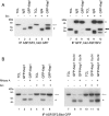
HEK-293 cells were transfected with different kep1 constructs in the presence or absence of activated Src. (A) Cell lysates were subjected to a phosphotyrosine immunoprecipitation (I.P.) (Py99) followed by a GFP Western blot. Lanes 1–3, kinase dead Src; lanes 4–6, activated Src. (B) Cell lysates were subjected to a Kep1 immunoprecipitation followed by a phosphotyrosine Western blot. TCL (total cell lysate), 5% of the cleared transfected cell lysates. IgG, mouse immunogloblin used as a negative control for immunoprecipitations. NRS, normal rabbit serum obtained from the preimmune rabbit. Arrows indicate the position of the transfected Kep1 proteins. The asterisk indicates the position of the IgG heavy chain.
Figure 3
Figure 3. In vivo status of the Kep1 protein
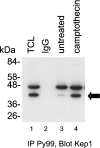
Ovaries from w1118 Drosophila were dissected into Schneider S2 medium and left untreated or treated with camptothecin for 6 h prior to a phosphotyrosine immunoprecipitation (IP). The arrow indicates the position of the endogenous Kep1 protein. TCL, total cell lysate.
Figure 4
Figure 4. In vitro interaction between GST–Kep1 and Drosophila ASF
cDNAs for different members of the SR protein family were transcribed and translated in vitro in the presence [35S]methionine. Reactions were incubated with either GST or GST–Kep1 and subjected to a pull-down. TCL lane is the input control for the splicing factors ASF, SRp54, Sc35 and B52 containing 10% of the rabbit reticulocyte lysate.
Figure 5
Figure 5. Kep1–ASF interactions in the presence of activated Src
(A) COS-7 cells were co-transfected with GFP–Kep1 and either kinase dead Ser (lanes 1–3 and 7–9) or activated Src (lanes 4–6 and 10–12). Total cell lysates (lanes 1, 4, 7 and 10; TCL), IgG controls (lanes 2, 5, 8 and 11), ASF immunoprecipitations (lanes 3 and 6) and GFP immunoprecipitations (lanes 9 and 12) are shown. Open arrow indicates the position of GFP–Kep1, whereas the closed arrow indicates the position of ASF. (B) COS-7 cells were transfected with GFP–Kep1 (lanes 1–6) or GFP–Kep1 I to N which no longer binds homopolymeric RNA (lanes 7–10). To examine the effect of phosphorylation, samples were co-transfected with activated Src (lanes 4–6 and 10–12). For RNase A treatment, cell lysates were incubated with 10 μg of RNase A for 30 min at 37 °C prior to performing the immunoprecipitation (IP) experiments. Open arrow denotes the position of the different GFP–Kep1 fusion proteins. *, heavy chain; and <, light chain.
Figure 6
Figure 6. Modulation of CD44 exon 5 inclusion in cells transfected with Kep1
(A) NIH 3T3 cells were co-transfected with the CD44v5 reporter construct and either an empty expression plasmid, Myc-pCDNA, or with Myc–Kep1. Isolated RNA was subjected to real-time RT–PCR. (A) shows the ratio of exon 5 included over exon 5 excluded from the amplified transcripts (I/E). (B) Western blotting of the above cell lysates demonstrating the presence of Myc–Kep1 expression. (C) Src 3T3 cells were transfected as above and the ratio of exon 5 included over exon 5 excluded was determined by real-time RT–PCR.

Similar articles

Cited by

References

    1. Schwerk C., Schulze-Osthoff K. Regulation of apoptosis by alternative re-mRNA splicing. Mol. Cell. 2005;19:1–13. - PubMed
    1. Wang L., Miura M., Bergeron L., Zhu H., Yuan J. Ich-1, an Ice/ced-3-related gene, encodes both positive and negative regulators of programmed cell death. Cell. 1994;78:739–750. - PubMed
    1. Boise L. H., Gonzalez-Garcia M., Postema C. E., Ding L., Lindsten T., Turka L. A., Mao X., Nunez G., Thompson C. B. bcl-x, a bcl-2-related gene that functions as a dominant regulator of apoptotic cell death. Cell. 1993;74:597–608. - PubMed
    1. Shaham S., Horvitz H. R. An alternatively spliced C. elegans ced-4 RNA encodes a novel cell death inhibitor. Cell. 1996;86:201–208. - PubMed
    1. Srinivasula S. M., Ahmad M., Guo Y., Zhan Y., Lazebnik Y., Fernandes-Alnemri T., Alnemri E. S. Identification of an endogenous dominant-negative short isoform of caspase-9 that can regulate apoptosis. Cancer Res. 1999;59:999–1002. - PubMed

Publication types

MeSH terms