. 2006 Aug 15;15(16):2421-37.
doi: 10.1093/hmg/ddl165. Epub 2006 Jul 11.

Nf1+/- mast cells induce neurofibroma like phenotypes through secreted TGF-beta signaling

Affiliations

Nf1+/- mast cells induce neurofibroma like phenotypes through secreted TGF-beta signaling

Feng-Chun Yang et al. Hum Mol Genet. .

Abstract

Neurofibromas are common tumors found in neurofibromatosis type 1 (NF1) patients. These complex tumors are composed of Schwann cells, mast cells, fibroblasts and perineurial cells embedded in collagen that provide a lattice for tumor invasion. Genetic studies demonstrate that in neurofibromas, nullizygous loss of Nf1 in Schwann cells and haploinsufficiency of Nf1 in non-neuronal cells are required for tumorigenesis. Fibroblasts are a major cellular constituent in neurofibromas and are a source of collagen that constitutes approximately 50% of the dry weight of the tumor. Here, we show that two of the prevalent heterozygous cells found in neurofibromas, mast cells and fibroblasts interact directly to contribute to tumor phenotype. Nf1+/- mast cells secrete elevated concentrations of the profibrotic transforming growth factor-beta (TGF-beta). In response to TGF-beta, both murine Nf1+/- fibroblasts and fibroblasts from human neurofibromas proliferate and synthesize excessive collagen, a hallmark of neurofibromas. We also establish that the TGF-beta response occurs via hyperactivation of a novel Ras-c-abl signaling pathway. Genetic or pharmacological inhibition of c-abl reverses fibroblast proliferation and collagen synthesis to wild-type levels. These studies identify a novel molecular target to inhibit neurofibroma formation.


Conflict of interest statement

Conflict of Interest statement. None declared.

Figures

Figure 1
Figure 1
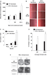
Measurement of bioactivity of MCCM on fibroblast cellular functions. (A) Bioactivity of WT and Nf1+/− MCCM on collagen production was examined by [3H]proline incorporation. Results represent the mean ± SEM from three replicate dishes in one of five independent experiments with similar results. *P < 0.001 for comparison of Nf1+/− versus WT fibroblast collagen production mediated by Nf1+/− MCCM using the Student’s t-test. **P < 0.05 for comparison of WT fibroblast collagen synthesis in response to Nf1+/− MCCM versus WT MCCM using the Student’s t-test. In five independent experiments, Nf1+/− MCCM promotes a 3.1 ± 1-fold increase in [3H]proline incorporation in Nf1+/− fibroblasts versus WT fibroblasts, whereas WT MCCM does not increase [3H]proline incorporation above background (1.1 ± 0.3). (B) Quiescent, mitotic inactivated WT and Nf1+/− fibroblasts were scraped with a pipette tip, and then stimulated with WT or Nf1+/− MCCM. The number of migrating cells were scored by counting cells in 10 high-power fields/dish. (C) Quantitative evaluation of migrating Nf1+/− and WT fibroblasts in response to MCCM. Data represent the mean ± SEM of three replicate dishes in one of five independent experiments with similar results. *P < 0.01 for comparison of the number of WT versus Nf1+/− fibroblast migration mediated by Nf1+/− MCCM using the Student’s t-test. **P < 0.05 for comparison of the number of WT fibroblasts migrating to Nf1+/− MCCM versus that of WT fibroblasts in response to WT MCCM. In five independent experiments, Nf1+/− MCCM promotes a 3.1 ± 1-fold increase in migration of Nf1+/− fibroblasts. WT MCCM does not increase the migration of WT or Nf1+/− fibroblasts above background levels (1.2 ± 0.2). (D) 105 WT or Nf1+/− fibroblasts and 105 WT or Nf1+/− mast cells were admixed in a three-dimensional collagen lattice and lattice contraction was measured. Data represent the mean ± SEM of three replicate dishes from one of five independent experiments with similar results. *P < 0.01 comparing collagen contraction of Nf1+/− fibroblasts in lattices with Nf1+/− mast cells versus WT mast cells. **P < 0.01 comparing collagen contraction of Nf1+/− fibroblasts with WT fibroblasts in response to the same stimulus. (E) Representative lattices containing the indicated populations and a ring indicating the area of the contracted lattice is shown. The baseline area of each lattice is 961 mm2.
Figure 2
Figure 2
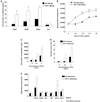
Quantification of the concentrations of profibrotic growth factors in MCCM and the effect of TGF-β on fibroblast proliferation, collagen synthesis and migration. (A) The concentration of murine bFGF, PDGF-AB, TGF-β and IL-6 in WT and Nf1+/− MCCM was determined by ELISA. Results represent the mean ± SEM of five independent cultures. *P < 0.001 for Nf1+/− versus WT MCCM by Student’s t test. (B) About 104 WT or Nf1+/− fibroblasts were starved, stimulated with indicated concentrations TGF-β and [3H]thymidine incorporation was measured. Results represent the mean ± SEM of three replicates from one of five independent experiments with similar results. *P < 0.01 for Nf1+/− versus WT fibroblast proliferation. (C) Collagen synthesis of WT and Nf1+/− fibroblasts was examined by [3H]proline incorporation in response to 1 ng/ml TGF-β. Results represent the mean ± SEM of three replicates from one of five independent experiments with similar results. *P < 0.001 for comparison of Nf1+/− versus WT fibroblasts. In five independent experiments, Nf1+/− fibroblasts had a 2.7 ± 0.4-fold increase in [3H]proline incorporation at the 0.1 ng/ml concentration of TGF-β and a 2.2 ± 0.1-fold increase in [3H]proline incorporation in response to 1.0 ng/ml of TGF-β when compared with WT controls. (D) TGF-β-mediated fibroblast migration was measured by a wound healing assay. Data represent mean ± SEM of three replicates from one of five independent experiments with similar results. *P < 0.01 comparing migration of Nf1+/− fibroblasts with WT fibroblasts in response to TGF-β. In five independent experiments, Nf1+/− fibroblasts had a 2.8 ± 0.4 and a 2.3 ± 0.3-fold increases in migration at the 0.1 and 1 ng/ml concentrations of TGF-β when compared with WT fibroblasts. (E) About 5 × 10⁴ WT and Nf1+/− fibroblasts were pre-incubated with a neutralizing antibody to TGF-β or vehicle, stimulated with 1 ng/ml of TGF-β or MCCM and [3H]proline incorporation was measured. Results represent the mean ± SEM of three replicates from one of five independent experiments with similar results. *P < 0.01 comparing TGF-β-stimulated proline incorporation of Nf1+/− fibroblast with WT fibroblasts in the absence of TGF-β neutralizing antibody. **P < 0.01 comparing proline incorporation of Nf1+/− fibroblasts or WT fibroblasts in the presence of TGF-β neutralizing antibodies with genotype equivalent controls treated with vehicle only following TGF-β stimulation. ***P < 0.01 comparing proline incorporation of Nf1+/− fibroblasts with proline incorporation of WT fibroblasts in response to Nf1+/− MCCM. ****P < 0.01 comparing Nf1+/− MCCM-mediated proline incorporation of Nf1+/− fibroblasts in cultures containing TGF-β neutralizing antibodies versus vehicle control. In five independent experiments, there was a 2.4–2.5 ± 0.3 fold increase in [3H]proline incorporation in Nf1+/− fibroblasts when compared with WT fibroblasts following TGF-β stimulation or Nf1+/− MCCM stimulation. The increase in Nf1+/− MCCM-mediated [3H]proline incorporation was reduced to background levels in the presence of the anti-TGF-β antibody (1.1 ± 0.1).
Figure 3
Figure 3
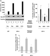
TGF-β-mediated Ras activity and collagen synthesis in Nf1+/− fibroblasts expressing the GRDs of NF1. (A) Nf1+/− fibroblasts were transduced with retroviral constructs expressing a selectable marker and the GRDs of NF1, the GRDs containing a point mutation that inactivates GAP activity (1276) or a construct encoding only the reporter transgene (Pac). WT cells were transduced with the Pac construct. Ras activity in the indicated populations at basal levels and 5 min following stimulation with TGF-β (left panel) and mean activation from three independent experiments (right panel) are shown. (B) Nf1+/− and WT fibroblasts were transduced with the indicated constructs. Following antibiotic selection, transduced cells were stimulated with TGF-β and proline incorporation was measured. Results represent the mean ± SEM of three replicates from four independent experiments. *P < 0.01 for comparison of Nf1+/− fibroblasts transduced with the Pac construct in response to TGF-β versus WT fibroblasts. **P < 0.01 comparing TGF-β-mediated proline incorporation of Nf1+/− fibroblasts transduced with the GRD construct with Nf1+/− fibroblasts expressing the Pac or 1276 constructs using the Student’s t-test.
Figure 4
Figure 4
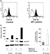
Evaluation of TGF-β-mediated Ras activity and collagen synthesis in fibroblasts from neurofibromas. (A) Fibroblasts from cutaneous neurofibromas and fibroblasts from normal volunteers were examined using an antibody to the surface antigen of Col Ia by using fluorescence cytometry. The population indicated by solid bars represents Col Ia-positive cells and the population indicated by open bars indicates the fluorescence in cells following staining with an isotype control antibody. (B) Ras activity in NF1 and control fibroblasts was examined at basal levels and 5 min following TGF-β stimulation. A representative experiment (left panel) and the mean data from four independent experiments (right panel) are indicated. *P < 0.001 comparing Ras-GTP activity in NF1 patients with unaffected controls using the Student’s t-test. (C) Fibroblasts from NF1 patients or healthy controls were transduced and collagen synthesis was measured following a 24 h stimulation with TGF-β. Results represent the mean ± SEM of three replicates from one of four independent experiments with similar results. *P < 0.01 comparing proline incorporation of NF1 Pac transduced cells with control Pac transduced fibroblasts. **P < 0.01 comparing proline incorporation of NF1 GRD transduced fibroblasts with NF1 pac and NF1 1276 transduced fibroblasts using the Student’s t-test. (D) NF1 fibroblasts were transduced with the indicated constructs and wound healing assays were performed following stimulation with TGF-β. Left panel: results represent the mean ± SEM of three replicate dishes from one of four independent experiments with similar results. *P < 0.05 comparing the number of unaffected control or NF1 fibroblasts that migrate in response to TGF-β versus vehicle. **P < 0.01 comparing migration of NF1 pac transduced fibroblasts with Pac control fibroblasts using the Student’s t-test. ***P < 0.01 comparing the migration of NF1 GRD transduced fibroblasts with NF1 pac or NF1 1276 transduced fibroblasts using the Student’s t-test. Right panel: summary of the relative TGF-β-mediated migration of human NF1 fibroblasts expressing NF1 GAP sequences from four independent experiments.
Figure 4
Figure 4
Evaluation of TGF-β-mediated Ras activity and collagen synthesis in fibroblasts from neurofibromas. (A) Fibroblasts from cutaneous neurofibromas and fibroblasts from normal volunteers were examined using an antibody to the surface antigen of Col Ia by using fluorescence cytometry. The population indicated by solid bars represents Col Ia-positive cells and the population indicated by open bars indicates the fluorescence in cells following staining with an isotype control antibody. (B) Ras activity in NF1 and control fibroblasts was examined at basal levels and 5 min following TGF-β stimulation. A representative experiment (left panel) and the mean data from four independent experiments (right panel) are indicated. *P < 0.001 comparing Ras-GTP activity in NF1 patients with unaffected controls using the Student’s t-test. (C) Fibroblasts from NF1 patients or healthy controls were transduced and collagen synthesis was measured following a 24 h stimulation with TGF-β. Results represent the mean ± SEM of three replicates from one of four independent experiments with similar results. *P < 0.01 comparing proline incorporation of NF1 Pac transduced cells with control Pac transduced fibroblasts. **P < 0.01 comparing proline incorporation of NF1 GRD transduced fibroblasts with NF1 pac and NF1 1276 transduced fibroblasts using the Student’s t-test. (D) NF1 fibroblasts were transduced with the indicated constructs and wound healing assays were performed following stimulation with TGF-β. Left panel: results represent the mean ± SEM of three replicate dishes from one of four independent experiments with similar results. *P < 0.05 comparing the number of unaffected control or NF1 fibroblasts that migrate in response to TGF-β versus vehicle. **P < 0.01 comparing migration of NF1 pac transduced fibroblasts with Pac control fibroblasts using the Student’s t-test. ***P < 0.01 comparing the migration of NF1 GRD transduced fibroblasts with NF1 pac or NF1 1276 transduced fibroblasts using the Student’s t-test. Right panel: summary of the relative TGF-β-mediated migration of human NF1 fibroblasts expressing NF1 GAP sequences from four independent experiments.
Figure 5
Figure 5
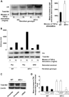
Effect of c-abl activation on Nf1+/− fibroblast function. (A) WT and Nf1+/− fibroblasts were stimulated with TGF-β and c-abl activity was examined using a kinase assay. Left panel: data are representative of one of four independent experiments using different primary cell lines. Right panel: mean activation of c-abl in WT and Nf1+/− fibroblasts (n = 4). (B) Nf1+/− and WT fibroblasts were transduced with the indicated retroviral constructs and TGF-β-mediated c-abl activity was examined. Results are representative of one of four independent experiments. (C) Nf1+/− and WT fibroblasts were transfected with siRNAs to c-abl or with scramble siRNA sequences, and 48 h following transfection, western blotting was performed to measure endogenous c-abl. (D) Nf1+/− and WT fibroblasts were transfected with siRNA sequences to c-abl, scramble siRNA sequences or the vehicle, then stimulated with TGF-β, and [3H]proline incorporation was measured. Results show the mean ± SEM from five independent experiments. *P < 0.01 comparing proline incorporation of Nf1+/− fibroblasts with vehicle or scramble siRNA to Nf1+/− fibroblasts containing c-abl siRNA. (E) Nf1+/− and WT fibroblasts were cultured in the presence of TGF-β and imatinib mesylate or TGF-β and the vehicle for 24 h prior to measuring proline incorporation. Results represent mean ± SEM from one of five independent experiments. *P < 0.001 comparing Nf1+/− fibroblasts with WT fibroblasts following TGF-β stimulation. **P < 0.001 comparing Nf1+/− fibroblasts treated with TGF-β versus Nf1+/− fibroblasts treated with TGF-β and imatinib mesylate. In five independent experiments, TGF-β mediated a 2.1 ± 0.3-fold increase in [3H]proline incorporation of Nf1+/− fibroblasts compared with WT fibroblasts and increased incorporation was inhibited by imatinib mesylate. (F) Human fibroblasts from neurofibromas or normal donors were stimulated with TGF-β ± imatinib mesylate. *P < 0.001 comparing Nf1+/− fibroblasts with WT fibroblasts following TGF-β stimulation. **P < 0.001 comparing fibroblasts from an NF1 patient treated with TGF-β versus NF1 patient derived fibroblasts treated with TGF-β and imatinib mesylate. (G) Nf1+/− and WT fibroblasts were stimulated with TGF-β ± imatinib mesylate and c-abl activity was measured. Data are representative of one of three independent experiments with similar results. (H) Nf1+/− mast cells and fibroblasts were admixed in three-dimensional collagen lattices together with imatinib mesylate or the vehicle. Matrix contraction was measured 24 h after initiation of cultures. Data represent mean ± SEM of five independent experiments with similar results. *P < 0.01 comparing collagen contraction of Nf1+/− fibroblasts with WT fibroblasts incubated with WT mast cells. **P < 0.01 comparing collagen contraction of Nf1+/− fibroblasts with WT fibroblasts incubated with TGF-β. ***P < 0.001 comparing collagen contraction of cultures containing imatinib mesylate added to the cultures with genotypic equivalent cultures incubated in the vehicle. (I) Representative collagen lattices of the indicated genotypes and experimental treatments.
Figure 5
Figure 5
Effect of c-abl activation on Nf1+/− fibroblast function. (A) WT and Nf1+/− fibroblasts were stimulated with TGF-β and c-abl activity was examined using a kinase assay. Left panel: data are representative of one of four independent experiments using different primary cell lines. Right panel: mean activation of c-abl in WT and Nf1+/− fibroblasts (n = 4). (B) Nf1+/− and WT fibroblasts were transduced with the indicated retroviral constructs and TGF-β-mediated c-abl activity was examined. Results are representative of one of four independent experiments. (C) Nf1+/− and WT fibroblasts were transfected with siRNAs to c-abl or with scramble siRNA sequences, and 48 h following transfection, western blotting was performed to measure endogenous c-abl. (D) Nf1+/− and WT fibroblasts were transfected with siRNA sequences to c-abl, scramble siRNA sequences or the vehicle, then stimulated with TGF-β, and [3H]proline incorporation was measured. Results show the mean ± SEM from five independent experiments. *P < 0.01 comparing proline incorporation of Nf1+/− fibroblasts with vehicle or scramble siRNA to Nf1+/− fibroblasts containing c-abl siRNA. (E) Nf1+/− and WT fibroblasts were cultured in the presence of TGF-β and imatinib mesylate or TGF-β and the vehicle for 24 h prior to measuring proline incorporation. Results represent mean ± SEM from one of five independent experiments. *P < 0.001 comparing Nf1+/− fibroblasts with WT fibroblasts following TGF-β stimulation. **P < 0.001 comparing Nf1+/− fibroblasts treated with TGF-β versus Nf1+/− fibroblasts treated with TGF-β and imatinib mesylate. In five independent experiments, TGF-β mediated a 2.1 ± 0.3-fold increase in [3H]proline incorporation of Nf1+/− fibroblasts compared with WT fibroblasts and increased incorporation was inhibited by imatinib mesylate. (F) Human fibroblasts from neurofibromas or normal donors were stimulated with TGF-β ± imatinib mesylate. *P < 0.001 comparing Nf1+/− fibroblasts with WT fibroblasts following TGF-β stimulation. **P < 0.001 comparing fibroblasts from an NF1 patient treated with TGF-β versus NF1 patient derived fibroblasts treated with TGF-β and imatinib mesylate. (G) Nf1+/− and WT fibroblasts were stimulated with TGF-β ± imatinib mesylate and c-abl activity was measured. Data are representative of one of three independent experiments with similar results. (H) Nf1+/− mast cells and fibroblasts were admixed in three-dimensional collagen lattices together with imatinib mesylate or the vehicle. Matrix contraction was measured 24 h after initiation of cultures. Data represent mean ± SEM of five independent experiments with similar results. *P < 0.01 comparing collagen contraction of Nf1+/− fibroblasts with WT fibroblasts incubated with WT mast cells. **P < 0.01 comparing collagen contraction of Nf1+/− fibroblasts with WT fibroblasts incubated with TGF-β. ***P < 0.001 comparing collagen contraction of cultures containing imatinib mesylate added to the cultures with genotypic equivalent cultures incubated in the vehicle. (I) Representative collagen lattices of the indicated genotypes and experimental treatments.
Figure 6
Figure 6
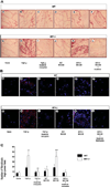
TGF-β-mediated invasion of fibroblasts in matrigel plugs. (A) Representative hematoxylin and eosin stained sections of matrigel plugs from WT and Nf1+/− mice that contained vehicle, TGF-β or MCCM as a stimulus are indicated. Sections from mice that were administered plugs containing TGF-β or MCCM as a stimulus and then fed imatinib mesylate are also indicated. (B) Merged image of PE conjugated anti-CollA positive cells and DAPI counterstain to identify the nuclei. Treatment groups are indicated. (C) Quantitative scoring of fibroblasts within matrigel plugs of the indicated genotypes and experimental treatments. Data represent the mean ± SEM of 10 individual sections from five mice per experimental group. *P < 0.01 comparing the number of fibroblasts per high-power field in plugs containing TGF-β harvested from WT mice with the number of fibroblasts in plug from WT mice containing vehicle. **P < 0.001 comparing the number of fibroblasts per high-power field in the plug harvested from Nf1+/− mice with the number of fibroblasts in plugs from WT mice containing TGF-β. ***P < 0.001 comparing fibroblasts/high-power field from Nf1+/− mice that were administered matrigel plugs containing TGF-β and then fed 200 mg/kg/day of imatinib mesylate daily with mice that were administered TGF-β containing matrigel plugs and fed the vehicle control.

Similar articles

Cited by

References

    1. Knudson AG. Two genetic his (more or less) to cancer. Nat. Rev. Cancer. 2001;2:157–162. - PubMed
    1. Cook WD, McCaw BJ. Accommodating haploinsufficient tumor suppressor genes in Knudson’s model. Oncogene. 2000;19:3434–3438. - PubMed
    1. Venkatachalam S, Shi YP, Jones SN, Vogel H, Bradley A, Pinkel D, Donehower LA. Retention of wild-type p53 in tumors from p53 heterozygous mice: reduction of p53 dosage can promote cancer formation. EMBO J. 1998;17:4657–4667. - PMC - PubMed
    1. Viskochil D, Buchberg AM, Xu G, Cawthon RM, Stevens J, Wolff RK, Culver M, Carey JC, Copeland NG, Jenkins NA, et al. Deletions and a translocation interrupt a cloned gene at the neurofibromatosis type 1 locus. Cell. 1990;62:187–192. - PubMed
    1. Wallace MR, Marchuk DA, Andersen LB, Letcher R, Odeh HM, Saulino AM, Fountain JW, Brereton A, Nicholson J, Mitchell AL, et al. Type 1 neurofibromatosis gene: identification of a large transcript disrupted in three NF1 patients. Science. 1990;249:181–186. - PubMed

Publication types

MeSH terms