. 2006 Nov 1;108(9):2989-97.
doi: 10.1182/blood-2006-05-022137. Epub 2006 Jul 11.

STAT5 acts as a repressor to regulate early embryonic erythropoiesis

Affiliations

STAT5 acts as a repressor to regulate early embryonic erythropoiesis

Matthew Schmerer et al. Blood. .

Abstract

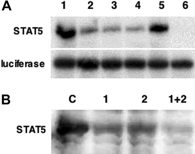
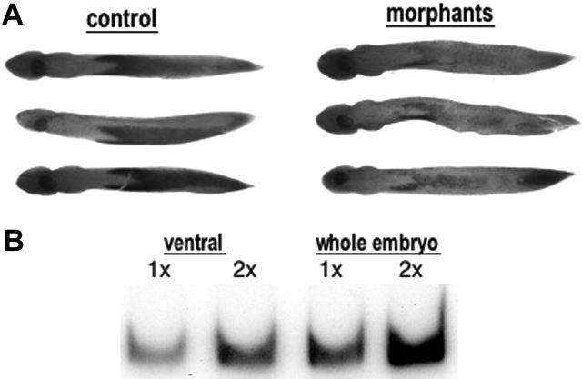
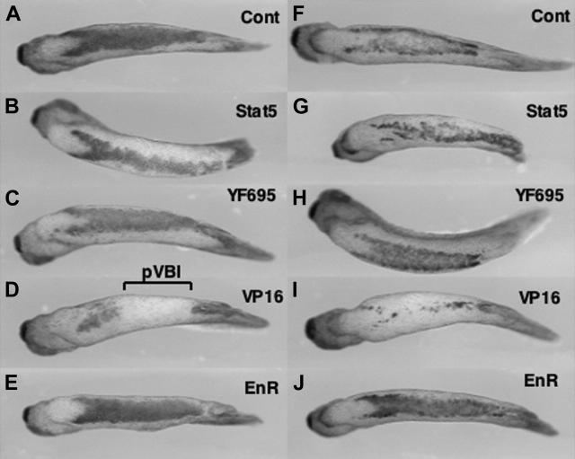
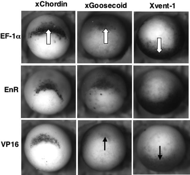
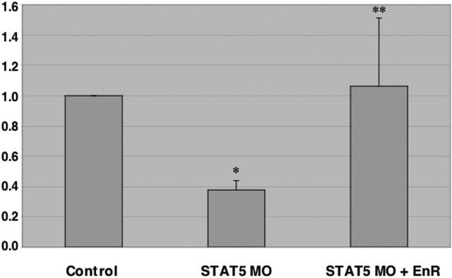
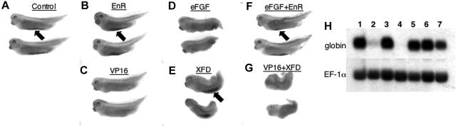
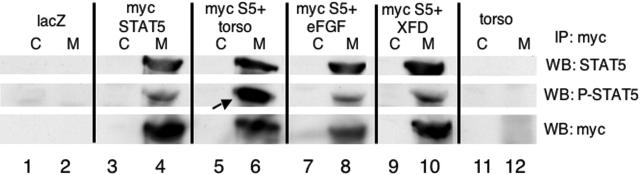
STAT5 regulates definitive (adult stage) erythropoiesis through its ability to transduce signals from the erythropoietin receptor. A function for STAT-dependent signaling during primitive (embryonic) erythropoiesis has not been analyzed. We tested this in the Xenopus system, because STAT5 is expressed at the right time and place to regulate development of the embryonic primitive ventral blood island. Depletion of STAT5 activity results in delayed accumulation of the first globin-expressing cells, indicating that the gene does regulate primitive erythropoiesis. Our results suggest that in this context STAT5 functions as a repressor, since forced expression of an activator isoform blocks erythropoiesis, while embryos expressing a repressor isoform develop normally. The erythroid phenotype caused by the activator isoform of STAT5 resembles that caused by overexpression of fibroblast growth factor (FGF). We show that STAT5 isoforms can function epistatic to FGF and can be phosphorylated in response to hyperactivated FGF signaling in Xenopus embryos. Therefore, our data indicate that STAT5 functions in both primitive and definitive erythropoiesis, but by different mechanisms.


Figures

Figure 1.
Figure 1.
Characterization of STAT5 morpholinos. (A) Two distinct morpholinos (MO1 and MO2) were designed to target sequences around the ATG initiation site of xSTAT5. The ability of each to inhibit STAT5 translation was tested first in vitro. STAT5 protein (top panel) or as a control luciferase protein (bottom panel) was transcribed and translated in vitro using rabbit reticulocyte lysates either alone or in the presence of MO1, MO2, or both. Lanes are 1) DNA template alone, 2) 100 ng MO1, 3) 100 ng MO2, 4) 100 ng MO1 + 100 ng MO2, 5) 200 ng nonspecific control morpholino, and 6) 250 ng MO1 + 250 ng MO2. Translation lysates were analyzed by sodium dodecyl sulfate–polyacrylamide gel electrophoresis (SDS-PAGE) followed by fluorography. (B) The activity of the morpholinos was next tested in vivo. Shown is a representative Western blot for xSTAT5, following injection of RNA encoding xSTAT5 alone (control, C) or co-injected with either MO1 or MO2 alone (1 or 2, 10 ng) or both MO1 and MO2 together (1 + 2, 10 ng each). Under these conditions the morpholinos reproducibly result in a substantial inhibition of STAT5 expression.
Figure 2.
Figure 2.
STAT5 activity is required for normal blood island development. (A) Shown are representative embryos that were either uninjected (control) or injected with both MO1 and MO2 (20 ng each) into both blastomeres at the 2-cell stage. Embryos were harvested at stage 25 and processed by whole mount in situ hybridization to detect transcript levels for embryonic α-globin, which are substantially reduced in the morphant embryos. Embryos are positioned ventral side up and anterior to the left. This experiment was repeated 3 times, and although the number of affected embryos varies, the statistics provided under “Results” are representative. (B) RNA was isolated at stage 25 from presumptive VBI regions (ventral) or whole embryos and tested for the presence of xSTAT5 RNA using semiquantitative RT-PCR. Samples for the RT reaction contained either 0.25 μg(1×) or 0.5 μg(2×) of total RNA. The xSTAT5 RNA was detected in this example using 28 cycles of PCR and still showing dependence on the amount of RNA in the original RT reaction. This experiment was repeated 3 times with similar results.
Figure 3.
Figure 3.
Expression of an activator isoform of STAT5 inhibits primitive erythropoiesis in the VBI. Embryos were injected at the 4-cell stage into the 2 presumptive “ventral” blastomeres, to target RNA to the presumptive posterior VBI. Shown are representative embryos processed by in situ hybridization at stage 35 with probes for embryonic α-globin (left panels, A-E) or SCL (right panels, F-J). Embryos were injected with RNA encoding lacZ as a control (A, F), STAT5 (B, G), a mutated form of STAT5 that should be inactive (C, H), a constitutively activated isoform STAT5-VP16 (D, I), or a repressor isoform STAT5-EnR (E, J). The region of the pVBI that is inhibited for erythropoiesis by STAT5-VP16 is indicated in panel D. Views are ventral, with anterior to the left. The data are consistent with a normal function for STAT5 as a repressor of genes that inhibit globin expression. While the transcript patterns can vary somewhat from embryo to embryo, the phenotypes seen were reproducible in at least 3 independent experiments. The embryos shown here are derived from one experiment, where the expression pattern shown is most representative from a batch of (n) embryos: A, 12 of 12; B, 11 of 11; C, 10 of 15; D, 7 of 9; E, 13 of 13; F, 17 of 20; G, 10 of 13; H, 8 of 13; I, 9 of 12; J, 10 of 14.
Figure 4.
Figure 4.
Modulation of STAT5 activity does not correlate with altered BMP signaling. Shown are representative embryos processed by whole mount in situ hybridization at stage 10.5 for the dorsal-anterior mesoderm marker xChordin (left column panels), the Spemann Organizer marker xGoosecoid (middle column panels), or the ventral-posterior marker Xvent-1 (right column panels). All embryos are shown with a dorsal lip view, anterior at the top. Embryos were injected with EF-1α as a control (top row) or had been injected with RNA encoding STAT5-EnR (middle row) or STAT5-VP16 (bottom row), as indicated. The white arrows point out the normal expression domains for these early markers (seen in this experiment for the majority of at least 12 embryos). As indicated by the black arrows, STAT5-VP16 reduces modestly the expression levels for both xGoosecoid (seen in this experiment for 7 of 12 embryos) and Xvent-1 (seen in this experiment for 10 of 16 embryos) but not xChordin (13 embryos).
Figure 5.
Figure 5.
Globin transcript levels are dependent on STAT5 repressor activity. Embryos were injected with a control morpholino or the STAT5 morpholino (STAT5 MO) and, in addition, 1 ng RNA encoding STAT5-EnR, or RNA encoding lacZ as a control. Embryos were harvested at stage 24-25 and RNA processed for quantitative RT-PCR assays. Each independent sample consisted of a pool of 50 embryos. The median of each sample was normalized to its respective ODC control, and the median average from 4 independent experiments was graphed as fold change in RNA expression. Error bars indicate standard error of the mean. The asterisk indicates that the change in α-globin transcript levels comparing control injected and STAT5 MO injected (first and second samples) is statistically significant (P < .001). The double asterisk indicates that the rescue with STAT5-EnR RNA compared to MO co-injected with lacZ RNA (second and third samples) is statistically significant (P < .03).
Figure 6.
Figure 6.
STAT5 activity can interact with FGF signaling in Xenopus embryos. Shown are representative embryos fixed and stained with benzidine to detect differentiated globin-expressing erythroid cells. Embryos had been injected into both blastomeres at the 2-cell stage with RNA encoding (A) lacZ as a control, (B) STAT5-EnR, (C) STAT5-VP16, (D) eFGF, (E) the dominant-negative FGFR isoform XFD, (F) eFGF + STAT5-EnR, (G) STAT5-VP16 + XFD. Benzidine-positive blood islands are indicated by the black arrows. Note that STAT5-VP16 and eFGF each inhibit VBI development (C-D). Restoring STAT5 repressor activity by STAT5-EnR is sufficient to rescue at least partially the repressive effect of eFGF (F), while XFD is unable to rescue VBI development that is repressed by STAT5-VP16 (G). Views are lateral with anterior to the left. The patterns shown were reproducible in at least 3 independent experiments. The embryos shown here are most representative of the staining patterns from one experiment representing the phenotype of (n) embryos: A, 12 of 19; B, 14 of 27; C, 12 of 12; D, 8 of 19; E, 23 of 26; F, 18 of 32; G, 21 of 29. (H) Shown is a representative Northern blot probed for RNA encoding embryonic α-globin (top panel) or as a loading control, the same blot reprobed for the housekeeping gene EF-1α (bottom panel). Samples are derived from pools of at least 25 embryos that had been injected with RNA encoding (1) lacZ as a control, (2) STAT5-VP16, (3) STAT5-EnR, (4) eFGF, (5) XFD, (6) eFGF + STAT5-EnR, (7) XFD + STAT5-VP16. Note that STAT5 repressor activity rescues much of the inhibition of globin transcription by eFGF (lane 6), while STAT5-VP16 is still partially repressive even in the presence of XFD (lane 7).
Figure 7.
Figure 7.
Expression of an activated FGF receptor results in increased phosphorylation of STAT5 in Xenopus embryos. Shown are representative Western blotting experiments on samples that had been immunoprecipitated with antibodies specific for the myc epitope (M) or equivalent samples that were processed similarly using an isotype-matched control antibody (C). Lysates were derived from pools of 20 stage 12 embryos that had been injected with RNA encoding LacZ as a control (lanes 1, 2), myc-tagged xSTAT5 (myc STAT5, lanes 3, 4), the constitutively activate isoform of the FGFR1 (torso) + myc-STAT5 (myc S5, lanes 5, 6), eFGF + myc STAT5 (lanes 7, 8), myc-STAT5 + the dominant-negative FGFR1 (XFD, lanes 9, 10), or the constitutively active isoform of the FGFR1 (torso) alone (lanes 11, 12). Blotted samples were probed sequentially using antibodies for total STAT5 protein (top panel), STAT5 phosphorylated specifically at tyrosine 695 (P-STAT5, middle panel), or total myc-tagged proteins (myc, bottom panel). The phospho-STAT5 levels are increased over baseline upon expression of torso (arrow in lane 6), although the baseline levels of phospho-STAT5 are not reduced in the presence of XFD.

Similar articles

Cited by

References

    1. Evans T. Developmental biology of hematopoiesis. Hematol Oncol Clin North Am. 1997;11: 1115-1147. - PubMed
    1. Mangia F, Procicchiami G, Manelli H. On the development of the blood island in Xenopus laevis embryos: light and electron microscope study. Acta Embryol Exp (Palermo). 1970;2: 163-184. - PubMed
    1. Chen XD, Turpen JB. Intraembryonic origin of hepatic hematopoiesis in Xenopus laevis. J Immunol. 1995;154: 2557-2567. - PubMed
    1. Smith JC. Mesoderm-inducing factors and mesodermal patterning. Curr Opin Cell Biol. 1995;7: 856-861. - PubMed
    1. Dale L, Wardle FC. A gradient of BMP activity specifies dorsal-ventral fates in early Xenopus embryos. Semin Cell Dev Biol. 1999;10: 319-326. - PubMed

Publication types

MeSH terms