. 2006 Sep;17(9):3989-4001.
doi: 10.1091/mbc.e06-03-0239. Epub 2006 Jul 12.

Fab1 phosphatidylinositol 3-phosphate 5-kinase controls trafficking but not silencing of endocytosed receptors

Affiliations

Fab1 phosphatidylinositol 3-phosphate 5-kinase controls trafficking but not silencing of endocytosed receptors

Tor Erik Rusten et al. Mol Biol Cell. 2006 Sep.

Abstract

The trafficking of endocytosed receptors through phosphatidylinositol 3-phosphate [PtdIns(3)P]-containing endosomes is thought to attenuate their signaling. Here, we show that the PtdIns(3)P 5-kinase Fab1/PIKfyve controls trafficking but not silencing of endocytosed receptors. Drosophila fab1 mutants contain undetectable phosphatidylinositol 3,5-bisphosphate levels, show profound increases in cell and organ size, and die at the pupal stage. Mutant larvae contain highly enlarged multivesicular bodies and late endosomes that are inefficiently acidified. Clones of fab1 mutant cells accumulate Wingless and Notch, similarly to cells lacking Hrs, Vps25, and Tsg101, components of the endosomal sorting machinery for ubiquitinated membrane proteins. However, whereas hrs, vps25, and tsg101 mutant cell clones accumulate ubiquitinated cargo, this is not the case with fab1 mutants. Even though endocytic receptor trafficking is impaired in fab1 mutants, Notch, Wingless, and Dpp signaling is unaffected. We conclude that Fab1, despite its importance for endosomal functions, is not required for receptor silencing. This is consistent with the possibility that Fab1 functions at a late stage in endocytic receptor trafficking, at a point when signal termination has occurred.


Figures

Figure 1.
Figure 1.
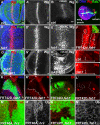
Drosophila fab1 structure, alleles, phenotypes, and failure of recruitment of GFP-Atg18 to endocytic structures in fab1 mutants. (A) Cartoons representing the Drosophila fab1 cDNA (exons are in black and introns in white) and predicted protein structure including the N-terminal FYVE domain, chaperonin-like domain, and C-terminal kinase domain. The vertical bars with associated numbers indicate the position of the point mutations leading to premature stop codons in the respective alleles, fab18 (Q1308Stop), fab131 (L1321Stop), and fab121 (Q1521Stop). The complementation table shows fab1 alleles over a small, Df(2R)w30, and larger deficiency, Df(2R)Pcl7b, which both uncover the fab1 locus. The lethal phase of the respective transheterozygotic combinations is indicated. pa, pharate adult; wL3, wandering L3 larva; nd, not determined. (B) fab1 mutant pharate adults and dissected legs are larger than in control animals of a comparable stage of development (ventral and dorsal views of pupae are shown with arrows indicating body length). (C and D) Heads and eyes display tissue overgrowth in animals whose head consists mainly of fab1 mutant cells (mutant cells in the eye lack red pigmentation; see Materials and Methods). The average ommatidium size was 1.25 times larger in fab1 mutant eyes (n = 828 ommatidia, 8 eyes) than in control eyes (n = 952, 8 eyes). (E–E″) Confocal image in which recruitment of GFP-Atg18 can be seen to punctate structures (arrowheads) and late endosomes and lysosomes (arrows) labeled with LysoTracker in a unfixed eye disk from an L3 larva. (F–F″) Confocal image of apical endosomes labeled with a 20-min uptake of TRD in a fixed eye imaginal disk. Note that structures can be seen with TRD alone (asterisks, probably representing early endosomes) or together with GFP-Atg18 (arrows). Some structures label with GFP-Atg18 alone (arrowhead). (G–G″ and H–H″) Overview confocal images in which internalized dextran can be observed in apical developing photoreceptor cell clusters in both control and fab1 mutant eye disks. Although GFP-Atg18 can be clearly seen concentrated on subcellular structures in control disks, no such subcellular accumulation can be seen in the mutant tissue; 12.6 ± 3.6% of GFP-Atg18 structures also labeled with TRD in control animals (n = 583 endosomes 5 eye imaginal disks), whereas no GFP-Atg18 structures (0%) were observed with fab1 mutant discs (n = 303 endosomes, 5 eye imaginal disks). (I) Cartoon showing the conversion of PtdIns into PtdIns(3)P by PI 3-kinase (PI3K) and the subsequent conversion into PtdIns(3,5)P2 by Fab1. The FYVE domain and the yeast protein Atg18 can specifically bind PtdIns(3)P or PtdIns(3,5)P2, respectively, and serve as probes for detecting these lipids in vivo. Bars, 1 μ (C–E) and 2 μ (F–H). Genotypes are fab131/Df(2R)w30 and + /Df(2R)w30 (B), yw, ey-flp, GMR-LacZ/Y; FRT42D, p [mini-w], l(2)clR11/FRT42D, fab121, yw, ey-flp, GMR-LacZ/Y; FRT42D, p [mini-w], l(2)clR11/+ (mutant and control heads in C and D), w1118; tGFP-Atg18/+ (E–G), and fab131/Df(2R)w30; tGFP-Atg18/+ (H).
Figure 2.
Figure 2.
Fab1 subcellular localization and effect on lysosomal acidity. (A and B) Punctate subcellular staining of Fab1 was observed in isolated Garland cells from wandering L3 control animals but not from Df(2R)w30/Df(2R)Pcl7b mutants that lack the fab1 genomic region. The dashed line indicates the cell membrane. (C and D) OGD was internalized in Garland cells for 5 min, and immunolocalization with Fab1 was scored after 0 (C) and 40 min (D). Fab1 overlaps extensively with OGD after 40 min, when OGD primarily labels late endosomes/lysosomes. (E–H) In apical confocal sections of epithelial cells of wing imaginal disks, extensive overlap is also observed with GFP-Rab5 on early and GFP-Rab7 on late endosomes, but Fab1 seems to be in proximity rather than in the same region as GFP-2xFYVE and Hrs on sorting endosomes/late endosomes. (I) Intensity of LysoTracker emission is a measure of acidity of intracellular late endosomes and lysosomes. Pictures are shown taken with the same confocal scanning settings of Garland cells from mutant animals in which the average acidity is dramatically decreased compared with the control. (J and K) In optical Z-sections of the epithelium from wing imaginal disks, more space and irregular organization between TOTO-3–labeled nuclei can be observed in mutant tissue compared with the control. The organization of apical adherence junctions where actin accumulates and the slightly more basally localized septate junctions containing Disk large (Dlg) seem normal. Bars, 5 μm (A and B), 2 μm (C and D), 1 μm (E–H), 50 μm (I), and 5 μm (J and K). Genotypes are w1118 (A, C, D, F, I, and J), w1118/+; ptc-Gal4/UAS-GFP-Rab5 (E), w1118/+;ptc-Gal4/UAS-myc-GFP-dbFYVE (G), w1118/+; ptc-Gal4/UAS-GFP-Rab7 (H), and fab131/Df(2R)w30 (I and K).
Figure 3.
Figure 3.
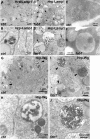
Altered fluid-phase transport and accumulation of cargo in a prelysosomal compartment in fab1 mutants. (A and B) OGD accumulated in small peripheral vesicles after 5 min of uptake and reached late endosomal/lysosomal compartment after a 40-min chase period (arrows). (C and D) In fab1 mutants, in contrast, OGD failed to reach acidified compartments after chase (note that detector gain of the red channel was increased to allow visualization of the more weakly acidified compartments in fab131/Df(2R)w30 cells). (E–J) Electron micrographs of control (E–G) and fab1 (H–J) Garland cells. (E and H) Low magnification overview shows normal-sized endosomes in control cells and greatly enlarged endosomes in the fab1 cells (arrows in both micrographs). To further characterize the endocytic pathway, we incubated cells with BSA-Gold (arrowheads in all micrographs) for 40-min continuous incubation. (F and G) In control cells, we observed BSA-gold (arrowheads in enlarged inset of F and G) both in endosomal structures and lysosomal compartments. (I) In fab1 mutants, BSA-gold localized to dramatically enlarged endosomes and was not observed in dense lysosomal structures. Note that only part of the endosome is depicted. (J) Enlarged view of boxed area in I shows gold particles associated with dense matter in endosome. n, nucleus; e, endosome; and l, lysosome. Bars, 2 μm (A–D), 1 μm (E and H), 100 nm (F), and 200 nm (G and I). Genotypes are w1118 (A, B, and E–G) and fab131/Df(2R)w30 (C, D, and H–J).
Figure 4.
Figure 4.
Cell autonomous accumulation of receptors and ligands in fab1 mutants. (A–C, and I) Wg is expressed in a central stripe of cells in the wing pouch and notum and taken up in neighboring cells where it can be observed in intracellular vesicles up to 20 μm away from the source (I, projection of 50 confocal stacks 1 μm apart). (D, E, G, H, and J) In fab1 mutant wing disks, Wg accumulation can be seen in enlarged intracellular vesicles both in the wing pouch and the notum visible up to 40 μm away from the source (J, projection like in I). This accumulation is specifically rescued in the wing pouch upon expression of full-length fab1 in the wing pouch under nub-Gal4 control (G and G′), and not in the notum (H). (F) Cell autonomous accumulation of Wg is seen in fab1 clones (dashed lines), labeled by the absence of GFP, outside the source of Wg production (overview shown in inset). (K) A similar accumulation of Notch in intracellular vesicles can be seen in fab1 mutant cells of the eye disks posterior to the morphogenetic furrow (arrowheads). (L) A similar accumulation is seen of the Notch ligand, Delta (Dl), in fab1 mutant follicle cells. (M and N) Ubiquitinated proteins accumulate in apical endosomes in hrs but not in fab1 mutant clones (outlined by dashed lines) in the wing disk. Bars, 10 μm (A, B, D, E, G, and H) and 50 μm (I and J). Genotypes are w1118 (A–C and I), fab131/Df(2R)w30 (D, E, and J), fab131,nub-Gal4/Df(2R)w30; p [mini-w, UAS-fab1]/+ (G and H), yw, hsp70-flp/+; FRT42D, p [mini-w, ubi-GFP]/FRT42D, fab121 (F, K, L, and N), and yw, hsp70-flp/+; FRT40A, p [mini-w, ubi-GFP]/FRT40A, hrs28D (M).
Figure 5.
Figure 5.
Receptors and ligands accumulate in enlarged late endosomal structures in fab1 mutants. Endosomal trafficking of Wg in receiving cells was followed in control and fab1 mutant wing disks. (A–D) Single confocal sections and separate channels of enlargement show localization of Wg in the lumen of enlarged Hrs-positive early endosomes and Rab7-positive MVBs in fab1 mutant wing disks. (E and F) Wg colocalizes with 2xFYVE in both control and mutant cells (arrows). An additional pool of larger Wg-vesicles is prominent in mutant disks (arrowhead). (G and H) The largest Wg-positive structures (2–5 μm) in mutant tissue colocalize with the luminal HRP moiety of the human Lamp1-HRP fusion protein, whereas little overlap is seen between Wg and HRP-Lamp1 in control tissue. (I) Endogenous Lamp1 is detected in punctae on the limiting membrane of the large Wg containing Lamp1-positive endosomes. (J) Like Wg, the large 2- to 5-μm-sized Notch (N) containing structures contain Lamp1 in what seems to be the limiting endosomal membrane in fab1 mutant clones (−/−), whereas little overlap is seen in heterozygous control cells (+/−). (K) The large Notch-containing structures colocalize with TRD. (L and M) Immunodetection of Wg and the HRP epitope in wing disks expressing Hrp-Wg under dpp-Gal4 control (overview to the left and enlargement with overlay and separate channels to the right) shows extensive colocalization of Wg and HRP in large structures many cell diameters away from the source in mutant tissue. In control tissue, HRP is mainly detected alone in small structures. Genotypes are w1118 (A), fab131/Df(2R)w30 (B, I, and K), w1118; pUAS-GFP-Rab7; dpp-Gal4/+ (C), fab131/Df(2R)w30, pUAS-GFP-Rab7; dpp-Gal4/+ (D), w1118; pUAS-myc-GFP-dbFYVE/+; dpp-Gal4/+ (E), fab131/Df(2R)w30, pUAS-UAS-myc-GFP-dbFYVE; dpp-Gal4/+ (F), w1118, dpp-Gal4/UAS-hrp-Lamp1 (G), w1118/+; fab131/Df(2R)w30; dpp-Gal4/UAS-hrp-Lamp1 (H), yw, hsp70-flp/+; FRT42D, p [mini-w, ubi-GFP]/FRT42D, fab121 (J), w1118/+; dpp-Gal4/UAS-hrp-Wg (L), and w1118/+; fab131/Df(2R)w30; dpp-Gal4/UAS-hrp-Wg (M). Bars, 5 μm (A–H, and K), 2 μm (I and J), and 1 μm (L and M).
Figure 6.
Figure 6.
Trafficking and sorting of HRP-Lamp1 and HRP-Wg in fab1 mutants. Electron micrographs showing localization of enzymatically converted HRP-Lamp1 and HRP-Wg expressed under dpp-Gal4 control within the wing disk epithelium. (A and B) HRP-Lamp1 is found in typical MVBs in control wing disks (arrowheads in A). (C and D) In fab1 mutant disks, HRP-Lamp1 accumulates in larger vesicles (arrowheads in C) of variable morphology. Some have irregular intraluminal vesicles of variable size (arrowhead in D), whereas others seem to be of more regular multivesicular morphology (arrow in D). (E and F) The intraluminal vesicles of MVBs in both wild-type (E) and fab1 mutant cells (F) become more evident in tissue prepared by cryosectioning. (G and H) In control wing disks, HRP-Wg is mainly observed in MVBs ranging in diameter between 200 and 600 nm (our unpublished data). (I and J) In fab1 mutant disks, HRP-Wg accumulates within much larger vesicles, indicating that ligand sorting from the limiting membrane had occurred. Genotypes are dpp-Gal4/UAS-hrp-Lamp1 (A and B), w1118/+; fab131/Df(2R)w30; dpp-Gal4/UAS-hrp-Lamp1 (C and D), w1118 (E), fab131/Df(2R)w30 (F), w1118; dpp-Gal4/UAS-hrp-Wg (G and H), and w1118/+; fab131/Df(2R)w30; dpp-Gal4/UAS-hrp-Wg (I and J).
Figure 7.
Figure 7.
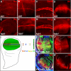
Wg target gene activation in fab1 and hrs mutant cells. Target gene expression activated by Wg signaling at different thresholds (summarized in I) was assessed in mosaic or mutant animals. (A–H and K) No discernible difference in target gene expression is observed in fab1 mutant cells compared with control cells. (J) Like previously reported (Piddini et al., 2005), hrs mutant cells display normal levels of Dll. Genotypes are w1118 (A–D), fab18/Df(2R)w30 (E–H), hsflp/+;FRT40A,Ubi-GFP/FRT40A, hrs28D (J), and hsflp/+;FRT42D, Ubi-GFP/FRT42D, fab131 (K).
Figure 8.
Figure 8.
Dpp and Notch target gene activation in fab1 mutant cells. (A and B) The range of two cell rows with nuclear phosphorylated pMAD (arrowheads, optical Z-section; B) activated by Dpp in follicle cells from a wild-type egg chamber. (C and D) The range of pMAD activation is not changed in fab1 mutant egg chambers (arrowheads, optical Z-section; D). (E and F) The same is true for spalt (Sal) activated in a gradient in response to Dpp signaling in the wing pouch (box in E enlarged in F). (G) No ectopic expression of the Notch target Eyg was observed in fab1 mutant clones in eye disks. Genotypes are w1118 (A and B), hsflp/+;FRT42D,Ubi-GFP/FRT42D, fab131 (C–F), and hsflp/+;FRT42D,Ubi-GFP/FRT42D, fab121 (G).

References

    1. Aldaz S., Morata G., Azpiazu N. The Pax-homeobox gene eyegone is involved in the subdivision of the thorax of Drosophila. Development. 2003;130:4473–4482. - PubMed
    1. Bache K. G., Slagsvold T., Stenmark H. Defective downregulation of receptor tyrosine kinases in cancer. EMBO J. 2004;23:2707–2712. - PMC - PubMed
    1. Bache K. G., Stuffers S., Malerød L., Slagsvold T., Raiborg C., Lechardeur D., Wälchli S., Lukacs G., Brech A., Stenmark H. The ESCRT-III subunit hVps24 is required for degradation but not silencing of the epidermal growth factor receptor. Mol. Biol. Cell. 2006;17:2513–2523. - PMC - PubMed
    1. Basler K., Struhl G. Compartment boundaries and the control of Drosophila limb pattern by hedgehog protein. Nature. 1994;368:208–214. - PubMed
    1. Berwick D. C., Dell G. C., Welsh G. I., Heesom K. J., Hers I., Fletcher L. M., Cooke F. T., Tavare J. M. Protein kinase B phosphorylation of PIKfyve regulates the trafficking of GLUT4 vesicles. J. Cell Sci. 2004;117:5985–5993. - PubMed

Publication types

MeSH terms