. 2006 Sep;17(9):4142-55.
doi: 10.1091/mbc.e06-02-0097. Epub 2006 Jul 12.

Dynamic regulation of mammalian numb by G protein-coupled receptors and protein kinase C activation: Structural determinants of numb association with the cortical membrane

Affiliations

Dynamic regulation of mammalian numb by G protein-coupled receptors and protein kinase C activation: Structural determinants of numb association with the cortical membrane

Sascha E Dho et al. Mol Biol Cell. 2006 Sep.

Abstract

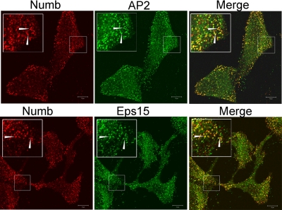
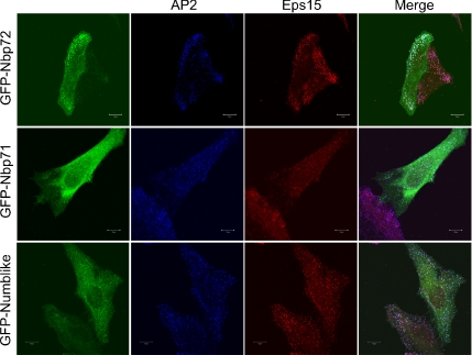
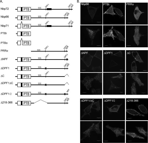
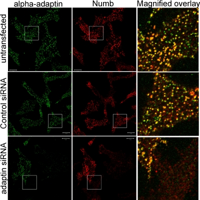
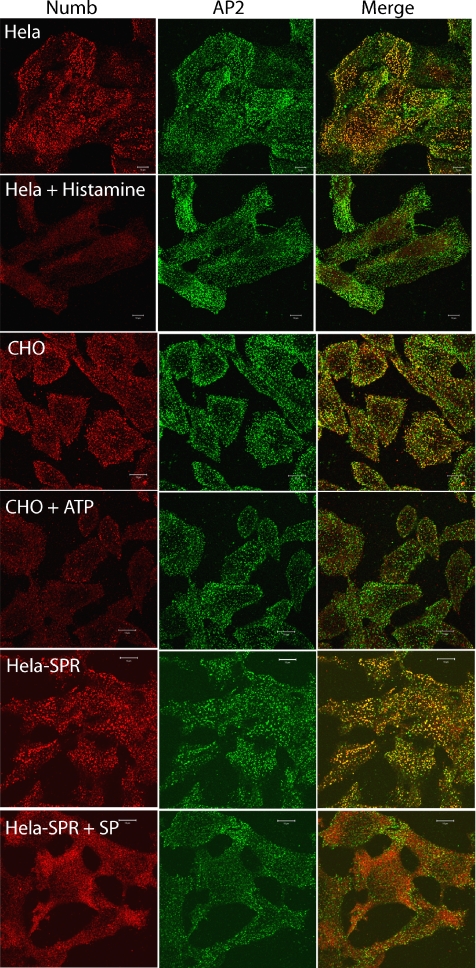
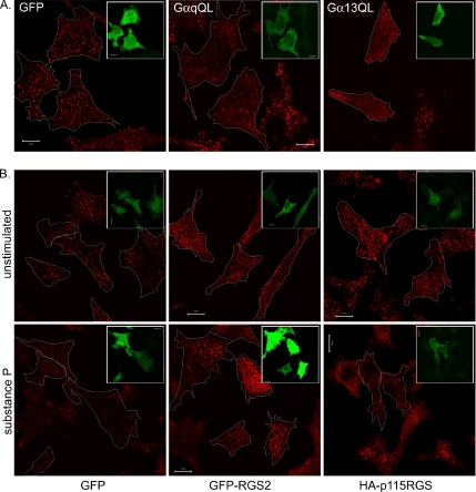
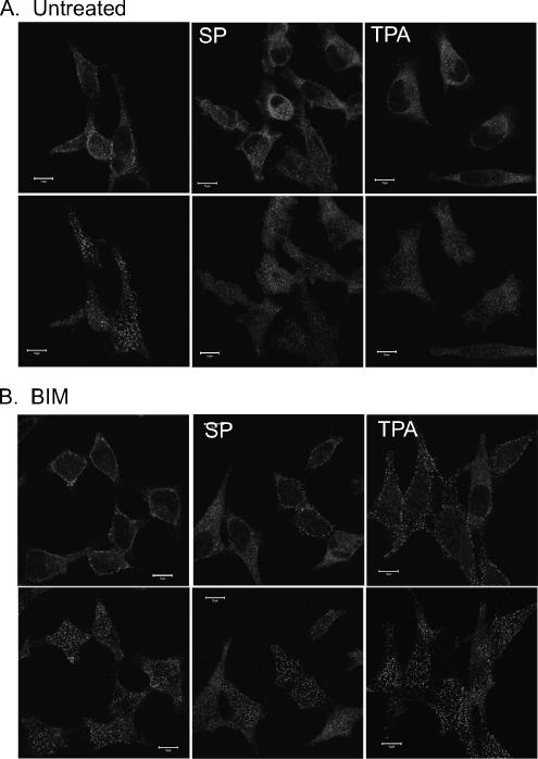
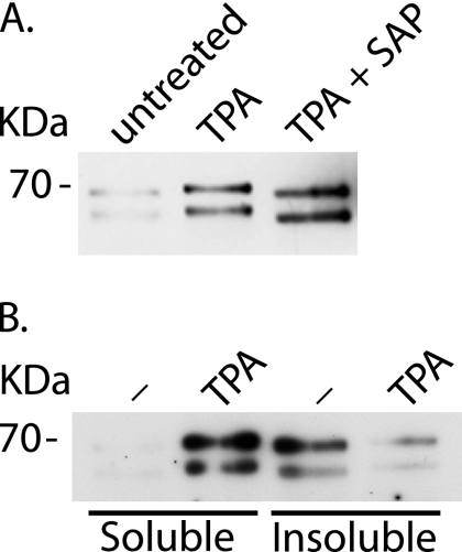
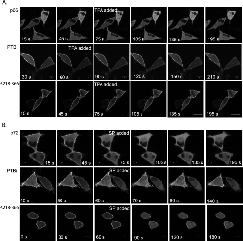
The cell fate determinant Numb is a membrane-associated adaptor protein involved in both development and intracellular vesicular trafficking. It has a phosphotyrosine-binding (PTB) domain and COOH-terminal endocytic-binding motifs for alpha-adaptin and Eps15 homology domain-containing proteins. Four isoforms of Numb are expressed in vertebrates, two of which selectively associate with the cortical membrane. In this study, we have characterized a cortical pool of Numb that colocalizes with AP2 and Eps15 at substratum plasma membrane punctae and cortical membrane-associated vesicles. Green fluorescent protein (GFP)-tagged mutants of Numb were used to identify the structural determinants required for localization. In addition to the previously described association of the PTB domain with the plasma membrane, we show that the AP2-binding motifs facilitate the association of Numb with cortical membrane punctae and vesicles. We also show that agonist stimulation of G protein-coupled receptors (GPCRs) that are linked to phospholipase Cbeta and protein kinase C (PKC) activation causes redistribution of Numb from the cortical membrane to the cytosol. This effect is correlated with Numb phosphorylation and an increase in its Triton X-100 solubility. Live-imaging analysis of mutants identified two regions within Numb that are independently responsive to GPCR-mediated lipid hydrolysis and PKC activation: the PTB domain and a region encompassing at least three putative PKC phosphorylation sites. Our data indicate that membrane localization of Numb is dynamically regulated by GPCR-activated phospholipid hydrolysis and PKC-dependent phosphorylation events.


Figures

Figure 1.
Figure 1.
Numb is localized in basal PM punctae and on vesicles associated with the cell cortex. (A) 293T cells were grown on glass coverslips, fixed, permeablized, and stained with anti-NbC. The images shown were taken at the basal, substratum-associated membrane (left) and a midsection through the cell (right). (B) MDCK cells grown for 3 d on polycarbonate filters were fixed, permeablized, and colabeled with anti-NbC (green) and either anti E-cadherin (red; left) or anti ZO-1 (red; right). Bars, 10 μm.
Figure 2.
Figure 2.
Numb colocalizes with α-adaptin and Eps15 in basal PM punctae. HeLa cells were fixed and colabeled with guinea pig anti-NbC (red) and either anti-adaptin or anti-Eps15 (green). Confocal microscopy was used to image the cell membrane attached to the glass coverslip. The boxed areas shown in each image are magnified to more clearly see punctae labeled with both anti-NbC and the indicated antibody (arrows indicate examples of colocalization). These images are representative of at least three separate experiments. Bars, 10 μm.
Figure 3.
Figure 3.
GFP-tagged Numb isoforms and Numblike exhibit differential membrane localization. GFP-Nbp72 (PTBi isoform), GFP-Nbp71 (PTBo isoform), and GFP-Nbl were transiently expressed in HeLa cells. Approximately 24 h after transfection, transfected cells were fixed and labeled with anti-adaptin (Cy3-labeled anti-mouse secondary) and anti-Eps15 (Cy5-labeled anti-rabbit secondary). Cells were imaged at the basal membrane by using confocal microscopy. Bars, 10 μm.
Figure 4.
Figure 4.
Adaptin-binding motifs facilitate the localization of Numb at the PM. (A) Diagrammatic representation of GFP-tagged constructs used to identify regions within Numb involved in localization and stimulated mobilization. (B) GFP-tagged Numb wild type and mutants were transiently expressed in SPR-HeLa cells. Approximately 24 h after transfection, live cells were imaged by confocal microscopy to assess the distribution of the transfected protein. Two optical sections are shown for each cell, one section acquired at the basal membrane attached to the glass coverslip (bottom image of each pair) and a second section acquired in the mid-region of the cell (top image of each pair). Cells expressing low to mid levels of expression were chosen, because high expression was often associated with apparent mislocalization of Numb to all regions of the PM. Bars, 10 μm.
Figure 5.
Figure 5.
PM α-adaptin is required for the localization of endogenous Numb in membrane punctae and vesicles. Endogenous Numb localization was assessed in SPR-HeLa cells treated with siRNA targeted against α-adaptin, control siRNA, or untreated as indicated. Cells were colabeled with anti-α-adaptin (red) and anti-NbC (green). The far right panels are the indicated boxed regions which were merged and magnified.
Figure 6.
Figure 6.
Endogenous Numb is rapidly lost from the PM after stimulation of G protein-coupled receptors. HeLa, CHO, and SPR-HeLa cells were grown on glass coverslips and stimulated for 5–10 min with the indicated GPCR agonist (200 μM histamine, 100 μM ATP, or 100 nM substance P). After fixation and permeablization, the cells were costained with anti-NbC (red) and anti-α adaptin (green). Bars, 10 μm.
Figure 7.
Figure 7.
GPCR-stimulated Numb mobilization is mediated by Gαq and Gα12/13. (A) SPR-HeLa cells transfected with either GFP, constitutively active forms of Gαq (GαqQL) or Gα13 (Gα13QL) were fixed and stained with anti-NbC (red). Transfected cells were identified using either anti-Gαq or anti-Gα13 and are shown in the insert of each image. Each transfected cell is outlined in the anti-NbC image. (B) SPR-HeLa cells transfected with GFP, GFP-RGS2, or HA-p115RGS were stimulated for 5 min with substance P, fixed, and stained with anti-NbC. Transfected cells are indicated as described in A and were identified by GFP fluorescence or labeling with anti-HA. Bars, 10 μm.
Figure 8.
Figure 8.
Substance P- and TPA-stimulated loss of Numb from the PM is mediated by activation of PKC. HeLa cells grown on glass coverslips were pretreated for 1 h with (B) or without (A) the PKC inhibitor BIM (3 μM). Cells were then stimulated with either 100 nM substance P (SP) or 0.5 μM TPA for 5 min and fixed and stained with anti-NbC. The images shown were taken at the basal, substratum-associated membrane (bottom) and a midsection through the same cells (top). Bars, 10 μm.
Figure 9.
Figure 9.
PKC activation is associated with phosphorylation of endogenous Numb and an increase in Numbs Triton X-100 solubility. SPR-HeLa cells were stimulated with 0.5 μM TPA for 5 min followed by lysis and immunoprecipitation of Numb by using anti-NbA as described in Materials and Methods. Numb immunoprecipitates were further treated with or without SAP (A) or fractionated into Triton X-100–soluble and –insoluble pools (B). The soluble fraction represents half of the cellular input relative to the insoluble fraction.
Figure 10.
Figure 10.
TPA stimulates a sustained loss of GFP-Nbp66 from the PM that is prevented by deletion of a serine-rich region (GFP-Δ218-366). SPR-HeLa cells were transfected with wild-type GFP-Numb (p66 or p72), GFP-PTBi, or GFP-Δ218-366; trypsinized 4 h after transfection, and seeded onto glass coverslips. Twenty hours later, live cells were imaged by confocal microscopy. TPA (0.5 μM; A) or substance P (100 nM; B) was added where indicated, and images were acquired at 15-s intervals in the continued presence of the stimulus. Animation may be viewed in Supplemental VideoNbp66TPA.mov, VideoPTBiTPA.mov, Videodelta218–366TPA.mov, VideoNbp72SP.mov, VideoPTBiSP.mov, and Videodelta218-366SP.mov.

References

    1. Aguilar R. C., Watson H. A., Wendland B. The yeast Epsin Ent1 is recruited to membranes through multiple independent interactions. J. Biol. Chem. 2003;278:10737–10743. - PubMed
    1. Balla T. Inositol-lipid binding motifs: signal integrators through protein-lipid and protein-protein interactions. J. Cell Sci. 2005;118:2093–2104. - PubMed
    1. Barr A. J., Brass L. F., Manning D. R. Reconstitution of receptors and GTP-binding regulatory proteins (G proteins) in Sf9 cells. A direct evaluation of selectivity in receptor G protein coupling. J. Biol. Chem. 1997;272:2223–2229. - PubMed
    1. Berdnik D., Torok T., Gonzalez-Gaitan M., Knoblich J. A. The endocytic protein alpha-adaptin is required for numb-mediated asymmetric cell division in Drosophila. Dev. Cell. 2002;3:221–231. - PubMed
    1. Betschinger J., Mechtler K., Knoblich J. A. The Par complex directs asymmetric cell division by phosphorylating the cytoskeletal protein Lgl. Nature. 2003;422:326–330. - PubMed

Publication types

MeSH terms