. 2006 Sep;17(9):4130-41.
doi: 10.1091/mbc.e05-11-1014. Epub 2006 Jul 12.

Neuronal calcium sensor-1 and phosphatidylinositol 4-kinase beta stimulate extracellular signal-regulated kinase 1/2 signaling by accelerating recycling through the endocytic recycling compartment

Affiliations

Neuronal calcium sensor-1 and phosphatidylinositol 4-kinase beta stimulate extracellular signal-regulated kinase 1/2 signaling by accelerating recycling through the endocytic recycling compartment

Yaara Kapp-Barnea et al. Mol Biol Cell. 2006 Sep.

Abstract

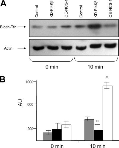
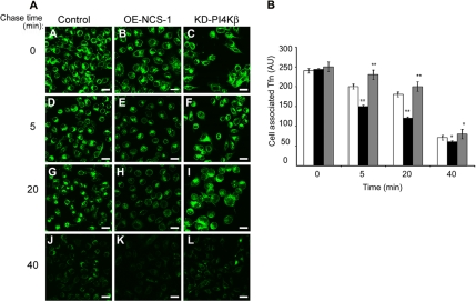
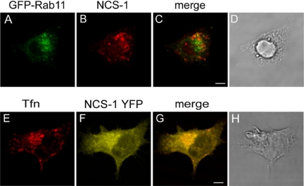
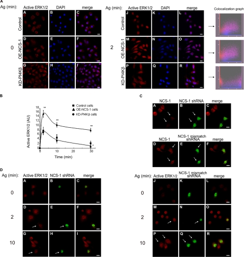
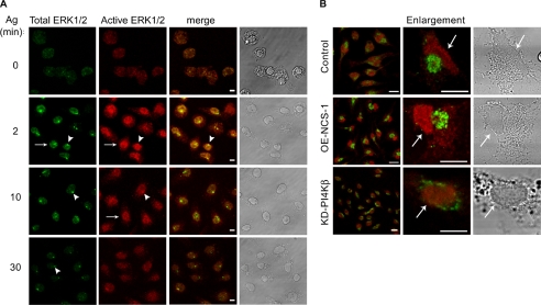
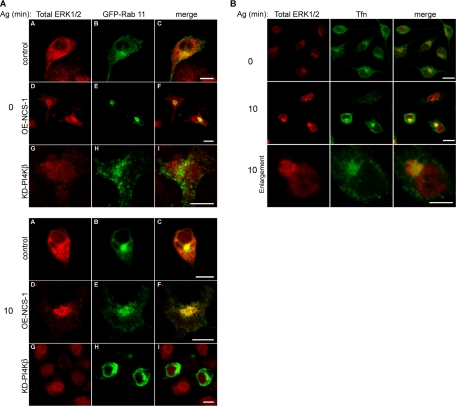
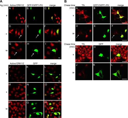
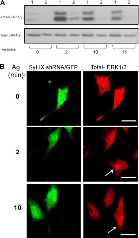
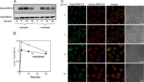
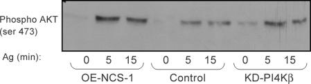
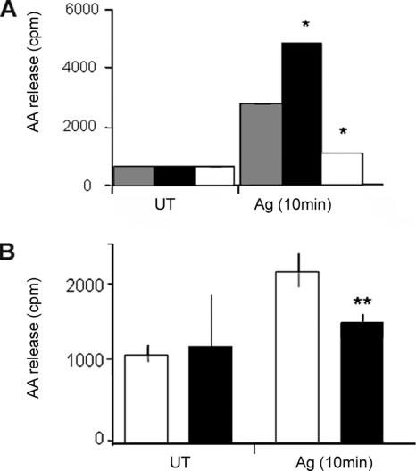
We demonstrate that recycling through the endocytic recycling compartment (ERC) is an essential step in Fc epsilonRI-induced activation of extracellular signal-regulated kinase (ERK)1/2. We show that ERK1/2 acquires perinuclear localization and colocalizes with Rab 11 and internalized transferrin in Fc epsilonRI-activated cells. Moreover, a close correlation exists between the amount of ERC-localized ERK1/2 and the amount of phospho-ERK1/2 that resides in the nucleus. We further show that by activating phosphatidylinositol 4-kinase beta (PI4Kbeta) and increasing the cellular level of phosphatidylinositol(4) phosphate, neuronal calcium sensor-1 (NCS-1), a calmodulin-related protein, stimulates recycling and thereby enhances Fc epsilonRI-triggered activation and nuclear translocation of ERK1/2. Conversely, NCS-1 short hairpin RNA, a kinase dead (KD) mutant of PI4Kbeta (KD-PI4Kbeta), the pleckstrin homology (PH) domain of FAPP1 as well as RNA interference of synaptotagmin IX or monensin, which inhibit export from the ERC, abrogate Fc epsilonRI-induced activation of ERK1/2. Consistently, NCS-1 also enhances, whereas both KD-PI4Kbeta and FAPP1-PH domain inhibit, Fc epsilonRI-induced release of arachidonic acid/metabolites, a downstream target of ERK1/2 in mast cells. Together, our results demonstrate a novel role for NCS-1 and PI4Kbeta in regulating ERK1/2 signaling and inflammatory reactions in mast cells. Our results further identify the ERC as a crucial determinant in controlling ERK1/2 signaling.


Figures

Figure 1.
Figure 1.
Effect of NCS-1 and KD-PI4Kβ on Tfn endocytosis. Control (mock-transfected), OE-NCS-1 or KD-PI4Kβ cells were serum starved for 1 h and incubated with 20 μg/ml biotin-Tfn for 1 h at 4°C. Unbound biotin-Tfn was removed by washing with ice-cold PBS, and the cells delivered to 37°C (time 0) for 10 min to allow endocytosis. At the end of incubation, cells were placed on ice, and the amount of intracellular biotin-Tfn was determined by subjecting cell lysates to SDS-PAGE, immunoblotting, and probing with HRP-conjugated streptavidin. (A) Blots were visualized by enhanced chemiluminescence (A). The cellular level of actin was determined to judge for equal loading. Blots of three similar experiments were quantified by densitometry and analyzed using one-tailed Mann–Whitney t test. The average ± SEM is presented. Shaded columns, control cells; filled columns, OE-NCS-1 cells; and open columns, KD-PI4Kβ cells; **p < 0.01 (B).
Figure 2.
Figure 2.
Effect of NCS-1 and KD-PI4Kβ on Tfn recycling. Control (mock-transfected), OE-NCS-1, or KD-PI4Kβ cells were grown on glass coverslips, serum starved for 1 h, and incubated with 50 μg/ml Alexa-488-Tfn for 1 h at 37°C. Cells were subsequently placed on ice (A–C) or washed and chased with unlabeled 100 μg/ml Tfn in the presence of 100 μM defroxamine mesylate for the indicated times (D–L). Cells were processed and visualized by laser confocal microscopy. Bar, 20 μm (A). To compare the rate of recycling from the ERC, the fluorescence intensity for each cell type at time 0 was normalized, and the fluorescence of 30–80 cells was measured for each time point. The average fluorescence per cell was calculated and is presented as arbitrary units (AU). Open columns, control cells; filled columns, OE-NCS-1 cells; and shaded columns, KD-PI4Kβ cells (B).
Figure 3.
Figure 3.
Subcellular localization of NCS-1. OE-NCS-1 cells, transiently transfected with GFP-Rab 11 cDNA (A–D) or RBL cells transiently transfected with NCS-1-YFP cDNA (E–H) were grown on glass coverslips for 24 h. Cells were subsequently either labeled with rabbit polyclonal anti NCS-1 antibodies followed by Cy3-conjugated anti rabbit IgG (A–C) or loaded with TR-conjugated Tfn (E–G). Cells were visualized by laser confocal microscopy. Bar, 4 μm.
Figure 4.
Figure 4.
Effect of NCS-1 and KD-PI4Kβ on ERK1/2 phosphorylation and nuclear translocation. (A) IgE-sensitized control (mock-transfected), OE-NCS-1, or KD-PI4Kβ cells were grown on glass coverslips and either left untreated (time 0) or incubated with antigen (Ag, DNP-HAS; 50 ng/ml) for 2 min. Cells were subsequently double stained with monoclonal anti active ERK1/2 antibodies followed by Cy3-conjugated anti-mouse IgG (red) and DAPI for nuclear staining (blue). To compare the extent of nuclear localization of phospho-ERK1/2, the intensity of ERK1/2 immunofluorescence measured was normalized, and double color colocalization analysis was performed for magnified cells. (B) IgE-sensitized control (mock-transfected), OE-NCS-1, or KD-PI4Kβ cells were incubated with antigen (Ag, DNP-HAS; 50 ng/ml) for the indicated times. Cells were subsequently lysed as described under Materials and Methods, resolved by SDS-PAGE, subjected to Western blot analysis, and probed with anti-active ERK1/2 antibodies. The intensities of the bands corresponding to pp42 and pp44 (ERK1/2) were quantified by densitometry. The data points presented are means ± SEM of three determinations of three independent experiments. Statistical analysis was performed using one-tailed Mann–Whitney t test with *p < 0.05 and **p < 0.01. (C) RBL cells transiently transfected with YFP-NCS-1 shRNA (A–C) or control YFP-NCS-1 mismatched shRNA (D–F) were grown on glass coverslips. Twenty-four hours later, cells were stained with anti NCS-1 antibodies followed by Cy3-conjugated anti-rabbit IgG. Cells were visualized by laser confocal microscopy. The arrows point to transfected cells. Bar, 8 μm. (D) RBL cells transiently transfected with YFP-NCS-1 shRNA (A–I) or control YFP-NCS-1 mismatched shRNA (J–R) were grown on glass coverslips, sensitized with DNP-specific IgE, and activated with DNP-HSA (Ag, 50 ng/ml) for the indicated times. Cells were then stained with monoclonal anti-active ERK1/2 followed by Cy3-conjugated anti-mouse IgG. Cells were visualized by laser confocal microscopy. The arrows point to transfected cells. Bar, 8 μm.
Figure 5.
Figure 5.
Cellular localization of active and total ERK1/2 in antigen-treated cells. IgE-sensitized RBL (A and B), OE-NCS-1 (B), or KD-PI4Kβ (B) cells were grown on glass coverslips and subsequently either left untreated (time 0) or incubated with antigen (Ag, DNP-HSA, 50 ng/ml) for the indicated times. Cells were then gently permeabilized with 100 μg/ml digitonin as described under Materials and Methods and double stained with polyclonal anti ERK1/2 (total ERK1/2) and monoclonal anti active ERK1/2 followed by fluorescein isothiocyanate-conjugated anti rabbit and Cy3-conjugated anti-mouse IgGs. Cells were visualized by laser confocal microscopy. Bars, 8 μm (A) and 10 μm (B). The arrows point to the nucleus (A and B), and arrowheads point to the perinuclear enzymes. Shown on the right are the corresponding phase contrast images.
Figure 6.
Figure 6.
Antigen-induced translocation of ERK1/2 to the ERC. (A) IgE sensitized control (mock-transfected), OE-NCS-1, or KD-PI4Kβ cells were transiently transfected with GFP-Rab 11 cDNA. Cells were subsequently grown on glass coverslips and either left untreated (time 0) or incubated with antigen (Ag, DNP-HSA, 50 ng/ml) for 10 min. Cells were then gently permeabilized with 100 μg/ml digitonin as described under Materials and Methods and stained with polyclonal anti ERK1/2 (Total ERK1/2) followed by Cy3-conjugated anti-rabbit IgG. Cells were visualized by laser confocal microscopy. Bar, 10 μm. (B) IgE sensitized control cells were grown on glass coverslips, serum starved for 1 h, and incubated with 50 μg/ml Alexa-488-Tfn for 45 min at 37°C. Cells were subsequently left untreated (time 0) or incubated with antigen (Ag, DNP-HSA, 50 ng/ml) for 10 min. Cells were then gently permeabilized with 100 μg/ml digitonin and stained with polyclonal anti ERK1/2 (total ERK1/2). Bar, 10 μm.
Figure 7.
Figure 7.
Effect of FAPP1-PH domain on ERK1/2 activation and Tfn recycling. (A) RBL cells transiently transfected with GFP-FAPP1-PH (A–I) or EGFP (J–R) cDNA were grown on glass coverslips, sensitized with DNP-specific IgE, and activated with DNP-HSA (Ag, 50 ng/ml) for the indicated times. Cells were then stained with monoclonal anti-active ERK1/2 followed by Cy3-conjugated anti-mouse IgG. Cells were visualized by laser confocal microscopy. The arrows point to transfected cells. Bar, 20 μm. (B) RBL cells transiently transfected GFP-FAPP1-PH (A–F) or enhanced green fluorescent protein (EGFP) cDNA (G–L) were grown on glass coverslips, serum starved for 1 h, and incubated with 50 μg/ml TR Tfn for 1 h at 37°C. Cells were then placed on ice or washed and chased with 100 μg/ml unlabeled Tfn in the presence of 100 μM defroxamine mesylate for 30 min, as indicated. Cells were visualized by laser confocal microscopy. The arrow points to a nontransfected cell. Bar, 20 μm.
Figure 8.
Figure 8.
Effect of Syt IX shRNA on ERK1/2 activation and localization. (A) OE-NCS-1 cells, nontransfected (1), or transiently transfected with Syt IX shRNA (2) were sensitized with DNP-specific IgE and activated with DNP-HSA (Ag, 50 ng/ml) for the indicated times. Cell lysates were resolved by SDS-PAGE, subjected to Western blot analysis, and probed with anti-active ERK1/2 antibodies, and reprobed with anti total ERK1/2, as indicated. (B) OE-NCS-1 cells were transiently transfected with Syt IX shRNA and EGFP vector (5:1). Cells were then grown on glass coverslips, sensitized with DNP-specific IgE and, activated with DNP-HSA (Ag, 50 ng/ml) for the indicated times. Cells were subsequently gently permeabilized with 100 μg/ml digitonin and stained with polyclonal anti ERK1/2 (total ERK/2), followed by Cy3-conjugated anti-rabbit IgG, and visualized by laser confocal microscopy. The arrows indicate the position of the ERC in nontransfected cells. Bar, 10 μm.
Figure 9.
Figure 9.
Effect of monensin on ERK1/2 activation and localization. (A) IgE-sensitized RBL cells were either left untreated or treated with 1 μM monensin for 2 h before activation with DNP-HSA (Ag, 50 ng/ml) for the indicated times. Cells were subsequently lysed, and cell lysates were resolved by SDS-PAGE and immunoblotted with anti-active ERK1/2. Blots were then reprobed with antibodies directed against ERK1/2 (total ERK1/2). (B) Densitometric analysis of the results presented in A. (C) IgE sensitized RBL cells were treated with 1 μM monensin for 2 h, washed, and incubated with DNP-HSA (Ag, 50 ng/ml) for the indicated times. Cells were analyzed as described under Figure 5A. Bar, 8 μm.
Figure 10.
Figure 10.
Effect of NCS-1 or KD-PI4Kβ on Akt/PKB phosphorylation. Control, OE-NCS-1, or KD-PI4Kβ cells were IgE-sensitized and activated by DNP-HSA (Ag, 50 ng/ml) for the indicated times. Cells were subsequently lysed, and cell lysates were resolved by SDS-PAGE and immunoblotted with anti-phospho (Ser 473)-Akt antibodies.
Figure 11.
Figure 11.
Effect of NCS-1 and KD-PI4Kβ on AA/metabolites release. (A) IgE-sensitized control (mock-transfected, shaded columns), OE-NCS-1 (filled columns), or KD-PI4Kβ cells (open columns) were loaded with [3H]AA. Buffer (UT) or DNP-HSA (Ag, 50 ng/ml) was subsequently added, and the cells incubated for further 10 min. AA release was then determined as described under Materials and Methods. (B) RBL cells were transiently transfected with GFP-FAPP1-PH (filled columns) or with an empty EGFP vector (open columns). Transfected cells were then sorted and collected by FACS and loaded with [3H]AA as described under Materials and Methods. Cells were subsequently sensitized with DNP specific IgE and incubated for 10 min with either buffer (UT) or DNP-HSA (Ag, 50 ng/ml). AA release was determined as described under Materials and Methods. The data points presented are means ± SEM of three determinations of three independent experiments. Statistical analysis was performed using one-tailed Mann–Whitney t test with *p < 0.05 and **p <0.01.

Similar articles

Cited by

References

    1. Birkeland H. C., Stenmark H. Protein targeting to endosomes and phagosomes via FYVE and PX domains. Curr. Top. Microbiol. Immunol. 2004;282:89–115. - PubMed
    1. Bourne Y., Dannenberg J., Pollmann V., Marchot P., Pongs O. Immunocytochemical localization and crystal structure of human frequenin (neuronal calcium sensor 1). J. Biol. Chem. 2001;276:11949–11955. - PubMed
    1. Braunewell K. H., Gundelfinger E. D. Intracellular neuronal calcium sensor proteins: a family of EF-hand calcium-binding proteins in search of a function. Cell Tissue Res. 1999;295:1–12. - PubMed
    1. Burgoyne R. D., Weiss J. L. The neuronal calcium sensor family of Ca2+-binding proteins. Biochem. J. 2001;353:1–12. - PMC - PubMed
    1. Carpentier J. L., Dayer J. M., Lang U., Silverman R., Orci L., Gorden P. Down-regulation and recycling of insulin receptors. Effect of monensin on IM-9 lymphocytes and U-937 monocyte-like cells. J. Biol. Chem. 1984;259:14190–14195. - PubMed

Publication types

MeSH terms

LinkOut - more resources