. 2006 Jul 25;103(30):11195-200.
doi: 10.1073/pnas.0601257103. Epub 2006 Jul 12.

Molecular evidence for deep evolutionary roots of bilaterality in animal development

Affiliations

Molecular evidence for deep evolutionary roots of bilaterality in animal development

David Q Matus et al. Proc Natl Acad Sci U S A. .

Abstract

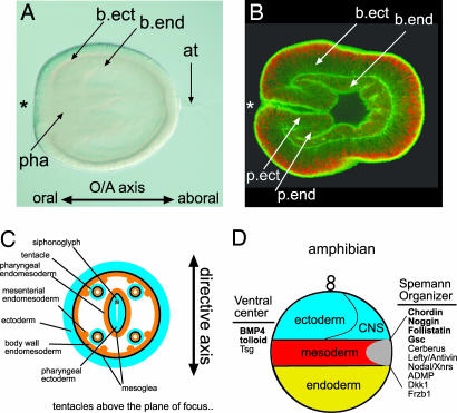
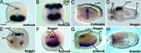
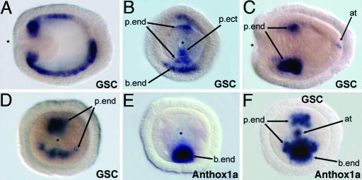
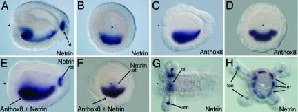
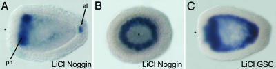
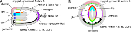
Nearly all metazoans show signs of bilaterality, yet it is believed the bilaterians arose from radially symmetric forms hundreds of millions of years ago. Cnidarians (corals, sea anemones, and "jellyfish") diverged from other animals before the radiation of the Bilateria. They are diploblastic and are often characterized as being radially symmetrical around their longitudinal (oral-aboral) axis. We have studied the deployment of orthologs of a number of family members of developmental regulatory genes that are expressed asymmetrically during bilaterian embryogenesis from the sea anemone, Nematostella vectensis. The secreted TGF-beta genes Nv-dpp, Nv-BMP5-8, six TGF-beta antagonists (NvChordin, NvNoggin1, NvNoggin2, NvGremlin, NvFollistatin, and NvFollistatin-like), the homeodomain proteins NvGoosecoid (NvGsc) and NvGbx, and the secreted guidance factor, NvNetrin, were studied. NvDpp, NvChordin, NvNoggin1, NvGsc, and NvNetrin are expressed asymmetrically along the axis perpendicular to the oral-aboral axis, the directive axis. Furthermore, NvGbx, and NvChordin are expressed in restricted domains on the left and right sides of the body, suggesting that the directive axis is homologous with the bilaterian dorsal-ventral axis. The asymmetric expression of NvNoggin1 and NvGsc appear to be maintained by the canonical Wnt signaling pathway. The asymmetric expression of NvNoggin1, NvNetrin, and Hox orthologs NvAnthox7, NvAnthox8, NvAnthox1a, and NvAnthox6, in conjunction with the observation that NvNoggin1 is able to induce a secondary axis in Xenopus embryos argues that N. vectensis could possess antecedents of the organization of the bilaterian central nervous system.


Conflict of interest statement

Conflict of interest statement: No conflicts declared.

Figures

Fig. 1.
Fig. 1.
Body plan of the anemone N. vectensis. (A and B) Micrographs of a lateral view of a planula-stage N. vectensis embryo. (A) Differential interference contrast micrograph. (B) Confocal micrograph with F-actin (green) and nuclei (red) stained with BODIPY-phalloidin and propidium iodide. The asterisk marks the blastopore and site of the future mouth. at, apical tuft; b.end, body wall endoderm; b.ect, body wall ectoderm; pha, pharynx; p.end, pharyngeal endoderm; p.ect, pharyngeal ectoderm. (C) Cross-sectional diagram of the N. vectensis body plan showing the different germ layers. (D) Diagram of a fate map for an amphibian embryo indicating the positions of ectodermal (including the CNS), mesodermal, and endodermal germ layers and some of the signaling molecules located on the ventral and dorsal (Spemann Organizer) sides involved in D-V polarity formation. Orthologs of some of the genes examined in this study are in bold. (Magnifications: ×100.)
Fig. 2.
Fig. 2.
Expression of four TGF-β antagonists during N. vectensis planula development. (A) NvAnthox8 is expressed asymmetrically along the directive axis, in body wall endoderm on one pole, and in a small cluster of cells in the pharyngeal endoderm on the opposite pole. (B) Double-label in situ hybridization showing asymmetric ectodermal expression of NvChordin on the side opposite of body wall endodermal Hox (NvAnthox8) expression. (C) NvFollistatin is expressed in pharyngeal endoderm in a ring in planula stages. (D and E) NvNoggin1 is expressed at the base and tips of growing tentacles (ten), in the endoderm below the apical tuft, and in the pharyngeal endoderm along one side of the directive axis, marking the site of the siphonoglyph. (F and G) Double-label in situ hybridization showing that the asymmetric pharyngeal endoderm expression of NvNoggin is on the side opposite of Hox gene expression (NvAnthox8). (G) NvNoggin1 is also expressed in the endoderm and ectoderm of the apical tuft at this stage of development. (H) NvGremlin is expressed in a ring of endodermal cells around the mouth on the oral pole on the polyp. The asterisk denotes the site of the blastopore and mouth. All embryo views are oral, except C, D, G, and H, which are lateral views, anterior to the left. (Magnifications: ×100.)
Fig. 3.
Fig. 3.
Gsc is asymmetrically expressed along the directive axis during development. (A and B) NvGoosecoid (gsc) is expressed initially in body wall endoderm (b.end) (A) and pharyngeal endoderm (p.end) and ectoderm (p.ect) (B). (C and D) During planula stages, NvGsc is expressed in pharyngeal endoderm, asymmetrically along the directive axis, and in the endoderm below the apical tuft (at) (C). (E) NvAnthox1a is expressed in body wall endoderm on one pole of the directive axis. (F) Double-label in situ hybridization showing that the larger domain of NvGsc pharyngeal endoderm expression is on the same pole of the directive axis as Hox expression (NvAnthox1a). The asterisk denotes the site of the blastopore and mouth. All embryo views are oral, except A and C, which are lateral views, anterior to the left. (Magnifications: ×100.)
Fig. 4.
Fig. 4.
Left/right expression of genes during Nematostella development defines a third body axis. (AC) NvGbx is expressed bilaterally in body wall endoderm (b.end) (A and B) and pharyngeal endoderm (p.end) (C). (D and E) Double-label in situ hybridization shows that NvGbx (purple) is expressed along the left-right axis, bilaterally, flanking NvAnthox8 (turqoise) expression in planula-stage embryos. (F) Double-label in situ hybridization showing the latest expression of NvChordin in polyp-stage embryos. NvChordin is expressed in two bilateral patches of cells to the left and right of the siphonoglyph NvNoggin1 expression. The asterisk denotes the mouth. All embryo views are lateral with anterior to the left, except B and E, which are oral views. (Magnifications: ×100.)
Fig. 5.
Fig. 5.
NvNetrin is expressed asymmetrically with Hox genes in the endoderm during planula development. (A and B) NvNetrin is expressed asymmetrically during planula development in the body wall endoderm, in a longitudinal stripe, and in a few ectodermal cells at the base of the apical tuft (at). (CF) Double-label in situ hybridization shows that NvNetrin1 is coexpressed with NvAnthox8 in the body wall endoderm. (G and H) During polyp development, NvNetrin expression is in a ring around the mouth (nr). The endodermal tips of the tentacles (tn) also express NvNetrin1 (G). Tentacles are out of the plane of focus in H. The asterisk denotes the site of the blastopore and mouth. All embryo views are lateral, with anterior to the left, except B, D, F, and H, which are oral views. (Magnifications: ×100.)
Fig. 6.
Fig. 6.
Lithium chloride treatment radializes expression of organizer genes. Ectopical activation of the canonical Wnt pathway by lithium chloride treatment radializes NvNoggin1 (A and B) and NvGsc (C) expression. NvNoggin1 is normally expressed in tentacle endoderm, at the base of the apical tuft, and asymmetrically along one side of the directive axis, marking the site of the future siphonoglyph. NvNoggin1 is expressed in an endodermal ring at the oral pole in lithium-treated embryos (A and B). NvGsc is normally expressed asymmetrically along the directive axis in the pharyngeal endoderm, with a larger domain of expression on one pole. NvGsc is expressed symmetrically in body wall endoderm and in the pharynx of lithium-treated embryos (C). (Magnifications: ×125.)
Fig. 7.
Fig. 7.
Summary of asymmetric gene expression patterns during development in N. vectensis. (A) Lateral view with the oral (anterior) pole toward the left. (B) Cross section through the pharynx. Asterisk marks the site of gastrulation and mouth. The mesoglea (yellow line) separates the ectodermal epidermis from the endodermal gastrodermis. Colored bars represent different gene expression patterns, with hash-marked color bars representing transient early gene expression. Tentacle and apical tuft expression is not show for clarity. A suite of bilaterian patterning genes (transcription factors, secreted ligands, and their antagonists) is dynamically expressed along both the oral/aboral axis and the directive axis in both germ layers. The asymmetric expression of NvChordin, NvDpp, NvNoggin, and NvAnthox8 in the pharynx on the side opposite the expression of NvNetrin, three Hox genes, the larger domain of NvGsc, and NvGDF5-like, suggests homology of the directive axis to the D-V axis of bilaterians. The bilateral expression of NvGbx and NvChordin marks, the left–right axis.

Similar articles

Cited by

References

    1. Wallberg A., Thollesson M., Farris J. S., Jondelius U. Cladistics. 2004;20:558–578. - PubMed
    1. Wainright P. O., Hinkle G., Sogin M. L., Stickel S. K. Science. 1993;260:340–342. - PubMed
    1. Odorico D. M., Miller D. J. Proc. Biol. Sci.; 1997. pp. 77–82. - PMC - PubMed
    1. Collins A. G., Cartwright P., McFadden C. S., Scheirwater B. Integr. Comp. Biol. 2005;45:585–594. - PubMed
    1. Collins A. G. J. Evol. Biol. 2002;15:418–432.

Publication types

Substances