. 2006 Jul 12;26(28):7328-36.
doi: 10.1523/JNEUROSCI.0559-06.2006.

Converging language streams in the human temporal lobe

Affiliations

Converging language streams in the human temporal lobe

Galina Spitsyna et al. J Neurosci. .

Abstract

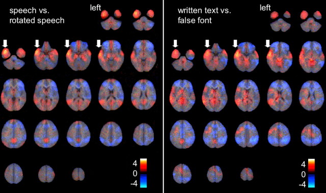
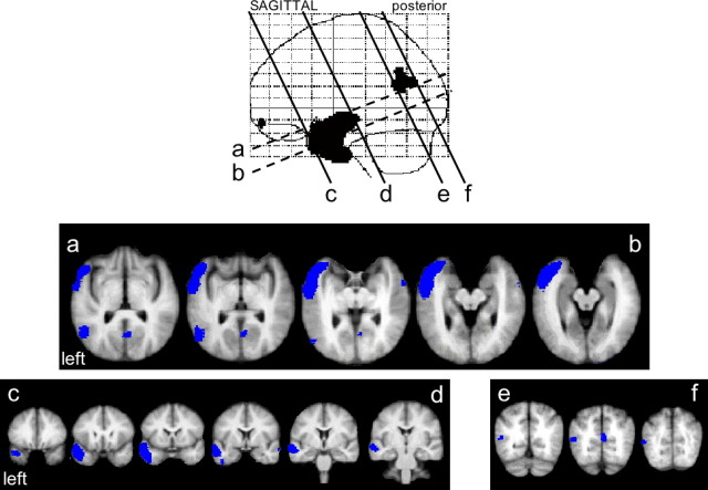
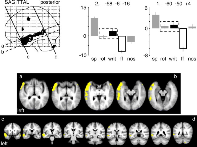
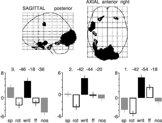
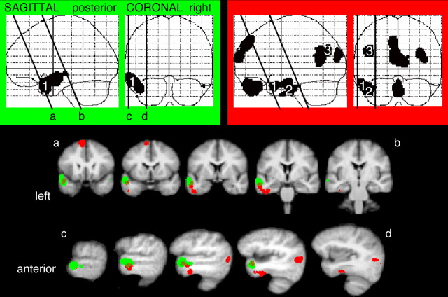
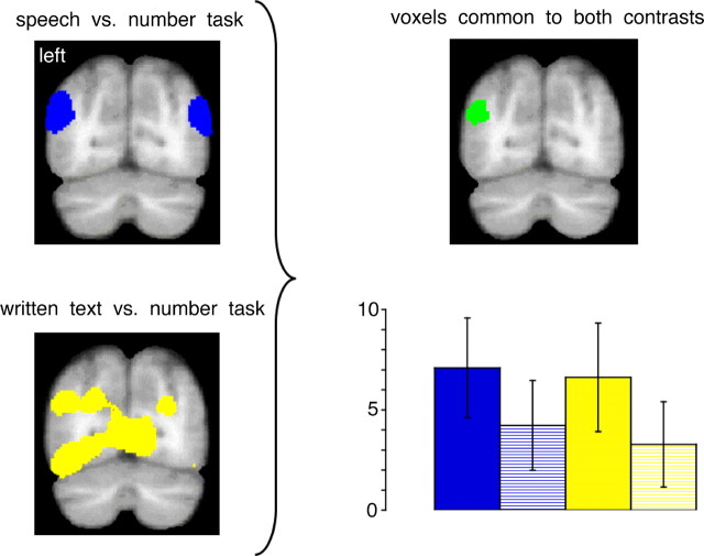
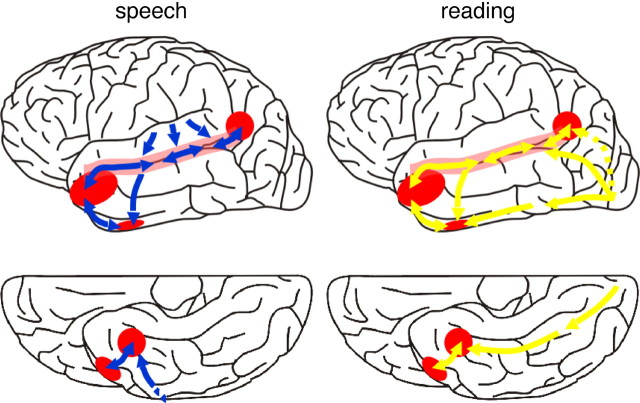
There is general agreement that, after initial processing in unimodal sensory cortex, the processing pathways for spoken and written language converge to access verbal meaning. However, the existing literature provides conflicting accounts of the cortical location of this convergence. Most aphasic stroke studies localize verbal comprehension to posterior temporal and inferior parietal cortex (Wernicke's area), whereas evidence from focal cortical neurodegenerative syndromes instead implicates anterior temporal cortex. Previous functional imaging studies in normal subjects have failed to reconcile these opposing positions. Using a functional imaging paradigm in normal subjects that used spoken and written narratives and multiple baselines, we demonstrated common activation during implicit comprehension of spoken and written language in inferior and lateral regions of the left anterior temporal cortex and at the junction of temporal, occipital, and parietal cortex. These results indicate that verbal comprehension uses unimodal processing streams that converge in both anterior and posterior heteromodal cortical regions in the left temporal lobe.


Figures

Figure 1.
Figure 1.
The results of analyses within wavelet space of two contrasts: speech with rotated speech, and written text with false font. The estimated effect sizes, both positive (red–yellow) and negative (blue) results, expressed as Z-scores without a cutoff threshold, are displayed on axial brain slices from ventral to dorsal. The strongest positive effect size for each contrast was observed within the left anterior temporal cortex (arrows).
Figure 2.
Figure 2.
The contrast of speech with rotated speech, displayed on a sagittal statistical parametric map (SPM), and coregistered (as a blue overlay) on to brain slices from a mean magnetic resonance imaging (MRI) template in Montreal Neurological Institute stereotactic space. The MRI slices were oriented along the plane of the STS, and consecutive axial (a–b) and coronal (c–d and e–f) slices, separated by 4 mm, are displayed with reference to their location on the SPM.
Figure 3.
Figure 3.
Written text contrasted with false font, using the same display design as in Figure 2 and a yellow overlay on the coregistered slices. The response profiles across all five conditions are shown for two peak voxels, identified within SPM99, at either end of the left STS. The size of effect (in arbitrary units) for the conditions used in the contrast, written text (writ; black bar) and false font (ff; white bar), are shown in relation to activity in response to speech (sp), rotated speech (rot), and the number task (nos), shown as gray bars. Mean activity (±SEM) for all five conditions was normalized around zero. Coordinates (in millimeters) show the location of peaks in Montreal Neurological Institute space, located within the posterior STS (1) and anterior STS (2). The dashed outlines draw attention to the equivalent response to rotated speech and written text at the two peak voxels.
Figure 4.
Figure 4.
A display of the statistical parametric map, both sagittal and axial views, for the contrast of intelligible written text (writ) with the two unintelligible baseline conditions, rotated speech (rot) and false font (ff). Activity across all five conditions is taken from peak voxels along the length of the left FG, using the same style of display as in Figure 3. At voxel 1, with stereotactic coordinates cited as the location of the visual word form area (Cohen and Dehaene, 2004), there was still a response to false font relative to rotated and intelligible speech, but this was considerably reduced or absent at voxels 2 and 3. The regions marked with asterisks show activity within extraocular muscles, active during the visual conditions.
Figure 5.
Figure 5.
A demonstration on sagittal and coronal views, using the same presentation format as Figures 2 and 3, of activation common to the language conditions contrasted with their modality-specific unintelligible baseline conditions (color-coded in green), and common to the language conditions separately contrasted with the number task (color-coded in red). Although there was overlap of the activations in the left lateral TP (1), the use of the number task as the baseline condition revealed activity in the anterior FG (2) and the left TOP junction (3).
Figure 6.
Figure 6.
Histograms of mean estimated effect sizes (in arbitrary units), with 95% confidence intervals, in two anterior temporal ROIs in each hemisphere: the lateral TP and the anterior FG. The language conditions are shown as columns for speech (sp) and written text (writ). The unintelligible baseline conditions are shown as columns for rotated speech (rot) and false font (ff). The mean effect sizes for all four conditions are relative to the common baseline condition of the number task.
Figure 7.
Figure 7.
Activity at the TOP junction associated with speech (top left, in blue) and written text (bottom left, in yellow), each contrasted with the number task, coregistered onto a mean anatomical coronal magnetic resonance image, 72 mm posterior to the anterior commissure. There were voxels common to these two contrasts at the left TOP junction (top right, in green). Histograms of mean estimated effect sizes (expressed in arbitrary units), with 95% confidence intervals, in this region are shown (bottom right). The language conditions are shown as a blue column for speech and a yellow column for written text. Their unintelligible baseline conditions are shown as a blue-hatched column for rotated speech and a yellow-hatched column for false font. The mean effect sizes for all four conditions are relative to the common baseline condition of the number task.
Figure 8.
Figure 8.
An anatomical summary of the results, overlaid on the lateral (top row) and ventral (bottom row) surfaces of the left cerebral hemisphere, showing the convergence of spoken and written language in the left STS (pink shading) and in posterior (the TOP junction) and anterior (lateral TP and anterior FG) cortex (red shading). The auditory streams of processing are shown in blue, and the visual streams are in yellow. In the nonhuman primate, the heteromodal cortex of the left STS is strongly interconnected along its length (Padberg et al., 2003), and we have assumed that the same is true for the human STS. A direct connection between the anterior STS and inferotemporal cortex is suggested by anatomical connectivity data from nonhuman primates (Saleem et al., 2000). The left TOP junction may receive direct projections from occipital cortex (dashed yellow line) or indirectly, via the STS.

References

    1. Alain C, Arnott SR, Hevenor S, Graham S, Grady CL (2001). “What” and “where” in the human auditory system. Proc Natl Acad Sci USA 98:12301–12306. - PMC - PubMed
    1. Alexander MP, Hiltbrunner B, Fischer RS (1989). Distributed anatomy of transcortical sensory aphasia. Arch Neurol 46:885–892. - PubMed
    1. Aston JA, Gunn RN, Hinz R, Turkheimer FE (2005). Wavelet variance components in image space for spatiotemporal neuroimaging data. NeuroImage 25:159–168. - PubMed
    1. Beauchamp MS, Argall BD, Bodurka J, Duyn JH, Martin A (2004). Unraveling multisensory integration: patchy organization within human STS multisensory cortex. Nat Neurosci 7:1190–1192. - PubMed
    1. Binder JR, Frost JA, Hammeke TA, Bellgowan PSF, Rao SM, Cox RW (1999). Conceptual processing during the conscious resting state: a functional MRI study. J Cogn Neurosci 11:80–93. - PubMed

Publication types

LinkOut - more resources