. 2006 Jul 12;26(28):7337-47.
doi: 10.1523/JNEUROSCI.0729-06.2006.

Role of the neurogranin concentrated in spines in the induction of long-term potentiation

Affiliations

Role of the neurogranin concentrated in spines in the induction of long-term potentiation

Anatol M Zhabotinsky et al. J Neurosci. .

Abstract

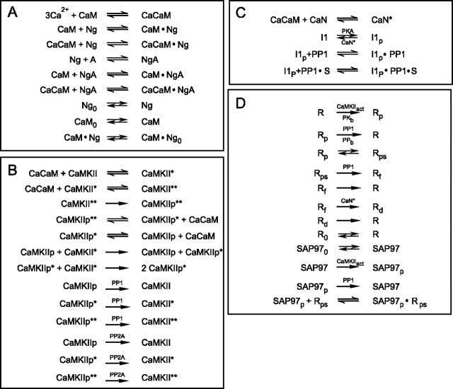
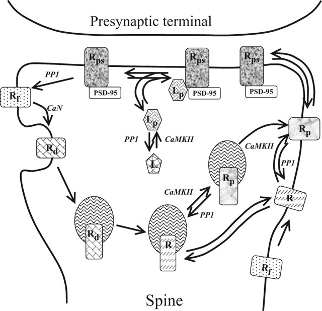
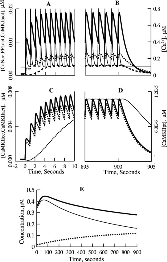
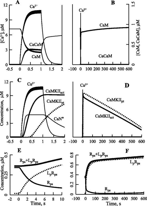
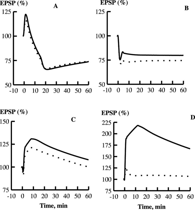
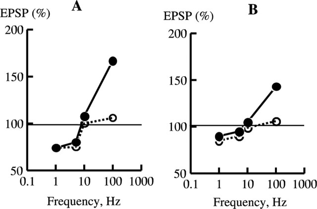
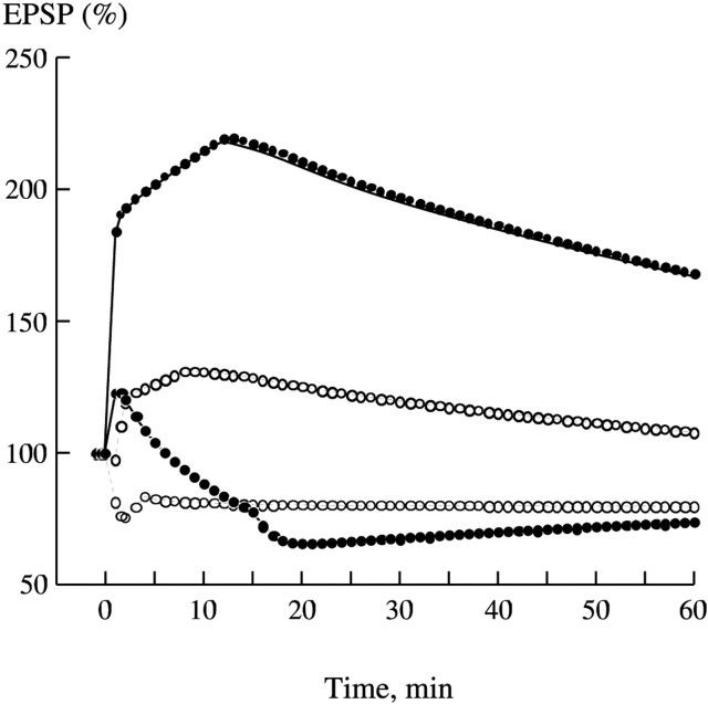
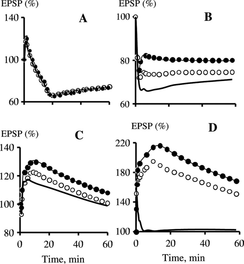
Synaptic plasticity in CA1 hippocampal neurons depends on Ca2+ elevation and the resulting activation of calmodulin-dependent enzymes. Induction of long-term depression (LTD) depends on calcineurin, whereas long-term potentiation (LTP) depends on Ca2+/calmodulin-dependent protein kinase II (CaMKII). The concentration of calmodulin in neurons is considerably less than the total concentration of the apocalmodulin-binding proteins neurogranin and GAP-43, resulting in a low level of free calmodulin in the resting state. Neurogranin is highly concentrated in dendritic spines. To elucidate the role of neurogranin in synaptic plasticity, we constructed a computational model with emphasis on the interaction of calmodulin with neurogranin, calcineurin, and CaMKII. The model shows how the Ca2+ transients that occur during LTD or LTP induction affect calmodulin and how the resulting activation of calcineurin and CaMKII affects AMPA receptor-mediated transmission. In the model, knockout of neurogranin strongly diminishes the LTP induced by a single 100 Hz, 1 s tetanus and slightly enhances LTD, in accord with experimental data. Our simulations show that exchange of calmodulin between a spine and its parent dendrite is limited. Therefore, inducing LTP with a short tetanus requires calmodulin stored in spines in the form of rapidly dissociating calmodulin-neurogranin complexes.


Figures

Figure 1.
Figure 1.
Block scheme of the model. A, Interactions of Ng, apoCaM, and CaCaM; exchange of Ng, CaM, and the CaM·Ng complex between the spine and parent dendrite; and accumulation of Ng in the spine; A is the Ng anchor. B, Activation of CaMKII by CaCaM, autophosphorylation of CaMKII, and dephosphorylation of CaMKIIP by PP1 and PP2A. C, Activation of CaN and control of PP1 by PKA and CaN via I1. D, Phosphorylation of the AMPAR·Sg complex (R) and SAP97 (SAP) by CaMKII and their dephosphorylation by PP1. Mass-action law reactions are shown by simple arrows, and enzymatic reactions are indicated by enzyme abbreviations above or below the arrows.
Figure 2.
Figure 2.
Traffic and postsynaptic retention of AMPARs dependent on GluR1, Sg, and SAP97. R is the nonphosphorylated form of the AMPAR·Sg complex formed via the GluR1–Sg interaction, Rp is the exstrasynaptic phosphorylated form, Rps is synaptic Rp, Rf is the immediate product of Rps dephosphorylation, and Rd is a nonphosphorylable form, which slowly converts to R; L is SAP97, and Lp is phosphorylated L.
Figure 3.
Figure 3.
Simulated kinetics of activation of CaN, PP1, and CaMKII, autophosphorylation of CaMKII, and phosphorylation of the AMPAR complex during (A, C) and immediately after (B, D) 1 Hz, 15 min stimulation. A, B, Kinetics of CaN bound to CaCaM (CaN*; ), active PP1 (PP1act; —▪—), and active CaMKII (CaMKIIact; — —) shown together with the Ca2+ pulses (—). C, D, Absence of autophosphorylation of CaMKII; CaMKII bound to CaCaM (CaMKIIcc; ), CaMKIIact (— —), and autophosphorylated CaMKII (CaMKIIpt; —). E, Kinetics of the postsynaptic AMPAR·Sg complexes: Rps (—), LpRps (— —), and Rps + LpRps ().
Figure 4.
Figure 4.
Kinetics of Ca2+, calmodulin, (Ca2+)3CaM, and activation of CaN, CaMKII, autophosphorylation of CaMKII, and phosphorylation of the AMPA receptor–stargazin complex during and after 100 Hz, 1 s stimulation. A, B, Kinetics of Ca2+, CaM, and CaCaM. C, Kinetics of Ca2+, CaN*, total activated CaMKII (CaMKIIact), and total autophosphorylated CaMKII (CaMKIIpt). D, Autophosphorylation of CaMKII is responsible for a prolonged activation of CaMKII. E, F, Kinetics of Rps, LpRps and Rps + LpRps, Lp is SAP97p.
Figure 5.
Figure 5.
Effect of Ng on LTP and LTD in the model. A, Kinetics of EPSP in response to 1 Hz, 15 min stimulation, at 60 min after the start of stimulation. WT, 74% of the basal transmission; Ng KO, 74% of the basal transmission. B, Response to 5 Hz, 3 min stimulation. WT, 80%; Ng KO, 75%. C, Response to 10 Hz, 90 s stimulation. WT, 108%; Ng KO, 100%. D, Response to 100 Hz, 1 s stimulation. WT, 167%; Ng KO, 106%. WT, (); Ng KO, (- - -).
Figure 6.
Figure 6.
Simulation of frequency–response curves from Huang et al. (2004). A, Frequency–response curves plotted from data taken at 60 min from the curves shown in Figure 5. B, Experimental curves plotted according to data from Huang et al. (2004). WT, •; KO, ○.
Figure 7.
Figure 7.
Synaptic plasticity depends weakly on affinity of Ng to (Ca2+)3CaM. LTD induced by 1 Hz, 15 min; 5 Hz, 3 min; and 10 Hz, 1.5 min protocols is practically indistinguishable whether affinity of (Ca2+)3CaM for Ng is negligible or crease in to that of apoCaM. LTP induced by a 100 Hz, 1 s protocol is slightly higher when Ng does not bind (Ca2+)3CaM. The curves appear in the following order (from bottom to top) after 20 min: 1, 5, 10, and 100 Hz stimulation. The lines correspond to Kd = 3 μm, and the circles correspond to 1/Kd = 0.
Figure 8.
Figure 8.
The spine–dendrite rate of exchange of CaM, Ng, and the CaM·Ng complex does not affect plasticity in WT. However, a high exchange rate could salvage LTP in KO. A, Kinetics of EPSP in response to 1 Hz, 15 min stimulation. B, Response to 5 Hz, 3 min stimulation. C, Response to 10 Hz, 90 s stimulation. D, Response to 100 Hz, 1 s stimulation. The curve designations are as follows: WT, the exchange rate constant (k), equals 10.0 s−1 (•); WT, k = 0.01 s−1 (—); KO, k = 10.0 s−1 (○); KO, k = 0.01 s−1 (—).

Similar articles

Cited by

References

    1. Aigner L, Arber S, Kapfhammer JP, Laux T, Schneider C, Botteri F, Brenner HR, Caroni P (1995). Overexpression of the neural growth-associated protein Gap-43 induces nerve sprouting in the adult nervous system of transgenic mice. Cell 83:269–278. - PubMed
    1. Alexander KA, Cimler BM, Meier KE, Storm DR (1987). Regulation of calmodulin binding to P-57—a neurospecific calmodulin binding protein. J Biol Chem 262:6108–6113. - PubMed
    1. Baudier J, Deloulme JC, Vandorsselaer A, Black D, Matthes HWD (1991). Purification and characterization of a brain-specific protein kinase C substrate, neurogranin (P17)—identification of a consensus amino-acid sequence between neurogranin and neuromodulin (GAP43) that corresponds to the protein kinase C phosphorylation site and the calmodulin-binding domain. J Biol Chem 266:229–237. - PubMed
    1. Beattie EC, Carroll RC, Yu X, Morishita W, Yasuda H, von Zastrow M, Malenka RC (2000). Regulation of AMPA receptor endocytosis by a signaling mechanism shared with LTD. Nat Neurosci 3:1291–1300. - PubMed
    1. Bhalla US, Iyengar R (1999). Emergent properties of networks of biological signaling pathways. Science 283:381–387. - PubMed

Publication types

MeSH terms

LinkOut - more resources