Bidirectional modifications of visual acuity induced by monocular deprivation in juvenile and adult rats
- PMID: 16837583
- PMCID: PMC6674195
- DOI: 10.1523/JNEUROSCI.0124-06.2006
Bidirectional modifications of visual acuity induced by monocular deprivation in juvenile and adult rats
Abstract
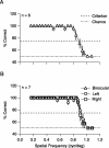
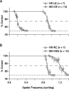
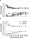
Recent electrophysiological studies of rodent visual cortex suggest that, in addition to deprived-eye depression, monocular deprivation (MD) also shifts ocular dominance by potentiation of open-eye responses. We used computer-based, two-choice discrimination tasks to assess the behavioral significance of these findings in rats. As expected, prolonged MD, from postnatal day 21 until adulthood (>150 d) markedly decreased visual acuity through the deprived eye. However, we also found that the acuity through the nondeprived eye was significantly enhanced compared with normally reared controls. Interestingly, when the deprived eye was opened in adults, there was a gradual but incomplete recovery of acuity in the deprived eye preceded by a loss of the enhanced acuity in the nondeprived eye. These changes were reversed by again reclosing the eye. These findings suggest that the bidirectional changes in visually evoked responses after MD are behaviorally meaningful and that significant plasticity is exhibited well into adulthood.
Figures


Similar articles
-
Visual deprivation reactivates rapid ocular dominance plasticity in adult visual cortex.J Neurosci. 2006 Mar 15;26(11):2951-5. doi: 10.1523/JNEUROSCI.5554-05.2006. J Neurosci. 2006. PMID: 16540572 Free PMC article.
-
Binocular Disparity Selectivity Weakened after Monocular Deprivation in Mouse V1.J Neurosci. 2017 Jul 5;37(27):6517-6526. doi: 10.1523/JNEUROSCI.1193-16.2017. Epub 2017 Jun 2. J Neurosci. 2017. PMID: 28576937 Free PMC article.
-
Adult visual experience promotes recovery of primary visual cortex from long-term monocular deprivation.Learn Mem. 2007 Aug 29;14(9):573-80. doi: 10.1101/lm.676707. Print 2007 Sep. Learn Mem. 2007. PMID: 17761542 Free PMC article.
-
Apoptosis in the mechanisms of neuronal plasticity in the developing visual system.Eur J Ophthalmol. 2003 Apr;13 Suppl 3:S36-43. doi: 10.1177/112067210301303s07. Eur J Ophthalmol. 2003. PMID: 12749676 Review.
-
Cross-Modal Plasticity in Brains Deprived of Visual Input Before Vision.Annu Rev Neurosci. 2022 Jul 8;45:471-489. doi: 10.1146/annurev-neuro-111020-104222. Annu Rev Neurosci. 2022. PMID: 35803589 Review.
Cited by
-
The "good" limb makes the "bad" limb worse: experience-dependent interhemispheric disruption of functional outcome after cortical infarcts in rats.Behav Neurosci. 2010 Feb;124(1):124-132. doi: 10.1037/a0018457. Behav Neurosci. 2010. PMID: 20141287 Free PMC article.
-
Mouse experimental myopia has features of primate myopia.Invest Ophthalmol Vis Sci. 2010 Mar;51(3):1297-303. doi: 10.1167/iovs.09-4153. Epub 2009 Oct 29. Invest Ophthalmol Vis Sci. 2010. PMID: 19875658 Free PMC article.
-
A sensitive period for the development of episodic-like memory in mice.Curr Biol. 2025 May 5;35(9):2032-2048.e3. doi: 10.1016/j.cub.2025.03.032. Epub 2025 Apr 10. Curr Biol. 2025. PMID: 40215964
-
Contrasting roles for parvalbumin-expressing inhibitory neurons in two forms of adult visual cortical plasticity.Elife. 2016 Mar 4;5:e11450. doi: 10.7554/eLife.11450. Elife. 2016. PMID: 26943618 Free PMC article.
-
The refinement of ipsilateral eye retinotopic maps is increased by removing the dominant contralateral eye in adult mice.PLoS One. 2010 Mar 29;5(3):e9925. doi: 10.1371/journal.pone.0009925. PLoS One. 2010. PMID: 20369001 Free PMC article.
References
-
- Dean P (1981). Visual pathways and acuity hooded rats. Behav Brain Res 3:239–271. - PubMed
-
- Fagiolini M, Pizzorusso T, Berardi N, Domenici L, Maffei L (1994). Functional postnatal development of the rat primary visual cortex and the role of visual experience: dark rearing and monocular deprivation. Vision Res 34:709–720. - PubMed
Publication types
MeSH terms
LinkOut - more resources
Full Text Sources
Research Materials
