

Time course of functional connectivity between dorsal premotor and contralateral motor cortex during movement selection
- PMID: 16837593
- PMCID: PMC6674199
- DOI: 10.1523/JNEUROSCI.1158-06.2006
Time course of functional connectivity between dorsal premotor and contralateral motor cortex during movement selection
Abstract
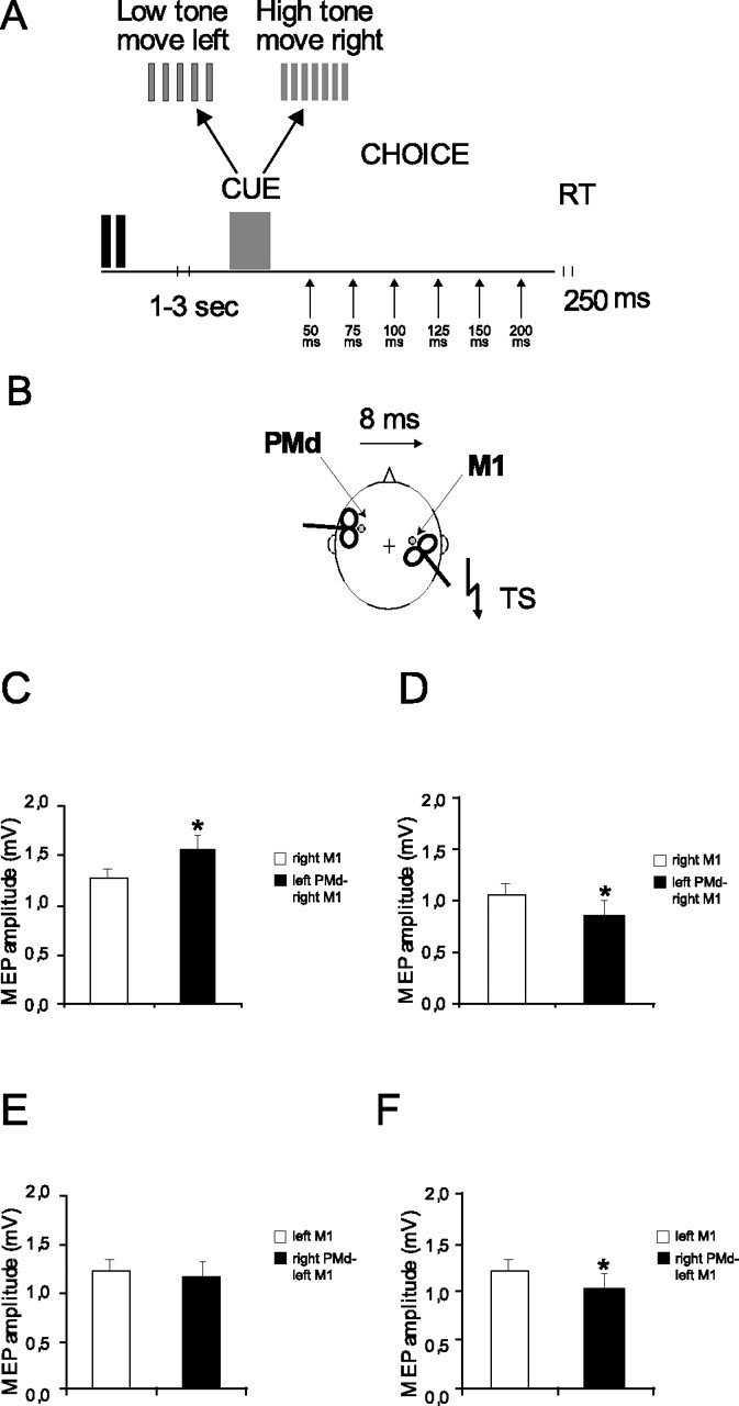
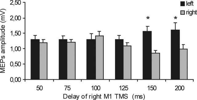
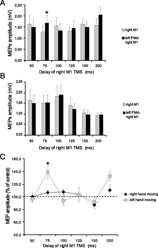
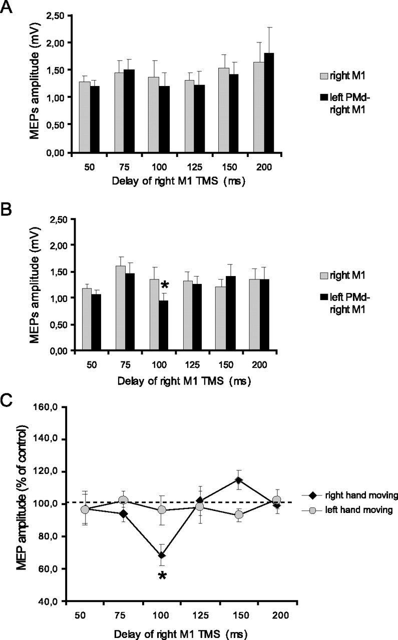
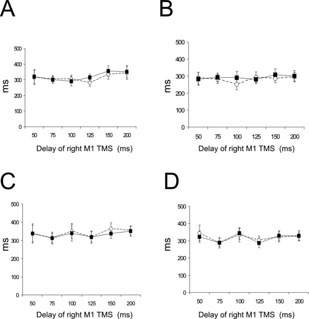
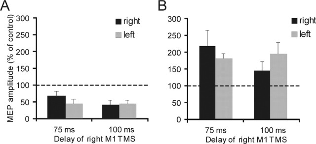
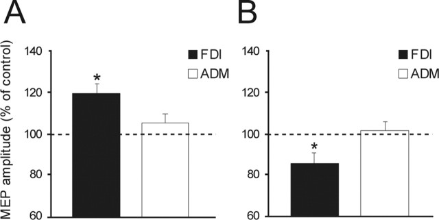
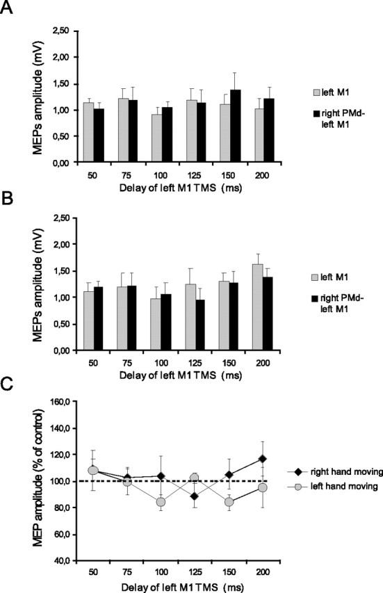
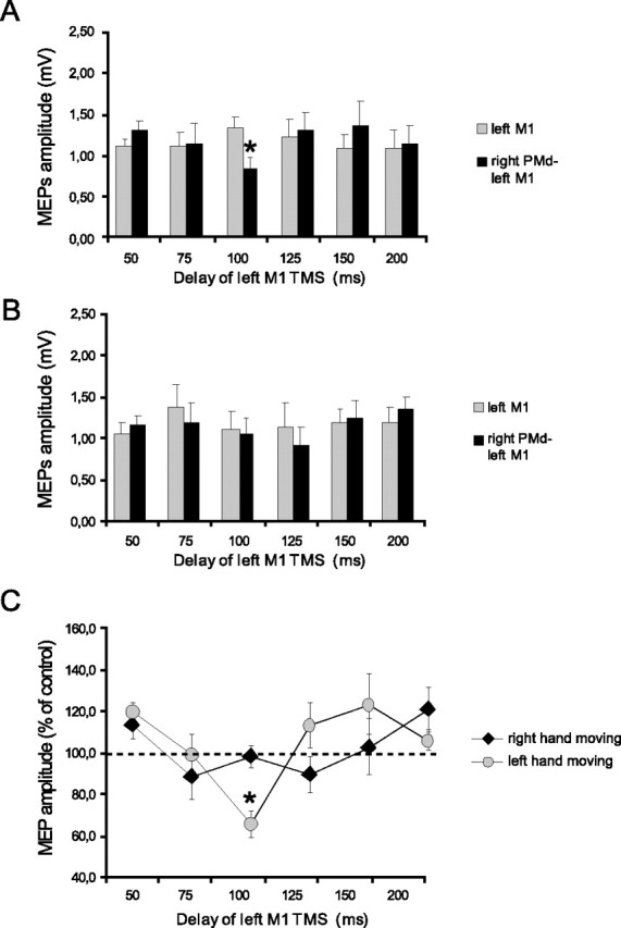
The left dorsal premotor cortex (PMd) is thought to play a dominant role in the selection of movements made by either hand. We used transcranial magnetic stimulation to study the functional connectivity of the left PMd and right primary motor cortex (M1) during an acoustic choice reaction time (RT) task involving contraction of the thumb and forefinger. The facilitatory and inhibitory pathways that can be demonstrated between left PMd and right M1 at rest were suppressed during most of the reaction period. However, they were activated briefly at the start of the reaction period, depending on whether the cue indicated that the forthcoming movement had to be made with the left or the right hand. The facilitatory pathway was active at 75 ms in those trials in which the subjects were required to move the left hand, whereas the inhibitory pathway was active at 100 ms in trials in which the subjects had to move the right hand. These changes in excitability did not occur in hand muscles not used in the task. There were no significant changes in the excitability of intracortical circuits [short intracortical inhibition (SICI) and intracortical facilitation (ICF)] in the right M1. Interhemispheric interactions between the right PMd and left M1 were mainly inhibitory at rest and showed the same temporal profile of interhemispheric inhibition as for left PMd-right M1, although no evidence was found for facilitatory interactions. The results illustrate the importance of PMd not only in facilitating cued movements but also in suppressing movements that have been prepared but are not used.
Figures
Similar articles
-
Neural mechanisms underlying the changes in ipsilateral primary motor cortex excitability during unilateral rhythmic muscle contraction.Behav Brain Res. 2013 Mar 1;240:33-45. doi: 10.1016/j.bbr.2012.10.053. Epub 2012 Nov 19. Behav Brain Res. 2013. PMID: 23174210
-
The human dorsal premotor cortex facilitates the excitability of ipsilateral primary motor cortex via a short latency cortico-cortical route.Hum Brain Mapp. 2012 Feb;33(2):419-30. doi: 10.1002/hbm.21221. Epub 2011 Mar 9. Hum Brain Mapp. 2012. PMID: 21391274 Free PMC article.
-
Performing two different actions simultaneously: The critical role of interhemispheric interactions during the preparation of bimanual movement.Cortex. 2016 Apr;77:141-154. doi: 10.1016/j.cortex.2016.02.007. Epub 2016 Feb 17. Cortex. 2016. PMID: 26963084
-
A novel dual-site transcranial magnetic stimulation paradigm to probe fast facilitatory inputs from ipsilateral dorsal premotor cortex to primary motor cortex.Neuroimage. 2012 Aug 1;62(1):500-9. doi: 10.1016/j.neuroimage.2012.05.023. Epub 2012 May 14. Neuroimage. 2012. PMID: 22626848
-
Repetitive stimulation of premotor cortex affects primary motor cortex excitability and movement preparation.Brain Stimul. 2009 Jul;2(3):152-62. doi: 10.1016/j.brs.2009.01.001. Epub 2009 Feb 28. Brain Stimul. 2009. PMID: 20633415 Clinical Trial.
Cited by
-
Stimulation of Different Sectors of the Human Dorsal Premotor Cortex Induces a Shift from Reactive to Predictive Action Strategies and Changes in Motor Inhibition: A Dense Transcranial Magnetic Stimulation (TMS) Mapping Study.Brain Sci. 2021 Apr 24;11(5):534. doi: 10.3390/brainsci11050534. Brain Sci. 2021. PMID: 33923217 Free PMC article.
-
Visual salience of the stop signal affects the neuronal dynamics of controlled inhibition.Sci Rep. 2018 Sep 24;8(1):14265. doi: 10.1038/s41598-018-32669-8. Sci Rep. 2018. PMID: 30250230 Free PMC article.
-
Transcranial Magnetic Mapping of the Short-Latency Modulations of Corticospinal Activity from the Ipsilateral Hemisphere during Rest.Front Neural Circuits. 2011 Oct 18;5:14. doi: 10.3389/fncir.2011.00014. eCollection 2011. Front Neural Circuits. 2011. PMID: 22022307 Free PMC article.
-
Transcranial direct current stimulation over the motor and premotor cortex with mirror therapy improves motor control, muscle function, and brain activity in chronic stroke: a double-blind randomized sham-controlled trial.J Neuroeng Rehabil. 2025 Apr 26;22(1):98. doi: 10.1186/s12984-025-01635-7. J Neuroeng Rehabil. 2025. PMID: 40287756 Free PMC article. Clinical Trial.
-
Functional interplay between posterior parietal and ipsilateral motor cortex revealed by twin-coil transcranial magnetic stimulation during reach planning toward contralateral space.J Neurosci. 2008 Jun 4;28(23):5944-53. doi: 10.1523/JNEUROSCI.0957-08.2008. J Neurosci. 2008. PMID: 18524898 Free PMC article.
References
-
- Buhmann C, Gorsler A, Bäumer T, Hidding U, Demiralay C, Hinkelmann K, Weiller C, Siebner HR, Münchau A (2004). Abnormal excitability of premotor–motor connections in de novo Parkinson’s disease. Brain 127:2732–2746. - PubMed
-
- Ceballos-Baumann AO, Passingham RE, Warner T, Playford ED, Marsden CD, Brooks DJ (1995). Overactive prefrontal and underactive motor cortical areas in idiopathic dystonia. Ann Neurol 37:363–372. - PubMed
-
- Cisek P, Kalaska JF (2005). Neural correlates of reaching decisions in dorsal premotor cortex: specification of multiple direction choices and final selection of action. Neuron 45:801–814. - PubMed
-
- Crammond DJ, Kalaska JF (1996). Differential relation of discharge in primary motor cortex and premotor cortex to movements versus actively maintained postures during a reaching task. Exp Brain Res 108:45–61. - PubMed
Publication types
MeSH terms
Grants and funding
LinkOut - more resources
Full Text Sources