. 2006 Jul 12;26(28):7502-12.
doi: 10.1523/JNEUROSCI.0096-06.2006.

Endogenous alpha-synuclein is induced by valproic acid through histone deacetylase inhibition and participates in neuroprotection against glutamate-induced excitotoxicity

Affiliations

Endogenous alpha-synuclein is induced by valproic acid through histone deacetylase inhibition and participates in neuroprotection against glutamate-induced excitotoxicity

Yan Leng et al. J Neurosci. .

Abstract

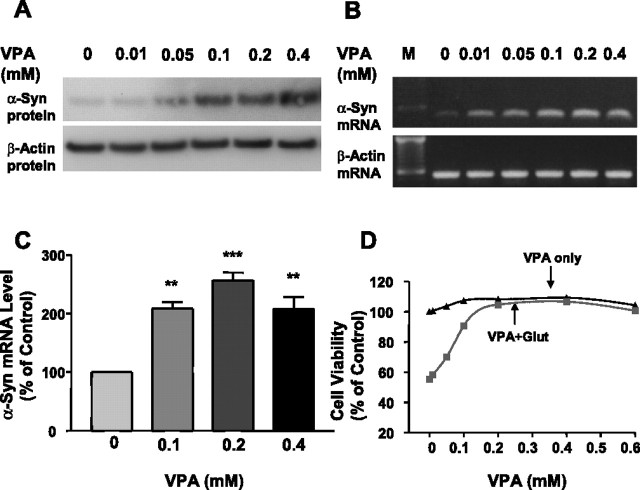
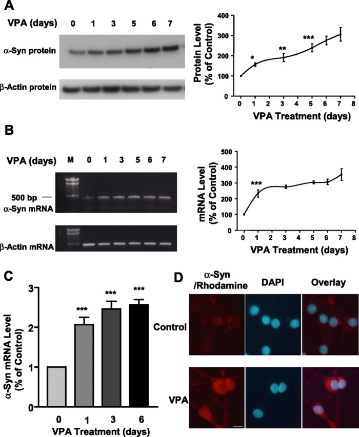
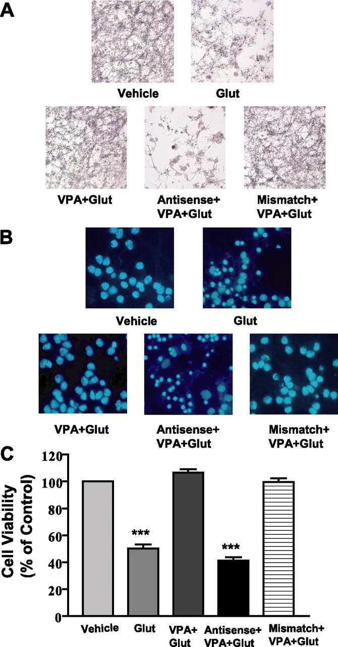
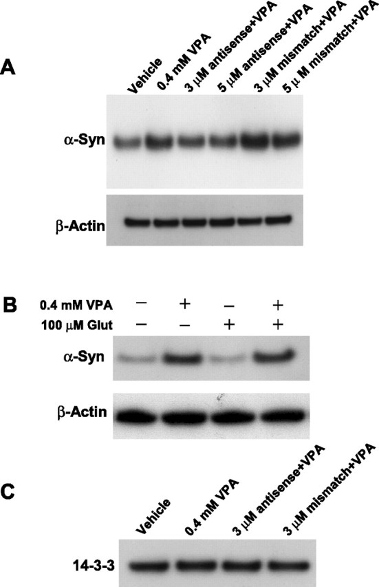
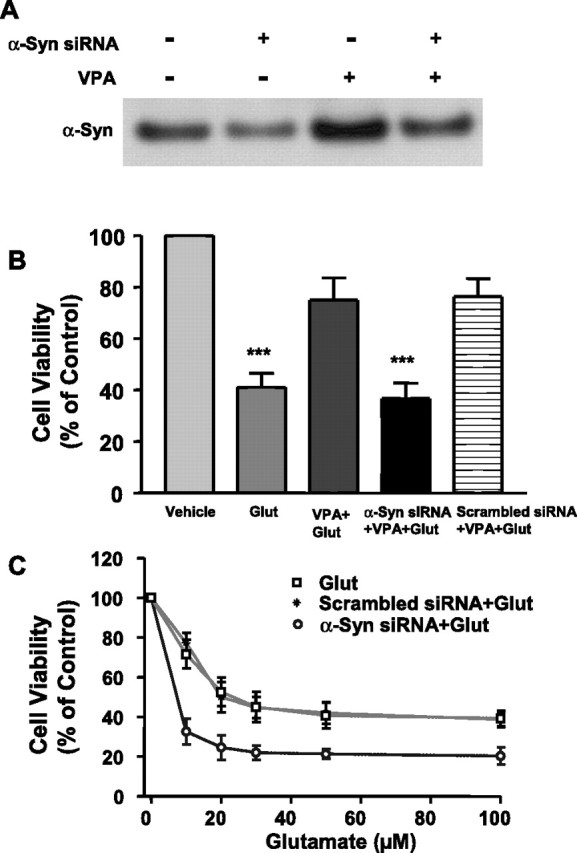
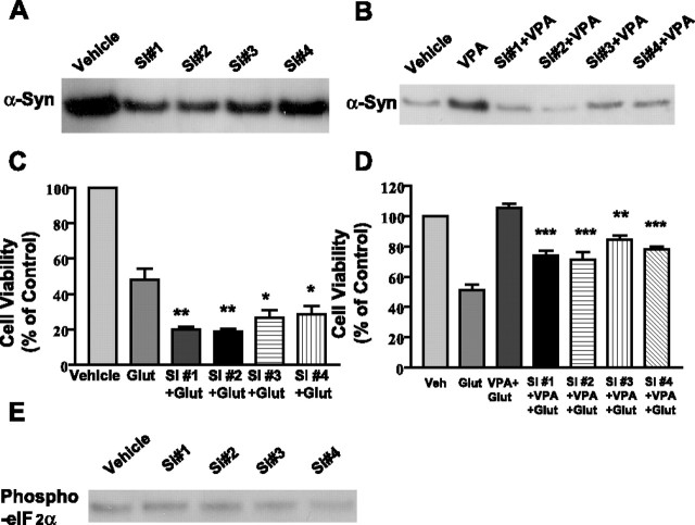
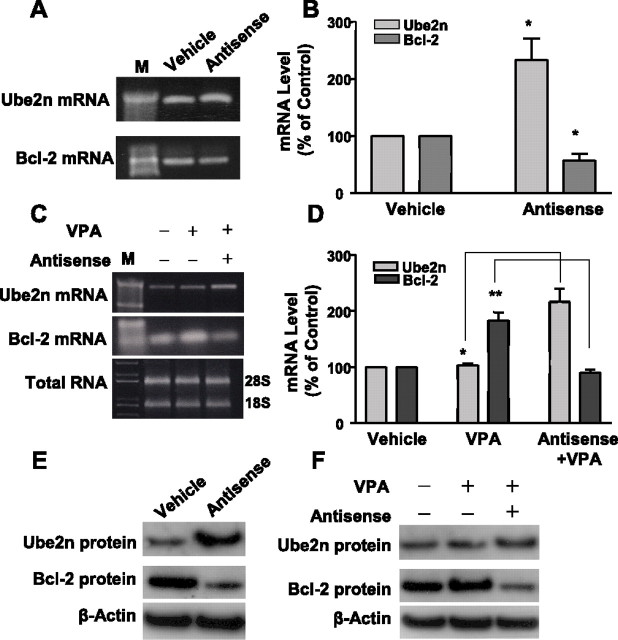
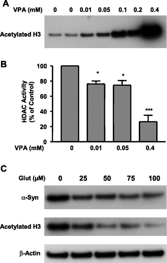
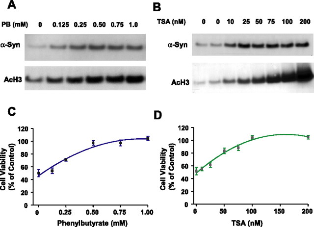
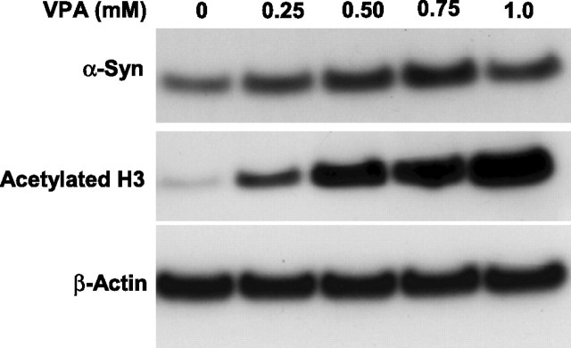
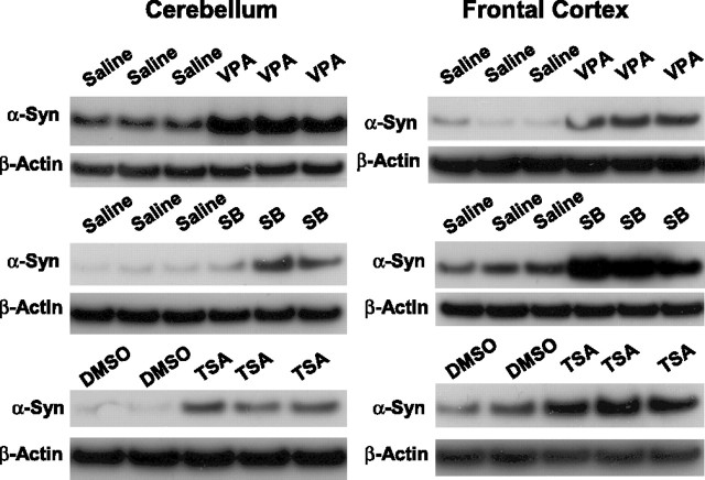
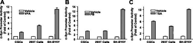
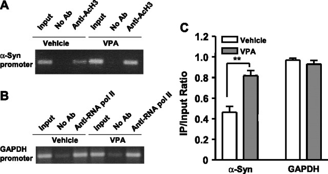
Emerging evidence suggests that alpha-synuclein (alpha-syn), which is traditionally thought to have a pathophysiological role in neurodegenerative diseases, can have neuroprotective effects. This study aimed to investigate whether endogenous alpha-syn in neurons can be induced by valproic acid (VPA), a mood-stabilizer, anticonvulsant and histone deacetylase (HDAC) inhibitor, and if so, whether the alpha-syn induction is neuroprotective. VPA treatment of rat cerebellar granule cells caused a robust dose- and time-dependent increase in levels of alpha-syn protein and mRNA and in the intensity of alpha-syn immunostaining. Knockdown of VPA-induced alpha-syn overexpression with alpha-syn antisense oligonucleotides or siRNA completely blocked VPA-induced neuroprotection. alpha-Syn knockdown also exacerbated glutamate neurotoxicity, stimulated the expression of the proapoptotic gene ubiquitin-conjugating enzyme E2N, and downregulated the expression of the anti-apoptotic gene Bcl-2. Induction of alpha-syn by VPA was associated with inhibition of HDAC activity, resulting in hyperacetylation of histone H3 in the alpha-syn promoter and a marked increase in alpha-syn promoter activity. Moreover, VPA-induced alpha-syn induction and neuroprotection were mimicked by HDAC inhibitors sodium 4-phenylbutyrate and trichostatin A (TSA). alpha-syn was also induced by VPA in rat cerebral cortical neurons. Additionally, treatment of rats with VPA, sodium butyrate, or TSA markedly increased alpha-syn protein levels in the cortex and cerebellum. Together, our results demonstrate for the first time that VPA induces alpha-syn in neurons through inhibition of HDAC and that this alpha-syn induction is critically involved in neuroprotection against glutamate excitotoxicity. Clinically, VPA may represent a suitable treatment for excitotoxicity-related neurodegenerative diseases.


Figures

Figure 1.
Figure 1.
VPA treatment increases the levels of α-syn protein and mRNA and protects against glutamate-induced excitotoxicity in CGC neurons. CGCs were treated with the indicated concentrations of VPA for 6 d starting from DIV 1. α-Syn protein levels were then determined by Western blotting (A) and α-syn mRNA levels by RT-PCR (B). The left lane in B shows the sizes of the DNA marker (M). Levels of β-actin protein and mRNA were used as the control in A and B, respectively. α-Syn mRNA levels were also quantified by real-time PCR in CGCs treated with vehicle and 0.1, 0.2, and 0.4 mm VPA for 6 d (C). Data are means ± SEM of percentage of control from three independent experiments. ∗∗p < 0.01; ∗∗∗p < 0.001 compared with the vehicle control. VPA-pretreated CGCs were also exposed to vehicle or 100 μm glutamate (glut) on DIV 7 for 24 h and then analyzed for cell viability by MTT assay (D). Data are presented as means ± SEM of percentage of untreated control from five to six independent cultures.
Figure 2.
Figure 2.
VPA-induced increase in the levels of α-syn protein and mRNA is duration dependent. CGCs were treated with 0.4 mm VPA for 1–6 d, and all cultures were harvested on DIV 7 for Western blotting of α-syn protein levels (A) and RT-PCR of α-syn mRNA levels (B). The left lane in B is the DNA marker (M). Levels of β-actin protein and mRNA were used as the control in A and B, respectively. Quantified results are shown in the respective right panels in A and B and are expressed as means ± SEM of four independent experiments. ∗p < 0.05; ∗∗p < 0.01; ∗∗∗p < 0.001 compared with the 0 time control. α-Syn mRNA levels were also quantified by real-time PCR in CGCs treated with 0.4 mm VPA for 1, 3, and 6 d (C). Data are means ± SEM of three independent experiments. ∗∗∗p < 0.001 compared with 0 time control. VPA-treated and untreated CGC cultures were also examined for α-syn immunofluorescence as described in Materials and Methods (D). α-Syn immunostaining was identified by incubation with goat anti-mouse Rhodamine Red-X-AffiniPure-conjugated secondary antibody and is shown in red, and the nuclear staining by DAPI is shown in blue. The overlay images are also shown. Scale bar, 10 μm.
Figure 3.
Figure 3.
Antisense, but not mismatch, oligonucleotides to α-syn block VPA-induced protection of CGCs from glutamate excitotoxicity. Cells were exposed to antisense or mismatch oligonucleotides (3 μm) for 24 h before pretreatment with 0.4 mm VPA (DIV 1 to DIV 7) and subsequent treatment with glutamate (Glut; 100 μm) for 24 h. All cultures were stained with MTT (A) or Hoechst dye 33258 (B) and then photographed. Cell viability was quantified by MTT assay and expressed as means ± SEM of vehicle-treated control from four independent cultures (C). ∗∗∗p < 0.001 compared with vehicle-treated control.
Figure 4.
Figure 4.
Antisense, but not mismatch, oligonucleotides to α-syn block VPA-induced increase in α-syn protein levels, and VPA prevents glutamate-induced α-syn downregulation. A, CGCs were exposed to 3 or 5 μm antisense or mismatch oligonucleotides for 24 h before treatment with 0.4 mm VPA for 6 d (DIV 1 to DIV 7). Cells were then harvested for Western blotting of α-syn. Levels of β-actin were used as the loading control. B, Cells were treated with 0.4 mm VPA for 6 d (DIV 1 to DIV 7), followed by treatment with 100 μm glutamate (Glut) for 24 h. Western blotting of α-syn and β-actin was then performed, and the blots of a typical experiment are shown. C, Cells were exposed to 3 μm antisense oligonucleotides to α-syn for 24 h before treatment with 0.4 mm VPA for 6 d (DIV 1 to DIV 7). Western blotting for 14-3-3 protein was then performed using an antibody against 14-3-3 (1:1000; BD Transduction Laboratories).
Figure 5.
Figure 5.
α-Syn siRNA pool suppresses VPA-induced increase of α-syn protein, blocks VPA-induced protection, and facilitates glutamate excitotoxicity. Cells were exposed to α-syn siRNA pool (100 nm) or scrambled siRNA (100 nm) for 24 h before pretreatment with 0.4 mm VPA for 6 d (DIV 1 to DIV 7). Cells were either harvested for Western blot (A) or subsequently treated with glutamate (Glut; 100 μm) for 24 h (B). Cells were also exposed to α-syn siRNA pool or its scrambled siRNA control for 6 d (DIV 1 to DIV 7) and then exposed to the indicated concentration of glutamate for 24 h (C). Cell viability was quantified by MTT assay and expressed as means ± SEM of vehicle-treated control from three independent cultures. ∗∗∗p < 0.001 compared with vehicle-treated control.
Figure 6.
Figure 6.
Individual siRNA decreases basal and VPA-stimulated α-syn protein levels, exacerbates glutamate-induced excitotoxicity, and attenuates VPA neuroprotection. Cells were exposed to 100 nm individual α-syn siRNA (S#1, S#2, S#3, or S#4) for 24 h before pretreatment with vehicle or 0.4 mm VPA for 6 d (DIV 1 to DIV 7). Cells were then harvested for Western blotting of α-syn protein (A, B) or further incubated without or with glutamate (Glut; 100 μm) for 24 h (C, D). Cell viability was quantified by MTT assay from three independent cultures. ∗p < 0.05; ∗∗p < 0.01 compared with glutamate group in C. ∗∗p < 0.01; ∗∗∗p < 0.001 compared with VPA plus glutamate group in D. Cell lysates from A were also used for immunoblotting of phospho-eIF 2α protein levels (E). Western blots are results from a typical experiment.
Figure 7.
Figure 7.
α-Syn antisense oligonucleotides upregulate Ube2n but downregulate Bcl-2 mRNA and differentially affect VPA-induced levels of these proteins. CGCs on DIV 0 were treated with 3 μm antisense oligonucleotides for α-syn. Four days later, cells were harvested and total RNA was extracted. Ube2n and Bcl-2 genes were analyzed for their mRNA levels by RT-PCR (A) and quantified by real-time PCR; ∗p < 0.05 compared with respective vehicle in B. CGCs were also treated with 3 μm antisense oligonucleotides for α-syn on DIV 0, followed by treatment with 0.4 mm VPA on DIV 1. Cells were harvested on DIV 4 for analysis of Ube2n and Bcl-2 mRNA levels by RT-PCR (C) and real-time RT-PCR (D). ∗p < 0.05; ∗∗p < 0.01 between indicated groups. M, DNA marker. Western blotting were also performed to verify the antisense-induced changes in Ube2n and Bcl-2 protein levels in CGCs without and with VPA treatment (E, F). The Western blots are from a typical experiment.
Figure 8.
Figure 8.
Histone H3 acetylation and α-syn levels are dose-dependently increased by VPA but decreased by glutamate treatment. CGCs were treated with the indicated concentration of VPA for 6 d (from DIV 1 to DIV 7). Cells were then harvested for Western blotting of acetylated histone H3 (A) and measurement of HDAC activity (B). CGCs were also treated with glutamate (100 μm; Glut) on DIV 7 for 24 h (C). Cells were then harvested for Western blotting of α-syn, acetylated histone H3, and β-actin. The quantified results are means ± SEM of vehicle-treated control from three independent experiments. ∗p < 0.05; ∗∗∗p < 0.001 compared with the vehicle-treated control.
Figure 9.
Figure 9.
HDAC inhibitors phenylbutyrate and TSA mimic VPA to induce α-syn, increase histone H3 acetylation (Ac-H3), and protect against glutamate excitotoxicity. CGCs were treated with indicated concentrations of sodium 4-phenylbutyrate (PB) for 6 d (A) or TSA for 2 d (B), starting from DIV 1 or DIV 5, respectively. Longer exposure of cells to high concentrations of TSA resulted in cytotoxicity. Cells were then harvested for Western blotting of α-syn and acetylation histone H3 levels. PB- or TSA-treated cells were also exposed to glutamate (100 μm) on DIV 7 for 24 h and then assayed for cell viability by MTT analysis (C, D). Quantified data expressed as percentage of untreated control are means ± SEM from three independent experiments.
Figure 10.
Figure 10.
VPA treatment increases levels of α-syn and acetylated histone H3 in rat cortical neuronal cultures. Cerebral cortical neurons were treated with the indicated concentrations of VPA for 4 d, starting from DIV 6. Cells were then harvested for Western blotting of levels of α-syn and acetylated histone H3, using β-actin as the control.
Figure 11.
Figure 11.
Repeated injections of rats with VPA, sodium butyrate, and TSA increase brain levels of α-syn protein. Male Sprague Dawley rats (250 ± 10 g) were subjected to daily subcutaneous injections with VPA (300 mg/kg in saline), sodium butyrate (SB) (300 mg/kg in saline), TSA (0.2 mg/kg in DMSO), and their respective vehicle. Four days later, animals were killed by decapitation. The brains were removed and dissected, followed by homogenization and sonication for 40 s in lysis buffer as described previously (Ren et al. 2004). An aliquot of 15 μg was used for Western blotting as described in Materials and Methods. The blot in each lane represents the result from an individual animal.
Figure 12.
Figure 12.
HDAC inhibitors induce a robust increase in the promoter activity of α-syn. CGCs, human embryonic kidney 293T cells, and human neuroblastoma SH-SY5Y cells were transfected with a 1.9 kb DNA fragment upstream of the human α-syn transcription initiation site (pASP-1.9). One day after transfection, these three cell types were treated for 24 h with 1 mm VPA (A), 2 mm phenylbutyrate (B; PB), or 50 nm TSA (C). The α-syn promoter activity was assayed in the luciferase reporter system. Data are means ± SEM from three independent experiments. Note that treatment with VPA induced a robust increase in the α-syn promoter activity in CGCs and HEK 293T cells and an even greater increase in SH-SY5Y cells. Similar increases in α-syn promoter activity were observed in all three cell types treated with PB or TSA.
Figure 13.
Figure 13.
VPA induces hyperacetylation of histone H3 in the α-syn promoter region in neuroblastoma SH-SY5Y cells. SH-SY5Y cells transfected with the pASP-1.9 construct were treated with VPA (1 mm) or vehicle for 24 h and then cross-linked with 1% formaldehyde, and the chromatin was prepared and sheared by sonication. The protein/DNA complex was incubated with or without an antibody (No Ab) against acetylated histone H3 for ChIP analysis, and PCR was performed to amplify α-syn promoter (A). The protein/DNA complex was also incubated with or without antibody against RNA pol II for ChIP analysis and PCR amplification of GAPDH promoter, which served as a positive reagent control (B). The quantified data are means ± SEM of the ratio of relative optical densities of bands within immunoprecipitated (IP) samples and input lanes derived from ethidium bromide-stained gels from four independent experiments (C). ∗∗p < 0.01 compared with the α-syn promoter in cells treated with the vehicle control.

Similar articles

Cited by

References

    1. Albani D, Peverelli E, Rametta R, Batelli S, Veschini L, Negro A, Forloni G (2004). Protective effect of TAT-delivered alpha-synuclein: relevance of the C-terminal domain and involvement of HSP70. FASEB J 18:1713–1715. - PubMed
    1. Alves da Costa CA, Ancolio K, Checler F (2000). Wild-type but not Parkinson’s disease-related Ala-53→Thr mutant α-synuclein protects neuronal cells from apoptotic stimuli. J Biol Chem 275:24065–24069. - PubMed
    1. Alves da Costa CA, Paitel E, Vincent B, Checler F (2002). α-synuclein lowers p53-dependent apoptotic response of neuronal cells. J Biol Chem 277:50980–50984. - PubMed
    1. Bantounas I, Phylactou LA, Uney JB (2004). RNA interference and the use of small interfering RNA to study gene function in mammalian systems. J Mol Endocrinol 33:545–557. - PubMed
    1. Borghi R, Marchese R, Negro A, Marinelli L, Forloni G, Zaccheo D, Abbruzzese G, Tabaton M (2000). Full length alpha-Synuclein is present in cerebrospinal fluid from Parkinson’s disease and normal subjects. Neurosci Lett 287:65–67. - PubMed

Publication types

MeSH terms

LinkOut - more resources