. 2006 Nov-Dec;27(6):868-79.
doi: 10.2164/jandrol.106.000349. Epub 2006 Jul 12.

Transgenerational effect of the endocrine disruptor vinclozolin on male spermatogenesis

Affiliations

Transgenerational effect of the endocrine disruptor vinclozolin on male spermatogenesis

Matthew D Anway et al. J Androl. 2006 Nov-Dec.

Abstract

The current study was designed to examine the actions of a model endocrine disruptor on embryonic testis development and male fertility. Pregnant rats (F0) that received a transient embryonic exposure to an environmental endocrine disruptor, vinclozolin, had male offspring (F1) with reduced spermatogenic capacity. The reduced spermatogenetic capacity observed in the F1 male offspring was transmitted to the subsequent generations (F2-F4). The administration of vinclozolin, an androgen receptor antagonist, at 100 mg/kg/day from embryonic day 8-14 (E8-E14) of pregnancy to only the F0 dam resulted in a transgenerational phenotype in the subsequent male offspring in the F1-F4 generations. The litter size and male/female sex ratios were similar in controls and the vinclozolin generations. The average testes/body weight index of the postnatal day 60 (P60) males was not significantly different in the vinclozolin-treated generations compared to the controls. However, the testicular spermatid number, as well as the epididymal sperm number and motility, were significantly reduced in the vinclozolin generations compared to the control animals. Postnatal day 20 (P20) testis from the vinclozolin F2 generation had no morphological abnormalities, but did have an increase in spermatogenic cell apoptosis. Although the P60 testis morphology was predominantly normal, the germ cell apoptosis was significantly increased in the testes cross sections of animals from the vinclozolin generations. The increase in apoptosis was stage-specific in the testis, with tubules at stages IX-XIV having the highest increase in apoptotic germ cells. The tubules at stages I-V also had an increase in apoptotic germ cells compared to the control samples, but tubules at stages VI-VIII had no increase in apoptotic germ cells. An outcross of a vinclozolin generation male with a wild-type female demonstrated that the reduced spermatogenic cell phenotype was transmitted through the male germ line. An outcross with a vinclozolin generation female with a wild-type male had no phenotype. A similar phenotype was observed in outbred Sprague Dawley and inbred Fisher rat strains. Observations demonstrate that a transient exposure at the time of male sex determination to the antiandrogenic endocrine disruptor vinclozolin can induce an apparent epigenetic transgenerational phenotype with reduced spermatogenic capacity.


Figures

Figure 1.
Figure 1.
Ratio of testes weights per body weight from adult (P60) male (A) Sprague Dawley and (B) Fisher rats from control and vinclozolin F1, F2, F3, and F4 generations, F2 generation male outcross (VOC) to wild-type females, and F2 generation female reverse outcross (RVOC) to wild-type male generations. Open bars (◻) represent control and solid bars (∎) represent vinclozolin generations. Statistically significant differences between control and vinclozolin generation rats are indicated by (*) for P < .05. Numbers of Sprague Dawley animals analyzed are F1 (n = 7 control, n = 7 vinclozolin); F2 (n = 8 control, n = 17 vinclozolin); F3 (n = 10 control, n = 8 vinclozolin); F4 (n = 5 control, n = 5 vinclozolin); and outcross VOC (n = 10), RVOC (n = 6) and wild-type as outcross controls (n = 5). Numbers of Fisher animals analyzed are F1 (n = 4 control, n = 10 vinclozolin); F2 (n = 4 control, n = 5 vinclozolin); and F3 (n = 11 control, n = 13 vinclozolin).
Figure 2.
Figure 2.
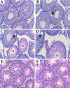
Testis morphology of control P60 (A) and P20 (E) and vinclozolin P60 (B–D) and P20 (F) F2 generation SD rats. Testis cross sections stained with H&E. Arrows indicated large vacules and asterisks are in the lumen of seminiferous tubules that have degenerated and have no spermatogenesis. (A–C) Magnification 100×; (D–F) magnification 200×. Micrographs are representative of a minimum of 8 different analyses of P20 and 17 analyses of P60 F2 vinclozolin animals.
Figure 3.
Figure 3.
Apoptosis (TUNEL) analyses in the testes P60 F2 generation SD rats from control (A) and vinclozolin (B) at 200× magnification and vinclozolin (C) and phase contrast (D) at 400× magnification. Apoptotic TUNEL positive cells labeled yellow. Arrows indicate possible spermatogonial cells along the basement membrane. Arrowheads indicate spermatocytes within the seminiferous tubule, and the lumen is depicted with a roman numeral indicating the approximate stage of the cycle of seminiferous epithelium. Micrographs are representative of a minimum of 10 different analyses of F2 vinclozolin generation animals.
Figure 4.
Figure 4.
Apoptotic germ cells from adult (P60) male Sprague Dawley rats from control (open bars, ◻) and vinclozolin (solid bars, ∎) F1, F2, F3, and F4 generations, F2 generation male outcross (VOC) to wild-type females, and F2 generation female reverse outcross (RVOC) to wild-type male generations. Data presented as the number tubules with +4 TUNEL positive cells per testicular cross section at stages IX–XIV (A), stages I–V (B), and stages VI–VIII (C) of the cycle of the seminiferous epithelium. Statistically significant differences between individual controls and vinclozolin generation rats are indicated by (*) for P < .05. The numbers of animals analyzed are F1 (n = 3 control, n = 3 vinclozolin); F2 (n = 4 control, n = 4 vinclozolin); F3 (n = 4 control, n = 4 vinclozolin); F4 (n = 3 control, n = 3 vinclozolin); and outcross VOC (n = 4), RVOC (n = 3) and wild-type as outcross controls (n = 3).
Figure 5.
Figure 5.
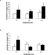
Apoptotic germ cells from adult (P60) male Fisher rats from control (open bars, ◻) and vinclozolin (solid bars, ∎) F1, F2, and F3 generations. Data presented as the number tubules with +4 TUNEL positive cells per testicular cross section at stages IX–XIV (A) and stages I–VIII (B) of the cycle of the seminiferous epithelium. Statistically significant differences between individual controls and vinclozolin generation rats are indicated by (*) for P < .05. The numbers of animals analyzed are F1 (n = 3 control, n = 3 vinclozolin); F2 (n = 4 control, n = 4 vinclozolin); and F3 (n = 4 control, n = 4 vinclozolin).
Figure 6.
Figure 6.
Apoptosis (TUNEL) analyses in the testes P20 F2 generation of Sprague Dawley rats from control (A) and vinclozolin (B) at 200× magnification. The lumen of the seminiferous tubule is indicated with an asterisk (*). (C) Presented as the total number of apoptotic germ cells from P20 male rats from control and vinclozolin F2 generation animals. The mean ± SEM is presented, and statistically significant differences between controls and vinclozolin generation rats are indicated by (*) for P < .05, F2 (n = 6 control, n = 8 vinclozolin).
Figure 7.
Figure 7.
Serum (A) testosterone levels from adult (P60) male Sprague Dawley rats from control and vinclozolin F1, F2, F3, and F4 generations, F2 generation male outcross (VOC) to wild-type females, and F2 generation female reverse outcross (RVOC) to wild-type males. (B) Testicular fluid (TF) testosterone levels from adult (P60) male rats from control and vinclozolin F2 generation. Open bars (◻) represent control and solid bars (∎) represent vinclozolin generation animals. Serum analyses from F1 (n = 7 control, n = 7 vinclozolin); F2 (n = 8 control, n = 17 vinclozolin); F3 (n = 10 control, n = 8 vinclozolin); F4 (n = 5 control, n = 5 vinclozolin); and outcross VOC (n = 10), RVOC (n = 6) and wild-type as outcross controls (n = 5). TF testosterone analyses F2 (n = 3 control, n = 4 vinclozolin). The mean ± SEM is presented, with no statistical difference detected.
Figure 8.
Figure 8.
Serum testosterone levels from adult (P60) male Fisher rats from control and vinclozolin F1, F2, and F3 generations. Open bars (◻) represent control and solid bars (∎) represent vinclozolin generation animals. Serum analyses from F1 (n = 6 control, n = 10 vinclozolin); F2 (n = 4 control, n = 5 vinclozolin) and F3 (n = 11 control, n = 13 vinclozolin). The mean ± SEM is presented, with no statistical difference detected.

References

    1. Anway MD, Cupp AS, Uzumcu M, Skinner MK. Epigenetic transgenerational actions of endocrine disruptors and male fertility. Science. 2005;308:1466–1469. - PMC - PubMed
    1. Barber R, Plumb MA, Boulton E, Roux I, Dubrova YE. Elevated mutation rates in the germ line of firstand second-generation offspring of irradiated male mice. Proc Natl Acad Sci U S A. 2002;99:6877–6882. - PMC - PubMed
    1. Basha MR, Wei W, Bakheet SA, Benitez N, Siddiqi HK, Ge YW, Lahiri DK, Zawia NH. The fetal basis of amyloidogenesis: exposure to lead and latent overexpression of amyloid precursor protein and beta-amyloid in the aging brain. J Neurosci. 2005;25:823–829. - PMC - PubMed
    1. Bayley M, Junge M, Baatrup E. Exposure of juvenile guppies to three antiandrogens causes demasculinization and a reduced sperm count in adult males. Aquat Toxicol (Amst). 2002;56:227–239. - PubMed
    1. Cooper RL, Goldman JM, Stoker TE. Neuroendocrine and reproductive effects of contemporary-use pesticides. Toxicol Ind Health. 1999;15:26–36. - PubMed

Publication types

LinkOut - more resources