. 2006 Jan-Mar;12(1-3):34-46.
doi: 10.2119/2006-00014.Hendricks.

Predifferentiated embryonic stem cells prevent chronic pain behaviors and restore sensory function following spinal cord injury in mice

Affiliations

Predifferentiated embryonic stem cells prevent chronic pain behaviors and restore sensory function following spinal cord injury in mice

Wesley A Hendricks et al. Mol Med. 2006 Jan-Mar.

Abstract

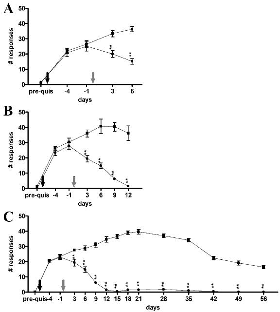
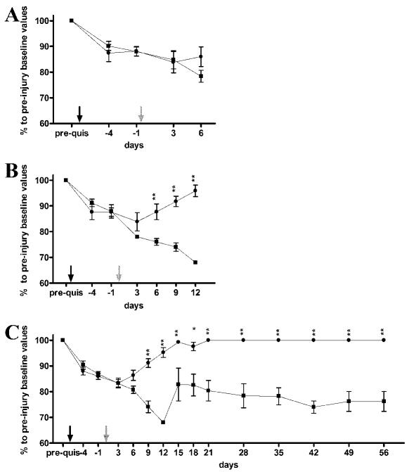
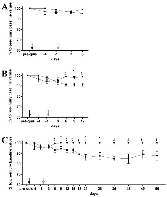
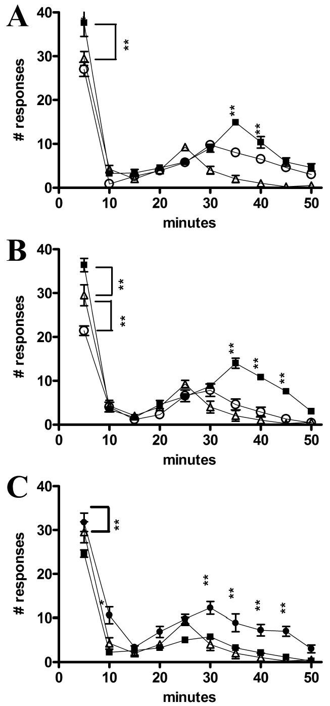
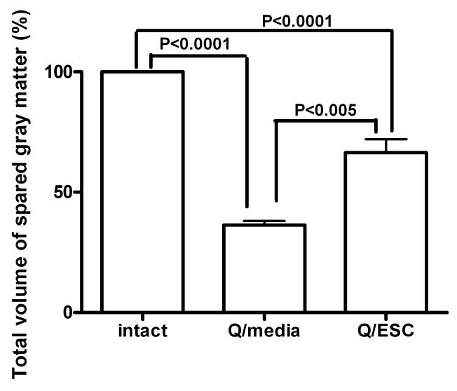
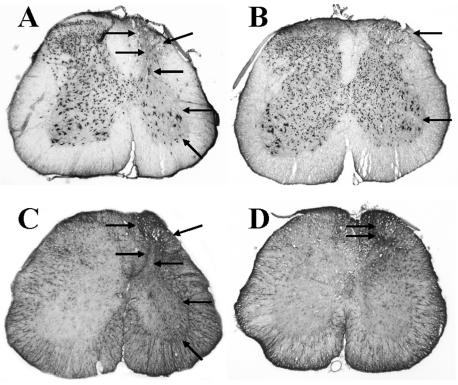
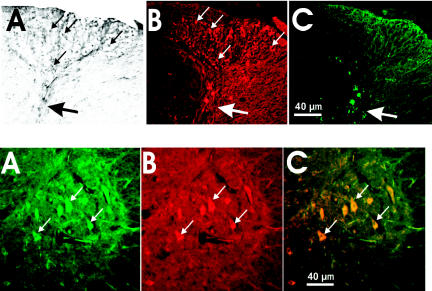
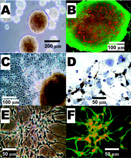
Embryonic stem (ES) cells have been investigated in repair of the CNS following neuronal injury and disease; however, the efficacy of these cells in treatment of postinjury pain is far from clear. In this study, we evaluated the therapeutic potential of predifferentiated mouse ES cells to restore sensory deficits following spinal cord injury (SCI) in mice. The pain model used unilateral intraspinal injection of quisqualic acid (QUIS) into the dorsal horn between vertebral levels T13 and L1. Seven days later, 60,000 predifferentiated ES cells or media were transplanted into the site of the lesion. Histological analysis at 7, 14, and 60 days post-transplantation revealed that animals receiving ES cell transplants suffered significantly less tissue damage than animals receiving media alone. Transplanted cells provided immediate effects on both spontaneous and evoked pain behaviors. Treatment with ES cells resulted in 0% (n = 28) excessive grooming behavior versus 60% (18 of 30) in media-treated animals. In the acetone test (to assess thermal allodynia), mice recovered to preinjury levels by 12 days after ES cell transplant, whereas control animals injected with media after SCI did not show any improvement up to 60 days. Similarly, the von Frey test (to assess mechanical allodynia) and the formalin test (to assess nociceptive hyperalgesia) showed that transplantation of predifferentiated ES cells significantly reduced these pain behaviors following injury. Here we show that predifferentiated ES cells act in a neuroprotective manner and provide antinociceptive and therapeutic effects following excitotoxic SCI.


Figures

Figure 1
Figure 1
An example of excessive grooming behavior displayed by 60% of the media-injected mice. Excessive grooming behavior was not observed in ES cell–transplanted mice.
Figure 2
Figure 2
Total number of responses to room temperature (23 °C) acetone. (A) Seven days after ES cell or media treatment. (B) Fourteen days after ES cell or media treatment. (C) Sixty days after ES cell or media treatment. Black arrow, QUIS injury; gray arrow, ES cell transplantation or media injection; •, ES cell–transplanted mice; ▪, media-injected mice. ** P < 0.001.
Figure 3
Figure 3
Allodynia to mechanical stimuli applied to the ipsilateral hindpaw for ES cell–transplanted and media-injected mice. (A) Seven days after ES cell (n = 7) or media (n = 10) treatment. (B) Fourteen days after ES cell (n = 10) or media (n = 10) treatment. (C) Sixty days after ES cell (n = 11) or media (n = 10) treatment. y-axis, percent of preinjury baseline values;•, ES cell–transplanted mice; ▪, media-injected mice; black arrows, QUIS injection; gray arrows, ES cell transplantation or media injection. *P < 0.01, **P < 0.001.
Figure 4
Figure 4
Allodynia to mechanical stimuli applied to the contralateral hind paw in ES cell transplanted and media injected mice. (A) Seven days after ES cell (n = 7) or media (n = 10) treatment. (B) Fourteen days after ES cell (n = 10) or media (n = 10) treatment. (C) Sixty days after ES cell (n = 11) or media (n = 10) treatment. y-axis, percent of preinjury baseline values; •, ES cell–transplanted mice; ▪, media-injected mice; black arrows, QUIS injection; gray arrows, ES cell transplantation or media injection. *P < 0.01, **P < 0.001.
Figure 5
Figure 5
Number of responses to formalin injection over a 50-min observation period. (A) Seven days after ES cell (n = 7) and media (n = 10) treatment and intact mice (n = 5). (B) Fourteen days after ES cell (n = 10) and media (n = 10) treatment and intact mice (n = 5). (C) Sixty days after ES cell (n = 11) and media (n = 10) treatment and intact mice (n = 5). •, ES cell–transplanted mice;▪, media-injected mice;▵, intact mice. *P < 0.01, ** P < 0.001.
Figure 6
Figure 6
Volume of spared gray matter in ES cell–transplanted and media-injected mice at 60 days.
Figure 7
Figure 7
Fluorescence microscopy of GFP-expressing cells 14 days after ES cell transplantation (A–C). (A) GFP cells fill epicenter of injury. Arrow indicates transplanted cells. (B) GFP cells migrating into surrounding tissue. Arrow indicates origin of migrating GFP-positive cells. (C) GFP cell with processes. Arrows indicate cell body and processes.
Figure 8
Figure 8
Immunohistochemical staining 14 days after media injection (A, C) and ES cell transplantation (B, D). (A) Visualization of NeuN-positive cells in media-injected spinal cord. Arrows indicate lesion and the absence of neuronal staining ipsilateral to injury. (B) Visualization of NeuN-positive cells in ES cell–transplanted spinal cord. Arrows indicate side ipsilateral to injury and cell transplant. (C) Visualization of GFAP-positive cells in media-injected spinal cord. Arrows indicate lesion ipsilateral to injury and increased intensity of GFAP immunostaining ipsilateral to injury. (D) Visualization of GFAP-positive cells in ES cell–transplanted spinal cord. Arrows indicate dorsal horn preservation on side ipsilateral to injury and cell transplant.
Figure 9
Figure 9
Immunohistochemical staining 60 days after media injection (A, C) and ES cell transplantation (B, D). (A) Visualization of NeuN-positive cells in media-injected spinal cord. Arrows indicate neuronal loss in all laminae ipsilateral to injury. (B) Visualization of NeuN-positive cells in ES cell–transplanted spinal cord. Arrows indicate side ipsilateral to injury. Note increased number of immunostained neuronal cells. (C) Visualization of GFAP-positive cells in media-injected spinal cord. Arrows indicate areas of high staining intensity in side ipsilateral to injury. (D) Visualization of GFAP-positive cells in ES cell–transplanted spinal cord. Arrows indicate increased intensity of GFAP staining in only a small area on side ipsilateral to injury and cell transplant.
Figure 10
Figure 10
Survival of ES cells ipsilateral to the lesion at 60 days. Upper panel: Immunodetection of GFP (A, DAB staining), NeuN (B, immunofluorescence), and GFAP (C, immunofluorescence) in the ipsilateral dorsal horn. Small white or black arrows indicate neurons. Big white or black arrow points at collapsed dorsal horn. Lower panel: Double immunofluorescence for GFP and MAP-2 (a neuronal marker). (A) GFP fluorescent cells. (B) MAP-2–positive cells. (C) Merge of 2 images. Small white arrows indicate neurons.
Figure 11
Figure 11
ES cells in vitro. Live culture pictures (A, C, E). (A) Free-floating embryoid bodies. (C) Differentiation induction, neural progenitors sprouting from embryoid body. (E) Neural network of differentiated cells. Immunocytochemical staining of cultured ES cells (B, D, F). (B) GFP-positive embryoid body viewed under fluorescence microscopy. Mitotic, histone H3–positive cells were visualized with Texas red. (D) Neural network. Staining against synapsin. (F) Double immunostaining of RA differentiated ES cells. Positive staining against Map-2 conjugated with FITC for green fluorescence indicates neuronal phenotype; LIM-2 conjugated with Texas red for red fluorescence allows further characterization of highly specialized dorsal interneurons.

Similar articles

Cited by

References

    1. France RD, Krishnan KRR. (1988) Chronic pain. American Psychiatric Press, Washington, DC, pp. xx, 561.
    1. Guttmann L. (1973) Spinal cord injuries: comprehensive management and research. Blackwell Scientific, Oxford, pp. xiii, 694 p.
    1. Windle WF. (1980) The spinal cord and its reaction to traumatic injury: anatomy-physiology-pharmacology-therapeutics. M. Dekker, New York, pp. xi, 384.
    1. Yezierski RP, Liu S, Ruenes GL, Kajander KJ, Brewer KL. Excitotoxic spinal cord injury: behavioral and morphological characteristics of a central pain model. Pain. 1998;75:141–55. - PubMed
    1. Siddall PJ, Loeser JD. Pain following spinal cord injury. Spinal Cord. 2001;39:63–73. - PubMed

Publication types

LinkOut - more resources