. 2006 Jan-Mar;12(1-3):17-24.
doi: 10.2119/2006-00001.Kim.

Phytosphingosine stimulates the differentiation of human keratinocytes and inhibits TPA-induced inflammatory epidermal hyperplasia in hairless mouse skin

Affiliations

Phytosphingosine stimulates the differentiation of human keratinocytes and inhibits TPA-induced inflammatory epidermal hyperplasia in hairless mouse skin

Sujong Kim et al. Mol Med. 2006 Jan-Mar.

Abstract

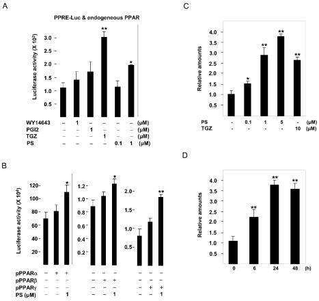
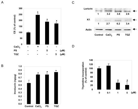
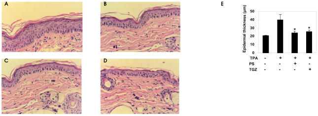
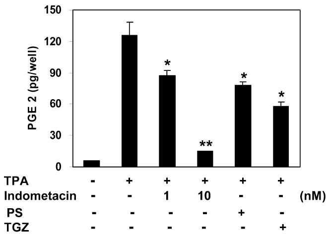
The binding of sphingoid bases to peroxisome proliferator-activated receptor (PPAR) has been detected in a solid-phase binding assay. However, sphingoid base-induced changes in PPAR transactivation activity have not been examined. In this report, we show by reporter gene analyses that phytosphingosine (PS), a natural sphingoid base, activates the transcriptional activity of PPARs in the immortalized human keratinocyte, HaCaT. Real-time PCR analyses showed that the mRNA level of PPARgamma was increased after PS treatment in HaCaT cells in a dose- and time-dependent manner. Because PPARs play important roles in skin barrier homeostasis by regulating epidermal cell growth, terminal differentiation, and inflammatory response, we examined the effect of PS on normal human epidermal keratinocytes (NHEKs) and mouse skin. PS increased the production of cornified envelope in NHEKs by approximately 1.8-fold compared with controls. Epidermal differentiation marker proteins such as involucrin, loricrin, and keratin1 were also increased in PS-treated NHEKs, by ELISA or Western blotting analysis. A [(3)H]thymidine incorporation assay showed that PS inhibited DNA synthesis in NHEKs to 20% compared with controls. The antiproliferative and anti-inflammatory effects of PS were examined in a mouse model of irritant contact dermatitis produced by topical application of 12-O-tetradecanoylphorbol-13-acetate (TPA). PS blocked epidermal thickening and edema and the infiltration of inflammatory cells into the dermis in the skin of TPA-treated hairless mice. The anti-inflammatory effects of PS were confirmed by the observation that PS blocked the TPA-induced generation of prostaglandin E(2) in peripheral mononuclear leukocytes. Taken together, our results provide an insight into the multiple regulatory roles of PS in epidermal homeostasis, and furthermore point to the potential use of PS as a therapeutic agent in the treatment of inflammatory and proliferative cutaneous diseases.


Figures

Figure 1
Figure 1
Structures of sphingoid bases.
Figure 2
Figure 2
The effect of phytosphingosine on the transcriptional activity of PPARs and the expression of PPARγ gene. PPRE-tk-Luc was transiently transfected (A) or cotransfected with 10 ng of the pCMX-mPPAR expression vector into HaCaT cells (B) as described in “Materials and Methods.” Transfected cells were treated with WY14643, prostacyclin (PGI2), troglitazone (TGZ), or phytosphingosine (PS) as indicated and assayed 24 h later for luciferase activity. The corresponding β-gal activity was used to normalize luciferase activity. HaCaT cells were treated with indicated concentrations of phytosphingosine (PS) or 10 μM troglitazone (TGZ) for 24 h (C) or treated with 5 μM PS for the indicated time periods (D). At the end of incubation, total RNA was isolated and mRNA levels of PPARγ were measured by quantitative real-time RT-PCR analysis as described in “Materials and Methods.” The mRNA level of PPARγ in vehicle control or control at 0 h was set at 1, and the relative expression levels are presented as fold induction compared with that of each control. Data represent the mean ± SD of 3 or 4 independent experiments. *Significant at P < 0.05. **Significant at P < 0.01.
Figure 3
Figure 3
The effect of phytosphingosine on the proliferation and differentiation of keratinocytes. (A) For the measurement of cornified envelope (CE) content, normal human epidermal keratinocytes were treated for 7 days with Ca2+ (1.5 mM), phytosphingosine (PS), or troglitazone (TGZ) at the concentrations indicated. CEs were extracted by exhaustive boiling followed by sonication and quantification by spectrophotometry at 310 nm. Data are means ± SD of 3 independent experiments. *Significant at P < 0.05. (B and C) To measure epidermal differentiation marker proteins, normal human epidermal keratinocytes were treated for 4 days with high Ca2+ (1.5 mM), 5 μM phytosphingosine (PS), or 5 μM troglitazone (TGZ). The level of involucrin was analyzed by ELISA. Data are means ± SD of 3 independent experiments. *Significant at P < 0.05. The expression of loricrin and keratin1 was assessed by immunoblotting with antiloricrin or antikeratin1 antibody. The relative densitometric ratios are indicated. The data are representative of 3 independent blots. (D) Normal human epidermal keratinocytes in 24-well plates were treated with the indicated concentrations of phytosphingosine (PS). [3H]thymidine (1 μCi) was added to the wells 20 h after the addition of PS, the plates were incubated for 4 h, and the amount of cell-associated radioactivity was quantified. The indicated DNA synthesis levels are the means of the levels of triplicate wells ± SD expressed as a percent of the control. The DNA synthesis rate for the control was 1,570,072 ± 141,322 cpm/well. *Significant at P < 0.01.
Figure 4
Figure 4
Inhibition of TPA-induced hyperplasia by phytosphingosine (H&E staining, ×400). Hairless mice were topically treated with TPA (10 nmoles in acetone) and then divided into 3 groups (n = 7). Each group of mice was treated with (A) vehicle (ethanol:propylene glycol, 7:3), (B) phytosphingosine (PS, 20 μmoles), or (C) troglitazone (20 μmoles) 30 min after TPA treatment. Three days after a single application, 4-mm punch biopsies were taken and stained with H&E. (D) Control group. (E) The epidermal thickness was determined using a computer-assisted digital morphometric analyzer, ImagePro-Plus software. Data are presented as means ± SEM (n = 7). *Significant at P < 0.05.
Figure 5
Figure 5
Inhibition of TPA-induced edema and MPO activity by phytosphingosine. The inner and outer surfaces of ears of ICR mice were treated with TPA (10 nmoles in acetone) and then vehicle (ethanol:propylene glycol, 7:3), aspirin (20 μmoles), phytosphingosine (PS, 20 μmoles), or troglitazone (TGZ, 20 μmoles). Edema was evaluated by measuring the weight of ears, and MPO activity was measured by the dianisidine-H2O2 assay. Data are presented as means ± SEM (n = 7). *Significant at P < 0.05.
Figure 6
Figure 6
Inhibition of TPA-induced PGE2 synthesis by phytosphingosine. Mouse peripheral mononuclear leukocytes were treated with TPA (16 nM) and indomethacin (1 nM and 10 nM), phytosphingosine (PS, 5 μM), or troglitazone (TGZ, 5μ M) for 16 h. PGE2 released into culture media was analyzed by enzyme immuno-assay. Data represent the mean ± SD of 3 independent experiments. *Significant at P < 0.05. **Significant at P < 0.01.

References

    1. Hanley K, et al. Keratinocyte differentiation is stimulated by activators of the nuclear hormone receptor PPARα. J Invest Dermatol. 1998;110:368–75. - PubMed
    1. Hanley K, et al. Fetal epidermal differentiation and barrier development in vivo is accelerated by nuclear hormone receptor activators. J Invest Dermatol. 1999;113:788–95. - PubMed
    1. Komuves LG, et al. Keratinocyte differentiation in hyperproliferative epidermis: topical application of PPARα activators restores tissue homeostasis. J Invest Dermatol. 2000;115:361–7. - PubMed
    1. Komuves LG, et al. Stimulation of PPARα promotes epidermal keratinocyte differentiation in vivo. J Invest Dermatol. 2000;115:353–60. - PubMed
    1. Kuenzli S, Saurat J-H. Peroxisome proliferator-activated receptors in cutaneous biology. Br J Dermatol. 2003;149:229–36. - PubMed

Publication types

MeSH terms

LinkOut - more resources