. 2006 Jan-Mar;12(1-3):8-16.
doi: 10.2119/2005-00046.Hewett.

Selective inhibition of the human tie-1 promoter with triplex-forming oligonucleotides targeted to Ets binding sites

Affiliations

Selective inhibition of the human tie-1 promoter with triplex-forming oligonucleotides targeted to Ets binding sites

Peter W Hewett et al. Mol Med. 2006 Jan-Mar.

Abstract

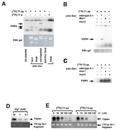
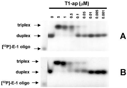
The Tie receptors (Tie-1 and Tie-2/Tek) are essential for angiogenesis and vascular remodeling/integrity. Tie receptors are up-regulated in tumor-associated endothelium, and their inhibition disrupts angiogenesis and can prevent tumor growth as a consequence. To investigate the potential of anti-gene approaches to inhibit tie gene expression for anti-angiogenic therapy, we have examined triple-helical (triplex) DNA formation at 2 tandem Ets transcription factor binding motifs (designated E-1 and E-2) in the human tie-1 promoter. Various tie-1 promoter deletion/mutation luciferase reporter constructs were generated and transfected into endothelial cells to examine the relative activities of E-1 and E-2. The binding of antiparallel and parallel (control) purine motif oligonucleotides (21-22 bp) targeted to E-1 and E-2 was assessed by plasmid DNA fragment binding and electrophoretic mobility shift assays. Triplex-forming oligonucleotides were incubated with tie-1 reporter constructs and transfected into endothelial cells to determine their activity. The Ets binding motifs in the E-1 sequence were essential for human tie-1 promoter activity in endothelial cells, whereas the deletion of E-2 had no effect. Antiparallel purine motif oligonucleotides targeted at E-1 or E-2 selectively formed strong triplex DNA (K(d) approximately 10(-7) M) at 37 degrees C. Transfection of tie-1 reporter constructs with triplex DNA at E-1, but not E-2, specifically inhibited tie-1 promoter activity by up to 75% compared with control oligonucleotides in endothelial cells. As similar multiple Ets binding sites are important for the regulation of several endothelial-restricted genes, this approach may have broad therapeutic potential for cancer and other pathologies involving endothelial proliferation/dysfunction.


Figures

Figure 1
Figure 1
Diagram showing the relative positions of the tandem ets transcription factor duplex target sequences (E-1 and E-2), SmaI and AccI restriction enzyme sites, and reported transcription start site (arrow) of the human tie-1 promoter (37). The sequences of the oligonucleotides encoding the E-1 and E-2 target sites that were annealed and used for electrophoretic mobility shift assays are indicated. The sequences of the antiparallel (relative to the purine strand of the target DNA sequence) triplex-forming oligonucleotides, T1-ap and T2-ap, targeted against E-1 and E-2, respectively, parallel control oligonucleotides (T1-p and T2-p), and parallel pyrimidine-rich (T1-(C/T)-p) oligonu-cleotide are shown.
Figure 2
Figure 2
The activity of the two tandem Ets transcription factor core DNA binding sites, E-1 and E-2, in the human tie-1 promoter. (A) The effect of 5′ deletion on the activity of tie-1 luciferase reporter constructs (ptie-1luc) in endothelial cells. ptie-1luc constructs were cotransfected with a CMV promoter–driven Renilla luciferase control (pRL-CMV) plasmid into bovine aortic endothelial cells (BAECs). After a 72-h expression period, promoter activity was quantified by dual luciferase assay and normalized to pRL-CMV activity. Results are the means (± 1 SE, bars) of duplicate samples from six separate experiments (n = 12) expressed as a percentage of the activity of the 830-bp ptie-1luc construct. (B) Mutations (mut-1 and mut-2) were introduced into the putative ets core DNA binding motifs (EBS, 5′-GGAA/T-3′) in E-1 by overlap PCR as described in “Materials and Methods.” The top strand of the E-1 sequence from the human tie-1 promoter is shown. (C) The effect of Ets binding site mutations in E-1 on the activity of the human tie-1 promoter. Wild-type (tie-1), mutated (mut-1 and mut-2), and reverse tie-1 control (tie-1rev) luciferase constructs were cotransfected with pRL-CMV into BAECs. Promoter activity was determined by dual luciferase assay after a 72-h expression period and represented as a percentage of wild-type 880-bp ptie-1luc plasmid (tie-1) following normalization to pRL-CMV activity. Results are means (± 1 SE, bars) of duplicate samples from at least 3 separate experiments (n ≥ 6).
Figure 3
Figure 3
Demonstration of specific triplex DNA formation with purine motif oligonucleotides (T1-ap and T2-ap) at the E-1 and E-2 tandem Ets transcription factor binding sites of the human tie-1 promoter by plasmid DNA binding assay. Plasmids containing the tie-1 promoter (ptie-1luc) (see Figure 2B) or 900 bp of the human tie-2 promoter (ptie-2luc) (37) were digested with SmaI alone or in combination with AccI (see Figure 1) and incubated overnight with radiolabeled oligonucleotides (T1-ap or T2-ap) at 37 °C in the presence of 10 mM Mg2+. Samples were separated on 1% agarose gels containing ethidium bromide (EtBr) and the DNA visualized under UV light (EtBr gel). The gels were dried and autoradiographed to detect triplex DNA formation. (A) Triplex DNA formation of the T1-ap oligonucleotide with intact ptie-1luc plasmid and SmaI or AccI ptie-1luc restriction fragments containing the E-1 site. No triplex DNA is observed with T1-ap against ptie-2luc SmaI restriction fragments or with the parallel control oligonucleotide T1-P. (B) Selective binding of T1-ap only to the 770-bp SmaI ptie-1luc restriction fragment containing wild-type E-1 sequence, but not the E-1 mutants (mut-1 and mut-2, see Figure 2B). (C) T2-ap binding to wild-type E-2 present in the same series of wild-type and E-1 mutant (mut-1 and mut-2) SmaI-digested ptie-1luc plasmid fragments. Effect of Mg2+ (D) and K+ (E) ion concentration on triplex formation with radiolabeled T1-ap and T2-ap.
Figure 4
Figure 4
Electrophoretic mobility shift assays showing triplex DNA formation at the E-1 tandem Ets binding motif sequence of the human tie-1 promoter. Radiolabeled oligonucleotide encoding the C/T-rich strand of the tie-1 promoter E-1 site ([32P]-E-1 oligo) was annealed to the cold complementary G/A strand (see Figure 1), and incubated with increasing concentrations of phosphodiester (A) and phosphodiester-phosphorothioate–linked (B) cold T1-ap (Figure 1), in binding buffer containing 10 mM MgCl and 150 mM KCl.
Figure 5
Figure 5
Selective inhibition of tie-1 promoter activity with the T1-ap triplex-forming oligonucleotide. Tie-1 or SV40 promoter-driven luciferase reporter constructs (ptie-1luc and pSV40luc, respectively) were incubated with phosphodiester (20 μM) (A) or partial phosphorothioate (PSO)-modified T1-ap (B), T2-ap and control oligonucleotides (T1-P, T2-P, and T1-(C/T)-P; see Figure 1) in binding buffer for 18 h and transfected into bovine aortic endothelial cells. After 72 h, reporter activity was determined by dual luciferase assay and normalized to the co-transfected pRL-CMV control plasmid. Results are means (± 1 SD, bars) of three to five experiments (n = 6–10) expressed as a percentage of the T1-P–treated control reporter plasmid.

Similar articles

Cited by

References

    1. Talks KL, Harris AL. The current status of anti-angiogenic factors. Br J Haematol. 2000;109:477–89. - PubMed
    1. Veikkola T, Karkkainen M, Claesson-Welsh L, Alitalo K. Regulation of angiogenesis via vascular endothelial growth factor receptors. Cancer Res. 2000;60:203–12. - PubMed
    1. Lauren J, Gunji Y, Alitalo K. Is angiopoietin-2 necessary for the initiation of tumor angiogenesis? Am J Pathol. 1998;153:1333–9. - PMC - PubMed
    1. Sato TN, et al. Distinct roles of the receptor tyrosine kinases Tie-1 and Tie-2 in blood vessel formation. Nature. 1996;376:70–4. - PubMed
    1. Saharinen P, et al. Multiple angiopoietin recombinant proteins activate the Tie1 receptor tyrosine kinase and promote its interaction with Tie2. J Cell Biol. 2005;169:239–43. - PMC - PubMed

Publication types

MeSH terms

LinkOut - more resources