. 2007 Jan;101(2):149-60.
doi: 10.1007/s10549-006-9281-1. Epub 2006 Jul 13.

The neonatal splice variant of Nav1.5 potentiates in vitro invasive behaviour of MDA-MB-231 human breast cancer cells

Affiliations

The neonatal splice variant of Nav1.5 potentiates in vitro invasive behaviour of MDA-MB-231 human breast cancer cells

William J Brackenbury et al. Breast Cancer Res Treat. 2007 Jan.

Abstract

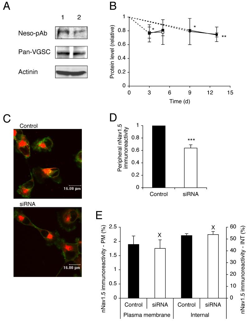
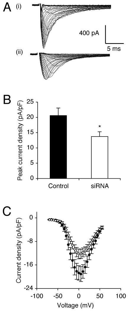
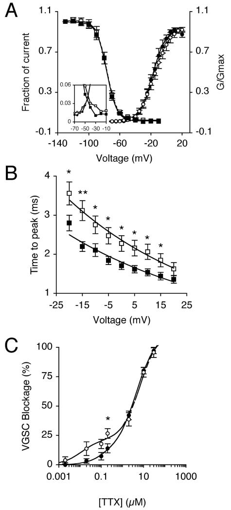
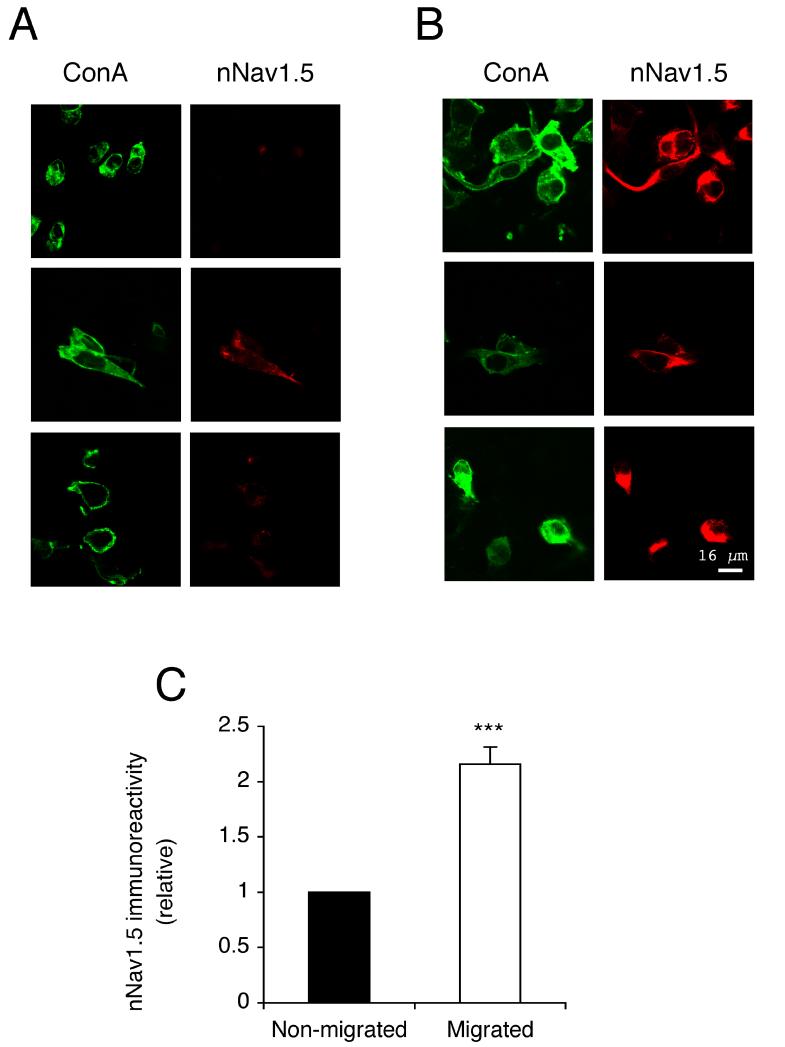
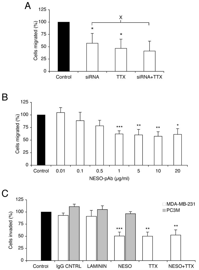
Upregulation of functional voltage-gated Na+ channels (VGSCs) occurs in metastatic human breast cancer (BCa) in vitro and in vivo. The present study aimed to ascertain the specific involvement of the "neonatal" splice variant of Nav1.5 (nNav1.5), thought to be predominant, in the VGSC-dependent invasive behaviour of MDA-MB-231 cells. Functional activity of nNav1.5 was suppressed by two different methods targeting nNav1.5: (i) small interfering RNA (siRNA), and (ii) a polyclonal antibody (NESO-pAb); effects upon migration and invasion were determined. nNav1.5 mRNA, protein and signalling were measured using real-time PCR, Western blotting, and patch clamp recording, respectively. Treatment with the siRNA rapidly reduced (by approximately 90%) the level of nNav1.5 (but not adult Nav1.5) mRNA, but the protein reduction was much smaller (approximately 30%), even after 13 days. Nevertheless, the siRNA reduced peak VGSC current density by 33%, and significantly increased the cells' sensitivity to nanomolar tetrodotoxin (TTX). Importantly, the siRNA suppressed in vitro migration by 43%, and eliminated the normally inhibitory effect of TTX. Migrated MDA-MB-231 cells expressed more nNav1.5 protein at the plasma membrane than non-migrated cells. Furthermore, NESO-pAb reduced migration by up to 42%, in a dose-dependent manner. NESO-pAb also reduced Matrigel invasion without affecting proliferation. TTX had no effect on cells already treated with NESO-pAb. It was concluded that nNav1.5 is primarily responsible for the VGSC-dependent enhancement of invasive behaviour in MDA-MB-231 cells. Accordingly, targeting nNav1.5 expression/activity may be useful in clinical management of metastatic BCa.


Figures

Figure 1
Figure 1. RNAi reduced nNav1.5, but not adult Nav1.5, or Nav1.7 mRNA level.
(A) Time course of relative nNav1.5 mRNA level after transfection with siRNA targeting nNav1.5. Squares, data compared with mock-transfected control (no siRNA). Circles, data compared with siControl non-targeting siRNA. (B) Relative level of adult Nav1.5 and Nav1.7 mRNA 5 days after transfection with siControl, or siRNA targeting nNav1.5. VGSC mRNA levels were normalised relative to Cytb5R by the 2−ΔΔCt method. Errors propagated through the 2−ΔΔCt analysis are shown. Significance: (*) P < 0.05, (**) P < 0.01, (***) P < 0.001.
Figure 2
Figure 2. RNAi reduced nNav1.5 protein level.
(A) Western blot with 40 μg of total protein per lane from cells 13 days after treatment with (1) siControl or (2) siRNA targeting nNav1.5. Antibodies used: NESO-pAb, a pan-VGSC antibody, and an actinin antibody as a control for loading. The same membrane was stripped and re-blotted. (B) Time courses of nNav1.5 (light symbols) and total VGSC (dark symbols) protein levels (relative to actinin controls) after transfection with siRNA targeting nNav1.5. Squares, compared with mock-transfected control (no siRNA). Circles, compared with siControl non-targeting siRNA. (C) Typical confocal images of cells double-immunolabelled with NESO-pAb (red) and concanavallin A plasma membrane marker (green) 13 days after transfection with siControl or siRNA targeting nNav1.5. (D) Relative peripheral nNav1.5 protein level in cells 13 days after transfection with siControl or siRNA targeting nNav1.5. (E) nNav1.5 protein distribution (immunoreactivity) along subcellular cross-sections (% of total). Left-hand bars, 1.5 μm sections measured inward from edge of concanavalin A staining; Right-hand bars, middle 30 % of cross-section. PM, plasma membrane; INT, internal. Data are presented as mean and SEM. Significance: (X) P > 0.05, (*) P < 0.05, (**) P < 0.01, (***) P < 0.001.
Figure 3
Figure 3. RNAi reduced peak VGSC current density after 13 days.
(A) Typical whole-cell VGSC currents elicited by 60 ms depolarising voltage pulses between −70 mV and +70 mV applied from a holding potential of −100 mV: (i) a siControl-treated cell; (ii) a cell treated with siRNA targeting nNav1.5. (B) Quantitative comparison of peak current densities recorded in siControl-treated cells, and cells treated with siRNA targeting nNav1.5. (C) Mean current-voltage relationships for siControl-treated cells (dark circles), and cells treated with siRNA targeting nNav1.5 (light circles). Data are presented as mean ± SEM. Significance: (*) P < 0.05.
Figure 4
Figure 4. RNAi altered VGSC kinetics and TTX sensitivity after 13 days.
(A) Mean availability-voltage (squares) and relative conductance (G/Gmax)-voltage relationships (circles) for siControl-treated cells (dark symbols) and cells treated with siRNA targeting nNav1.5 (light symbols). Control (solid lines) and siRNA data (dotted lines) are fitted with Boltzmann functions. The inset magnifies the voltage range (‘window’) in which current is activated and not fully inactivated. (B) Dependence of time to peak on membrane voltage for siControl-treated cells (dark squares), and cells treated with siRNA targeting nNav1.5 (light squares). Control (solid line) and siRNA data (dashed line) are fitted with single exponential functions. (C) Reduction of VGSC current by TTX for control-treated cells (dark circles), and cells treated with siRNA targeting nNav1.5 (light circles). Control (solid line) and siRNA data (dashed line) are fitted to double Langmuir adsoption isotherms. Data are presented as mean ± SEM. Significance: (*) P < 0.05, (**) P< 0.01.
Figure 5
Figure 5. Migrated cells had more nNav1.5 protein at the plasma membrane than non-migrated cells after 16 h.
(A and B) Typical confocal images of non-migrated and migrated cells in Transwell assays, respectively. Cells were non-permeabilized and double-immunolabelled with NESO-pAb (red) and concanavalin A plasma membrane marker (green). (C) Relative nNav1.5 protein level in migrated cells, normalised with respect to the level in non-migrated cells. Significance: (***) P < 0.001.
Figure 6
Figure 6. siRNA and NESO-pAb reduced VGSC-dependent metastatic cell behaviour.
(A) Relative number of cells migrating through a Transwell chamber over 7 h, 13 days after transfection with siControl, or siRNA targeting nNav1.5. Cells were treated with TTX (10 μM), or untreated during the migration assays. (B) Relative number of cells migrating through a Transwell chamber over 7 h, treated with 0.01-20 μg/ml NESO-pAb during the assay. (C) Relative number of cells invading through Matrigel chamber over 24 h. MDA-MB-231 (white bars) and PC-3M (grey bars) cells were treated with IgG (1 μg/ml), anti-laminin (1:100), NESO-pAb (1 μg/ml) with/without TTX (10 μM) during the invasion assay. In both (B) and (C), data shown are with respect to corresponding control, untreated cells (treated as 100 %). Data are presented as mean and SEM. Significance: (X) P > 0.05, (*) P < 0.05, (**) P < 0.01, (***) P < 0.001, relative to “control”, unless indicated.

Similar articles

Cited by

References

    1. Hille B. Ionic channels of excitable membranes. 2nd edn. Sinauer Associates Inc.; Sunderland (Massachusetts): 1992.
    1. Jurkat-Rott K, Lehmann-Horn F. Human muscle voltage-gated ion channels and hereditary disease. Curr Opin Pharmacol. 2001;1(3):280–287. - PubMed
    1. Viswanathan PC, Balser JR. Inherited sodium channelopathies: a continuum of channel dysfunction. Trends Cardiovasc Med. 2004;14(1):28–35. - PubMed
    1. Diss JK, Fraser SP, Djamgoz MB. Voltage-gated Na+ channels: multiplicity of expression, plasticity, functional implications and pathophysiological aspects. Eur Biophys J. 2004;33(3):180–193. - PubMed
    1. Fraser SP, Koyuturk M, Djamgoz MB. Ion channel activity and cancer cell proliferation: A short review with particular reference to prostate cancer. In: Rouzaire-Dubois B, Benoit E, Dubois JM, editors. Ion Channels and Physiopathologies of Nerve Conduction and Cell Proliferation. 1st edn. Research Signpost; 2002.

Publication types

MeSH terms