. 2006 Jul;2(7):e65.
doi: 10.1371/journal.ppat.0020065.

Live attenuated B. pertussis as a single-dose nasal vaccine against whooping cough

Affiliations

Live attenuated B. pertussis as a single-dose nasal vaccine against whooping cough

Nathalie Mielcarek et al. PLoS Pathog. 2006 Jul.

Abstract

Pertussis is still among the principal causes of death worldwide, and its incidence is increasing even in countries with high vaccine coverage. Although all age groups are susceptible, it is most severe in infants too young to be protected by currently available vaccines. To induce strong protective immunity in neonates, we have developed BPZE1, a live attenuated Bordetella pertussis strain to be given as a single-dose nasal vaccine in early life. BPZE1 was developed by the genetic inactivation or removal of three major toxins. In mice, BPZE1 was highly attenuated, yet able to colonize the respiratory tract and to induce strong protective immunity after a single nasal administration. Protection against B. pertussis was comparable to that induced by two injections of acellular vaccine (aPV) in adult mice, but was significantly better than two administrations of aPV in infant mice. Moreover, BPZE1 protected against Bordetella parapertussis infection, whereas aPV did not. BPZE1 is thus an attractive vaccine candidate to protect against whooping cough by nasal, needle-free administration early in life, possibly at birth.


Conflict of interest statement

Competing interests. The authors have declared that no competing interests exist.

Figures

Figure 1
Figure 1. Characterization of B. pertussis BPZE1
(A) TCT present in culture supernatants of BPSM and BPZE1 expressed as means of nM/OD540nm (± standard error) of three separate cultures for each strain. (B) Immunoblot analysis of PTX production in the culture supernatants of BPSM (lane 1) and BPZE1 (lane 2). The sizes of the molecular weight (Mr) markers are expressed in kDa and given in the left margin. (C) Southern blot analysis of the dnt locus in BPSM (lane 1) and BPZE1 (lane 2). The lengths of the size markers are indicated in base pairs (bp) are shown in the left margin.
Figure 2
Figure 2. Phenotypic Characterization of B. pertussis BPZE1
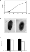
(A) Growth rates of BPSM (solid line) and BPZE1 (dotted line) in liquid culture. (B) Electron micrographs representative of BPSM (left) and BPZE1 (right) grown in liquid medium for 24 h. (C) In vitro adherence of BPSM (filled columns) and BPZE1 (open columns) to human pulmonary epithelial A549 cells (left) and murine macrophage-like J774 cells (right). The results are expressed as means (± standard error) of percentages of binding bacteria relative to the bacteria present in the inoculum from three different experiments.
Figure 3
Figure 3. In Vivo Characterization of B. pertussis BPZE1
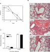
(A) Lung colonization by BPSM (solid lines) and BPZE1 (dotted lines) of adult mice infected intranasally with 106 CFU of BPZE1 or BPSM. The results are expressed as mean (± standard error) CFUs from three to four mice per group, and are representative of two separate experiments. The dashed line represents the limit of bacterial counts. (B) Histological analysis of lungs from BPZE1 (upper panel) or BPSM-infected (middle panel) adult mice compared to controls given PBS (lower panel). One week after infection, the lungs were aseptically removed and fixed in formaldehyde. Sections were stained with hematoxylin and eosin, and examined by light microscopy. (C) Susceptibility of BPZE1-infected mice to infection by M. tuberculosis. Balb/C mice were infected intranasally with 106 CFU of BPZE1 (filled columns) or received PBS (open columns) and were intranasally infected one week later with 5 × 10⁴ M. tuberculosis H37Rv. One week (left columns) and 5 wk (right columns) after M. tuberculosis infection, the M. tuberculosis CFUs present in the lungs were counted. The results are expressed as mean (± standard error) CFUs from four mice per group.
Figure 4
Figure 4. Protection against Bordetella Infection
Protection against B. pertussis in adult (A) and infant mice (B) and (C), or against B. parapertussis in infant mice (D). Mice immunized with BPZE1, aPV, or PBS (naive) were challenged with BPSM (A), (B), and (C), or B. parapertussis (D), and lung CFU counts were determined 3 h (open columns) or 7 d (filled columns) later. Results are expressed as mean (± standard error) CFUs from three to four mice per group and are representative of two separate experiments. (C) CFU counts 3 h after BPSM challenge in adult mice vaccinated with BPZE1 or aPV, compared to controls. The dashed lines in panels (A), (B), and (D) represent the limit of bacterial counts.
Figure 5
Figure 5. Immune Responses Induced in Infant Mice by BPZE1 or aPV Immunization
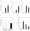
(A) Anti-FHA, (B) anti-PTX, (C) anti-B. pertussis IgG heavy and light chain (H+L) titers, and (D) anti-FHA IgG1/IgG2a ratios before (open columns) or 1 wk after BPSM challenge (filled columns) in BPZE1 or aPV immunized 3-wk-old mice, compared to controls. (E) IFN-γ to IL-5 ratios produced by FHA-, PTX- or ConA-stimulated splenocytes from 8-wk-old mice vaccinated 2 mo before with BPZE1 (filled columns) or aPV (open columns), compared to controls (gray columns). Antibodies and cytokines were measured in individual mice, and the results are expressed as mean values (± standard error) for four mice per group tested in triplicate.

Similar articles

Cited by

References

    1. World Health Organization [WHO] The world health report 2004—Changing history. Geneva: WHO; 2004. Available: http://www.who.int/whr/2004/en. Accessed 31 May 2006.
    1. Das P. Whooping cough makes global comeback. Lancet Infect Dis. 2002;2:322. - PubMed
    1. Tan T, Trindade E, Skowronski D. Epidemiology of pertussis. Pediatr Infect Dis J. 2005;24:S10–S18. - PubMed
    1. Centers for Disease Control and Prevention. Pertussis. Available: http://www.cdc.gov/nip/publications/pink/pert.pdf. Accessed 31 May 2006.
    1. Wirsing von König CH, Halperin S, Riffelmann M, Guiso N. Pertussis of adults and infants. Lancet Infect Dis. 2002;2:744–750. - PubMed

Publication types

MeSH terms