. 2006 Jul 13:7:15.
doi: 10.1186/1471-2172-7-15.

Structure function analysis of SH2D2A isoforms expressed in T cells reveals a crucial role for the proline rich region encoded by SH2D2A exon 7

Affiliations

Structure function analysis of SH2D2A isoforms expressed in T cells reveals a crucial role for the proline rich region encoded by SH2D2A exon 7

Stine Granum et al. BMC Immunol. .

Abstract

Background: The activation induced T cell specific adapter protein (TSAd), encoded by SH2D2A, interacts with and modulates Lck activity. Several transcript variants of TSAd mRNA exist, but their biological significance remains unknown. Here we examined expression of SH2D2A transcripts in activated CD4+ T cells and used the SH2D2A variants as tools to identify functionally important regions of TSAd.

Results: TSAd was found to interact with Lck in human CD4+ T cells ex vivo. Three interaction modes of TSAd with Lck were identified. TSAd aa239-256 conferred binding to the Lck-SH3 domain, whereas one or more of the four tyrosines within aa239-334 encoded by SH2D2A exon 7 was found to confer interaction with the Lck-SH2-domain. Finally the TSAd-SH2 domain was found to interact with Lck. The SH2D2A exon 7 encoding TSAd aa 239-334 was found to harbour information essential not only for TSAd interaction with Lck, but also for TSAd modulation of Lck activity and translocation of TSAd to the nucleus. All five SH2D2A transcripts were found to be expressed in CD3 stimulated CD4+ T cells.

Conclusion: These data show that TSAd and Lck may interact through several different domains and that Lck TSAd interaction occurs in CD4+ T cells ex vivo. Alternative splicing of exon 7 encoding aa239-334 results in loss of the majority of protein interaction motives of TSAd and yields truncated TSAd molecules with altered ability to modulate Lck activity. Whether TSAd is regulated through differential alternative splicing of the SH2D2A transcript remains to be determined.


Figures
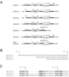
Figure 1
Figure 1
An overview of the polypeptides encoded by SH2D2A transcript variants. A. Schematic presentation of putative protein-interacting domains in the SH2D2A isoforms and mutants described in this study: The SH2D2A-1 encodes the full length TSAd, whereas the SH2D2A-2 and -3 variants represent different N-terminal sequences of TSAd. The SH2D2A-4 variant has a 10 aa insertion in the SH2-domain, whereas the fifth variant, SH2D2A-5, lacks aa239–334, containing the proline rich region including the four C-terminal tyrosines (Y = tyrosine). TSAd 4YF has the four C-terminal tyrosines exchanged with phenylalanine (F = phenylalanine). TSAd d239–256 has a deletion of amino acids 239–256 containing the motif PSQLLRPKPPIPAKPQLP. B. Comparison of the N-terminal amino acid sequences of the SH2D2A-1, -2 and -3 variants: The amino acid position is numbered according to full length TSAd (SH2D2A-1). The SH2D2A-2 and -3 variants differ from the SH2D2A-1 in the N-terminal 1–21 aa and 1–28 aa, respectively. C. Comparison of the N-terminus SH2 domain aa sequences of the SH2D2A-1 and -4 variants: The conserved arginine (R) at position 120 is marked. The SH2D2A-4 has an additional 10 aa (ins aa103–112) in the TSAd-SH2 domain. A SH2 consensus (con) sequence obtained from a Blast search, [42] is included for comparison. Conserved residues are marked in bold.
Figure 2
Figure 2
Expression of SH2D2A variants in primary CD4+ T cells. A. Expression of SH2D2A transcript variants in anti-CD3 stimulated CD4+ T cells: mRNA expression levels of SH2D2A variants relative to Zap-70 were assessed by RT-PCR in CD4+ T cells stimulated with CD3 ligation for 2 hours from four healthy blood donors. Results shown are median values ± SD of the estimated level of alternative SH2D2A transcripts relative to the total amount of SH2D2A transcripts. The SH2D2A-4 variant was not always observed. The median value of the SH2D2A-4 is 0,03% ± 0,2%. B: The relationship between the different SH2D2A transcripts does not vary significantly throughout anti-CD3 stimulation of CD4+ T cells: Primary CD4+ T cells were stimulated with anti-CD3 for 24 hours, and total amount of SH2D2A transcript (1–5) and mRNA levels of SH2D2A transcript variants 2, 3, 4 or 5 were assessed at different time points. After the initial two hours of anti-CD3 stimulation, the relationship between the five SH2D2A transcripts remained constant throughout the stimulation period. Results shown are median values of TSAd transcripts relative to Zap-70 transcripts. C: TSAd of 37 kDa is expressed in CD3 stimulated primary CD4+ T cells. Primary CD4+ T cells were stimulated with anti-CD3 for 2 and 4 hours and total sonicated cell lysates were separated by SDS-PAGE and immunoblotted with TSAd Abs and Zap-70 mAbs as a loading control. TSAd Abs detects two CD3 induced bands of 52 kDa and 37 kDa respectively. D. Expression of TSAd of 52 and 37 kDa is repressed by siRNA treatment of CD4+ T cells: Primary CD4+ T cells were transfected with control medium (0) or increasing concentration of TSAd siRNA (0,05, 0,5 or 5 μM p690) or with 5 μM control siRNA (C = TSAd p369) using Amaxa electroporator and stimulated with anti-CD3 beads for 24 hours. Total sonicated cell lysates were separated by SDS-PAGE and immunoblotted with TSAd Abs and Zap-70 mAbs as a loading control (not shown). Lysates of Jurkat TAg cells stably transfected with HA-tagged TSAd cDNA (3a3) was included as a blotting control. TSAd Abs detects two bands of 52 kDa and 37 kDa that can be inhibited by siRNA, and one non-specific band of 45 kDa, which is not affected by siRNA treatment.
Figure 3
Figure 3
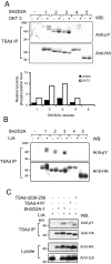
Differential Lck phosphorylation of variant TSAd molecules. A. Tyrosine phosphorylation of TSAd variants in Jurkat T cells: Jurkat T cells transiently transfected with pEF-HA or pEF-HA-SH2D2A-1-5 cDNAs, were stimulated (+) with anti-CD3 (OKT3) mAbs for 2.5 min or left unstimulated (-), lysed and subjected to immunoprecipitation with anti-TSAd Abs (TSAd IP). The precipitates were separated by SDS-PAGE and immunoblotted with anti-phoshotyrosine (pY) mAbs (upper panel) and anti-HA mAbs (lower panel). Relative level of tyrosine phosphorylation of TSAd variants is shown in the chart, and the relationships between the pY signal versus the HA signal of bands is normalised to that observed for the full length TSAd IP (SH2D2A-1) expressed in resting Jurkat T cells. B. Lck phosphorylates the SH2D2A-1, -2 and -3 variants of TSAd in 293T cells: 293T cells were transiently transfected with pEF-HA or pEF-HA-SH2D2A-1-5 cDNAs alone (-) or together (+) with pEF-Lck. The cells were lysed and treated as in A. C. Tyrosine phosphorylation of TSAd mutated for the four C-terminal tyrosines is attenuated when co-expressed with Lck in 293T cells. 293T cells were transiently transfected with pEF-HA, the pEF-HA-TSAd-4YF or pEF-HA-TSAd-d239–256 cDNAs together with pEF-Lck. The cells were lysed and treated as in A.
Figure 4
Figure 4
SH2D2A exon 7 encodes ligands for Lck-SH2 and SH3 domains. A. TSAd interacts with Lck: Primary CD4+ T cells were stimulated with anti-CD3/CD28 beads for one day. TSAd was precipitated with anti-TSAd Abs or irrelevant serum (NRS) and protein A/G sepharose beads from precleared lysates. Precipitates were separated by SDS-PAGE and immunoblotted with anti-Lck and anti-TSAd Abs as indicated. B. TSAd variants interact with Lck in Jurkat T cells: Jurkat T cells were transiently transfected with pEF-HA or one of the pEF-HA-SH2D2A-1-5cDNAs. Immunoprecipitation was performed using anti-TSAd Abs and protein G magnetic beads. Precipitates were separated by SDS-PAGE and immunoblotted with anti-Lck and anti-HA as indicated. C. The SH2 domain and aa239–334 of TSAd is important for interaction with the Lck SH2 domain: 293T cells were transiently transfected with one of the pEF-HA-SH2D2A-1-5 cDNAs alone (-) or together (+) with pEF-Lck. Cell lysates were subjected to pull down experiment with GST-Lck-SH2 Sepharose beads. Precipitates were immunoblotted with anti-HA mAbs (upper panel). An anti-HA immunoblot of precleared lysates before GST-Lck-SH2 pull down is included to verify expression of the different HA-tagged TSAd variants (lower panel). D. Aa239–334 contains ligands for the Lck-SH3 domain: 293T cells were transiently transfected with pEF-HA or one of the pEF-HA-SH2D2A-1-5 cDNAs together (+) with pEF-Lck. Only the pEF-HA-SH2D2A-1 cDNA were also co-transfected with pEF-HA (-). Cell lysates were subjected to pull down experiment with GST-Lck-SH3 Sepharose beads, and precipitates (upper panel) and precleared lysates (lower panel) were immunoblotted as in C. E. Identification of TSAd structures interacting with Lck domains: 293T cells were transiently transfected with pEF-HA, the pEF-HA-TSAd-4YF or pEF-HA-TSAd-d239–256 cDNAs together with pEF-Lck. Cell lysates were subjected to pull down experiment with GST-Lck-SH2 or GST-Lck-SH3 Sepharose beads. GST-Lck-SH2 (panel 1) and GST-Lck-SH3 (panel 2) precipitates were immunoblotted with anti-HA mAbs, and the precleared lysates were immunoblotted with anti-HA mAbs (panel 3) or anti-Lck mAbs (panel 4). F. The SH2 domain of TSAd precipitates Lck in 293T cells: 293T cells were transiently transfected with pEF-HA, pEF-Lck, pEF-HA-SH2D2A-1 or pEF-Lck and pEF-HA-SH2D2A-1 cDNAs together. Precleared cell lysates were subjected to pull down experiment with GST-TSAd-SH2 Sepharose beads. Precipitates were immunoblotted with anti-Lck mAbs (upper panel). An anti-Lck immunoblot of precleared lysates before GST-TSAd-SH2 pull down is included to verify expression of Lck (lower panel).
Figure 5
Figure 5
Tyrosines within aa239–334 determines the inhibitory effect of TSAd on Lck activity. The phosphorylation level of LAT and Y319 of Zap-70 in Jurkat T cells expressing the SH2D2A-5 variant and TSAd-4YF is normal upon TCR stimulation: Jurkat T cells were transiently transfected with either pEF-HA or one of the pEF-HA-SH2D2A-1-5 or pEF-HA-TSAd-4YF cDNAs. The cells were stimulated (+) with anti-CD3 (OKT3) mAbs for 2.5 min and lysed. The lysates were immunoblotted with Abs as indicated.
Figure 6
Figure 6
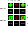
Nuclear translocation of TSAd depends on aa239–334. The SH2D2A-5 variant is excluded from the cell nucleus in Jurkat TAg cells: Confocal immunocytochemistry analysis of Jurkat TAg cells transiently transfected with pEF-HA-SH2D2A-2 and -5. The nuclear membrane protein LAP-2 was stained red (Cy™-3), whereas the HA-tagged recombinant proteins are stained green (Cy-2™). Cells were stimulated with anti-CD3 mAbs (OKT3) for 30 min. Horizontal sections are labelled Y, vertical sections are labelled Z (100× objective, 4× zoom).
Figure 7
Figure 7
Schematic presentation of TSAd functional domains and predicted sites. A Schematic presentation of functional regions of TSAd. B Predicted sites identified in high stringency Scansite search of SH2D2A-1 and SH2D2A-5 peptide sequences: Core amino acids are indicated. pY: tyrosine kinase phosphorylation site. SH2 and SH3: SH2 and SH3 binding sites respectively. ErkD: binding site for ErkD domain.

Similar articles

Cited by

References

    1. Lander ES, Linton LM, Birren B, Nusbaum C, Zody J, Baldwin MC, et al. Initial sequencing and analysis of the human genome. Nature. 2001;409:860–921. doi: 10.1038/35057062. - DOI - PubMed
    1. Leipzig J, Pevzner P, Heber S. The Alternative Splicing Gallery (ASG): bridging the gap between genome and transcriptome. Nucleic Acids Res. 2004;32:3977–3983. doi: 10.1093/nar/gkh731. - DOI - PMC - PubMed
    1. Smith CW, Valcarcel J. Alternative pre-mRNA splicing: the logic of combinatorial control. Trends Biochem Sci. 2000;25:381–388. doi: 10.1016/S0968-0004(00)01604-2. - DOI - PubMed
    1. Graveley BR. Alternative splicing: increasing diversity in the proteomic world. Trends Genet. 2001;17:100–107. doi: 10.1016/S0168-9525(00)02176-4. - DOI - PubMed
    1. Klemm JD, Schreiber SL, Crabtree GR. Dimerization as a regulatory mechanism in signal transduction. Annu Rev Immunol. 1998;16:569–592. doi: 10.1146/annurev.immunol.16.1.569. - DOI - PubMed

Publication types

MeSH terms