

Endostatin inhibits VEGF-A induced osteoclastic bone resorption in vitro
- PMID: 16839420
- PMCID: PMC1534045
- DOI: 10.1186/1471-2474-7-56
Endostatin inhibits VEGF-A induced osteoclastic bone resorption in vitro
Abstract
Background: Endostatin is a C-terminal fragment of collagen XVIII which is a component of basement membranes with the structural properties of both collagens and proteoglycans. Endostatin has a major role in angiogenesis which is intimately associated with bone development and remodeling. Signaling between the endothelial cells and the bone cells, for example, may have a role in recruitment of osteoclastic precursor cells. Our study aims at exploring a possibility that endostatin, either as a part of basement membrane or as a soluble molecule, may control osteoclastogenesis and osteoclastic bone resorption in vitro.
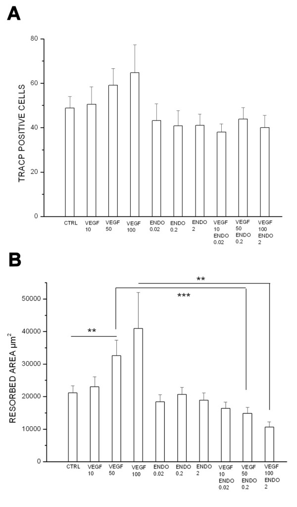
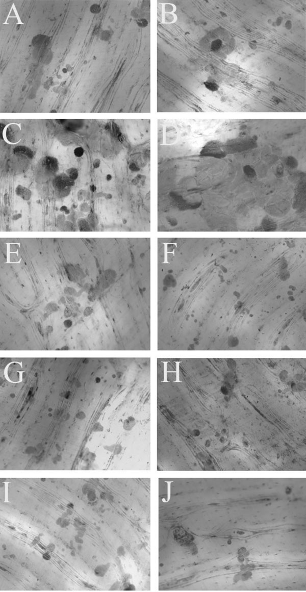
Methods: Rat pit formation assay was employed in order to examine the effect of endostatin alone or in combination with vascular endothelial growth factor-A (VEGF-A) on bone resorption in vitro. Effect of these agents on osteoclast differentiation in vitro was also tested. Osteoclastogenesis and the number of osteoclasts were followed by tartrate resistant acid phosphatase (TRACP) staining and resorption was evaluated by measuring the area of excavated pits.
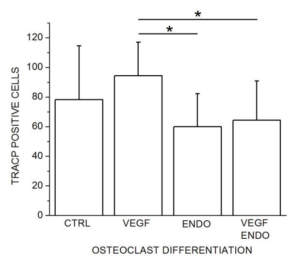
Results: Endostatin inhibited the VEGF-A stimulated osteoclastic bone resorption, whereas endostatin alone had no effect on the basal resorption level in the absence of VEGF-A. In addition, endostatin could inhibit osteoclast differentiation in vitro independent of VEGF-A.
Conclusion: Our in vitro data indicate that collagen XVIII/endostatin can suppress VEGF-A induced osteoclastic bone resorption to the basal level. Osteoclastogenesis is also inhibited by endostatin. The regulatory effect of endostatin, however, is not critical since endostatin alone does not modify the basal bone resorption.
Figures
Similar articles
-
Endostatin affects osteoblast behavior in vitro, but collagen XVIII/endostatin is not essential for skeletal development in vivo.Calcif Tissue Int. 2009 Nov;85(5):412-20. doi: 10.1007/s00223-009-9287-x. Epub 2009 Sep 10. Calcif Tissue Int. 2009. PMID: 19763371
-
Osteoclasts prefer aged bone.Osteoporos Int. 2007 Jun;18(6):751-9. doi: 10.1007/s00198-006-0298-4. Epub 2007 Jan 10. Osteoporos Int. 2007. PMID: 17216130
-
Effects of berberine on differentiation and bone resorption of osteoclasts derived from rat bone marrow cells.Zhong Xi Yi Jie He Xue Bao. 2009 Apr;7(4):342-8. doi: 10.3736/jcim20090408. Zhong Xi Yi Jie He Xue Bao. 2009. PMID: 19361364
-
Biological aspects of altered bone remodeling in multiple myeloma and possibilities of pharmacological intervention.Dan Med Bull. 2011 May;58(5):B4277. Dan Med Bull. 2011. PMID: 21535989 Review.
-
Tartrate-resistant acid phosphatase 5b (TRACP 5b) as a marker of bone resorption.Clin Lab. 2006;52(9-10):499-509. Clin Lab. 2006. PMID: 17078477 Review.
Cited by
-
Osteocytes as mechanosensors in the inhibition of bone resorption due to mechanical loading.Bone. 2008 Jan;42(1):172-9. doi: 10.1016/j.bone.2007.09.047. Epub 2007 Sep 26. Bone. 2008. PMID: 17997378 Free PMC article.
-
Inhibitory effect of polysulfated heparin endostatin on alkali burn induced corneal neovascularization in rabbits.Int J Ophthalmol. 2015 Apr 18;8(2):234-8. doi: 10.3980/j.issn.2222-3959.2015.02.04. eCollection 2015. Int J Ophthalmol. 2015. PMID: 25938033 Free PMC article.
-
Development of articular cartilage and the metaphyseal growth plate: the localization of TRAP cells, VEGF, and endostatin.J Anat. 2011 Jun;218(6):608-18. doi: 10.1111/j.1469-7580.2011.01377.x. Epub 2011 Apr 3. J Anat. 2011. PMID: 21457260 Free PMC article.
-
Activation of resorption in fatigue-loaded bone involves both apoptosis and active pro-osteoclastogenic signaling by distinct osteocyte populations.Bone. 2012 May;50(5):1115-22. doi: 10.1016/j.bone.2012.01.025. Epub 2012 Feb 9. Bone. 2012. PMID: 22342796 Free PMC article.
-
What is the role of bosentan in healing of femur fractures in a rat model?J Bone Miner Metab. 2015 Sep;33(5):496-506. doi: 10.1007/s00774-014-0622-6. Epub 2014 Oct 9. J Bone Miner Metab. 2015. PMID: 25298328
References
-
- Engsig MT, Chen QJ, Vu TH, Pedersen AC, Therkidsen B, Lund LR, Henriksen K, Lenhard T, Foged NT, Werb Z, Delaisse JM. Matrix metalloproteinase 9 and vascular endothelial growth factor are essential for osteoclast recruitment into developing long bones. J Cell Biol. 2000;151:879–889. doi: 10.1083/jcb.151.4.879. - DOI - PMC - PubMed
-
- Clauss M, Weich H, Breier G, Knies U, Rockl W, Waltenberger J, Risau W. The vascular endothelial growth factor receptor Flt-1 mediates biological activities. Implications for a functional role of placenta growth factor in monocyte activation and chemotaxis. J Biol Chem. 1996;271:17629–17634. doi: 10.1074/jbc.271.30.17629. - DOI - PubMed
Publication types
MeSH terms
Substances
LinkOut - more resources
Full Text Sources
