. 2006 Aug;80(15):7339-53.
doi: 10.1128/JVI.00309-06.

Promoter activation by the varicella-zoster virus major transactivator IE62 and the cellular transcription factor USF

Affiliations

Promoter activation by the varicella-zoster virus major transactivator IE62 and the cellular transcription factor USF

Min Yang et al. J Virol. 2006 Aug.

Abstract

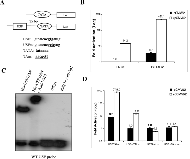
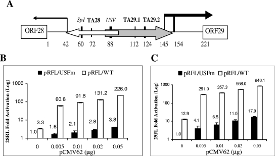
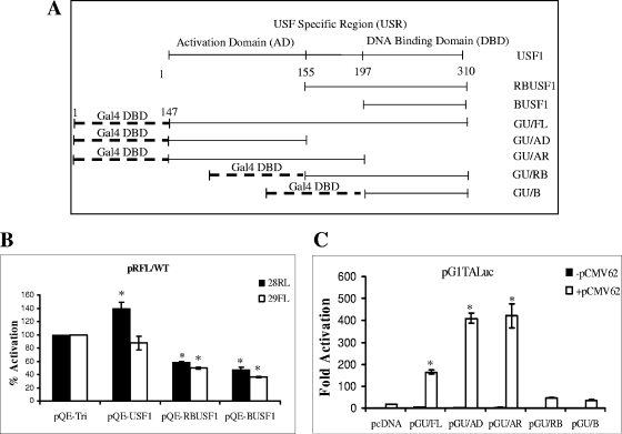
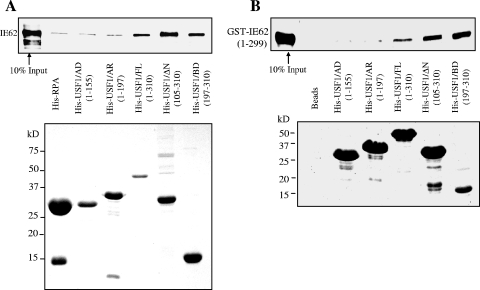
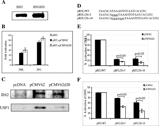
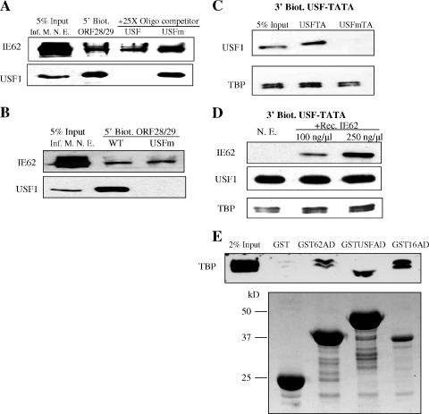
The varicella-zoster virus major transactivator, IE62, can activate expression from homologous and heterologous promoters. High levels of IE62-mediated activation appear to involve synergy with cellular transcription factors. The work presented here focuses on functional interactions of IE62 with the ubiquitously expressed cellular factor USF. We have found that USF can synergize with IE62 to a similar extent on model minimal promoters and the complex native ORF28/29 regulatory element, neither of which contains a consensus IE62 binding site. Using Gal4 fusion constructs, we have found that the activation domain of USF1 is necessary and sufficient for synergistic activation with IE62. We have mapped the regions of USF and IE62 required for direct physical interaction. Deletion of the required region within IE62 does not ablate synergistic activation but does influence its efficiency depending on promoter architecture. Both proteins stabilize/increase binding of TATA binding protein/TFIID to promoter elements. These findings suggest a novel mechanism for the observed synergistic activation which requires neither site-specific IE62 binding to the promoter nor a direct physical interaction with USF.


Figures

FIG. 1.
FIG. 1.
Synergistic IE62-USF activation of a model minimal promoter. (A) Schematic of the model luciferase reporter vector, pUSFTAluc, indicating the relative positions and sequences of the USF site and TATA element. Consensus binding motifs are shown in boldface. Mutations are underlined. (B) Results of luciferase assays. One microgram of each reporter plasmid including the basic pTALuc plasmid, which lacks the USF binding site, was cotransfected with or without 0.02 μg of the IE62-expressing plasmid, pCMV62, into MeWo cells. The luciferase activity expressed from the pTALuc reporter in the absence of IE62 was normalized to 1. The promoter activities resulting from the presence of USF, IE62, or both are reported as induction (n-fold) of the luciferase activity over the pTALuc level. The open and solid bars represent promoter activity in the absence and presence of IE62, respectively. (C) Results of EMSAs confirming that recombinant USFΔN binds to the consensus binding site inserted into pUSFTALuc and that the complex is supershifted by anti-USF1 antibody. Recombinant Sp1 and anti-Sp1 antibody were used as negative controls. (D) Control transfection assays showing the requirement of the TATA element for both USF and IE62 activation. These results were normalized to the activity observed with the pUSFTALuc reporter in the absence of IE62. Luciferase assay data in panels B and D represent the averages of triplicate transfections. The average values are shown above each bar, and the error bars represent standard deviations.
FIG. 2.
FIG. 2.
Synergistic IE62-USF activation of the VZV ORF28/29 regulatory element. (A) Schematic of the VZV ORF28/29 regulatory element showing authenticated transcription factor binding sites and TATA elements. The locations of the two overlapping minimal promoters are shown as open (ORF28) and gray (ORF29) arrows, respectively. The difference in thickness reflects their levels of transcription efficiency in the presence of IE62. The vertical lines capped by an arrow indicate the positions of transcription start sites. The difference in thickness of the two bold vertical lines over the ORF29 gene transcription start sites indicates that one is preferentially utilized (55). (B) Results of transfection experiments showing expression of Renilla luciferase (ORF28 position) activity from the wild-type (open bars) and mutant USFm (solid bars) dual luciferase reporter plasmids in the presence of increasing amounts of the pCMV62 expression plasmid. The level of Renilla luciferase activity observed with the pRFL/USFm reporter in the absence of IE62 was normalized to 1. (C) Results of transfection experiments showing expression of firefly luciferase (ORF29 position) activity from the wild-type (open bars) and USFm (solid bars) dual luciferase reporter plasmids in the presence of increasing amounts of the pCMV62 expression plasmid. The level of firefly luciferase activity observed with the pRFL/USFm reporter in the absence of IE62 was normalized to 1. Luciferase assay data in panels B and C represent the averages of triplicate transfections. The average values are shown above each bar, and the error bars represent standard deviations.
FIG. 3.
FIG. 3.
Identification of the region of USF1 that is involved in mediating IE62 activation. (A) Schematic depiction of the USF1 fragments expressed via pQE-tri plasmid and the Gal4-USF fusion proteins with the DNA binding domain (DBD) of Gal4 fused with different fragments of the USF1 protein. (B) Examination of the effect of ectopic expression of the full-length and truncated USF1 proteins on IE62 activation of the VZV ORF28/29 regulatory element. One microgram pRFL/WT reporter vector and 0.02 μg pCMV62 plasmid were cotransfected with 0.5 μg each of the USF1-expressing plasmids and the control vector pQE-tri. The solid bars represent the promoter activities in the direction of ORF28, and the open bars represent that in the direction of ORF29. The endogenous USF1-mediated IE62 activation of the individual luciferase reporter genes in the presence of the empty pQE-tri plasmid was normalized to 100%. (C) Analysis of the Gal4-USF1 fusion proteins in mediation of IE62 activation of the pG1TALuc vector. One microgram pG1TALuc reporter vector and 0.02 μg pCMV62 plasmid were cotransfected with 0.5 μg each of the Gal4-USF1 fusion protein-expressing plasmids and the control vector pcDNA as indicated in the figure. Open and closed bars represent activity in the presence and absence of IE62, respectively. Luciferase assay data in panels B and C represent the averages of triplicate transfections. The error bars represent standard deviations. *, P < 0.05.
FIG. 4.
FIG. 4.
GST pull-down analysis of the region of IE62 that interacts with USF1. (A) Preliminary mapping of the region of IE62 that interacts with USF. Truncated GST-tagged IE62 proteins were coupled to glutathione Sepharose beads and incubated with nuclear extracts of MeWo cells. GST alone was used as a control. The binding of USF to the GST-IE62 fusions was examined by Western blotting (upper panel). The lower panel shows a Western blot assay using anti-GST antibody documenting the levels of the GST and GST-IE62 fusion proteins eluted from the beads. (B) Fine mapping of the USF binding region of IE62 using a series of progressive 10-amino-acid C-terminal deletions of GST-IE62 (1-299). The upper panel shows an immunoblot analysis of the level of purified HIS-USF1 binding. The lower panel is a Coomassie blue stain showing the levels of the GST-IE62 fusions eluted from the glutathione beads. (C) Binding of recombinant full-length USF1 and USF1 present in MeWo cell nuclear extracts to GST-IE62 (1-299) and GST-IE62 (1-299d20). The top two panels are an immunoblot analysis of bound USF1. The bottom panel is a Coomassie blue stain showing the levels of the two fusion proteins eluted from the glutathione beads.
FIG. 5.
FIG. 5.
His-tagged protein affinity pull-down analysis of the region of USF1 that interacts with IE62. (A) E. coli-expressed full-length and truncated His-USF1 proteins were coupled to Ni-nitrilotriacetic acid magnetic agarose beads and incubated with recombinant IE62 protein. His-RPA14 was coexpressed with RPA32 in E. coli and used as a negative control. The binding of IE62 was examined by immunoblotting (upper panel). Coomassie blue staining shows the levels of the bound His-tagged proteins present on the beads (lower panel). (B) His-USF protein pull-down assays using purified bacterially expressed GST-IE62 (1-299) fusion protein. The binding of GST-IE62 (1-299) was examined by immunoblotting with anti-GST antibody (upper panel). Coomassie blue staining shows the levels of the bound His-tagged proteins present on the beads (lower panel).
FIG. 6.
FIG. 6.
Transactivation activity of IE62d20 mediated by USF. (A) Immunoblot analysis using polyclonal anti-IE62 antibody to detect expression of IE62 and IE62d20 in MeWo cells. Sixteen micrograms of pCMV62 and pCMV62d20 plasmids was transfected into MeWo cells in 100-mm petri dishes. Forty-eight hours posttransfection, cell extracts of the transfected MeWo cells were isolated and resolved by SDS-PAGE. (B) Results of transient-transfection assays. One microgram of pRFL/WT and pRFL/Sp1sub reporter vector was cotransfected with or without 0.02 μg pCMV62 and pCMV62d20 into MeWo cells. Striped bars represent the luciferase activities derived from the reporter vector alone, which were normalized to 1. Solid bars represent the wild-type IE62-mediated activities, and the open bars represent the IE62d20-mediated activities, both of which are reported as induction (n-fold) of the luciferase activity over the basal level. Data represent the averages of triplicate transfections. The error bars indicate standard deviations. (C) Results of coimmunoprecipitation experiments using anti-IE62 monoclonal antibody and extracts derived from cells transfected with pcDNA, pCMV62, and pCMV62d20. Detection of the levels of wild-type and mutant IE62 and USF1 was performed by immunoblotting using rabbit polyclonal antibodies against the respective proteins. (D) Positions and sequences of the 5- and 10-bp insertions between the ORF28 TATA element and the USF site within the ORF28/29 regulatory element. The TATA element and USF site are shown in italics. The insertions are shown in lowercase and underlined. The designations of the resulting dual luciferase reporter vectors are listed at the left. (E) Results of transfection assays showing the effects of the insertions on transactivation by IE62 (solid bars) and IE62d20 (open bars) in the context of the Renilla luciferase (ORF28) reporter. (F) Results of transfection assays showing the effects of the insertions on transactivation by IE62 (solid bars) and IE62d20 (open bars) in the context of the firefly luciferase (ORF29) reporter. Data in panels E and F represent the averages of triplicate transfections. The error bars indicate standard deviations. Statistical significance of the differences observed with IE62 and IE62d20 was determined by one-way analysis of variance followed by Tukey's post hoc test.
FIG. 7.
FIG. 7.
Analysis of the binding of IE62, USF, and TBP to promoters. (A) The effect of USF binding on IE62 recruitment to the ORF28/29 regulatory element. The 210-bp 5′-biotinylated ORF28/29 regulatory element (5′ Biot. ORF28/29) was conjugated to magnetic beads and incubated with nuclear extracts derived from VZV-infected MeWo cells (Inf. M. N. E.). Oligomers (22 bp) containing the wild-type or mutant USF binding site (USF or USFm, respectively) were used as competitors in the incubation. The presence or absence of IE62 and USF1 in eluates was determined by immunoblotting. (B) Immunoblot analysis of binding of IE62 present in infected cell nuclear extracts to the ORF28/29 regulatory element containing either the wild-type or mutant USF binding site (5′ Biot. ORF28/29 WT and USFm, respectively). (C) USF1 and TBP binding to the model USF-TATA promoter. Bead-immobilized 132-bp 3′-biotinylated USF-TATA promoter sequences containing the mutant or wild-type USF binding site (USFm or USF, respectively) were incubated with 250 μg nuclear extracts of uninfected MeWo cells. The levels of USF1 and TBP were determined by immunoblotting. (D) Effect of IE62 on TBP binding to the wild-type model promoter. Bead-immobilized 3′-biotinylated USF-TATA promoter was incubated with nuclear extracts of uninfected MeWo cells (N. E.) with or without preincubation with purified recombinant IE62 (Rec. IE62) present in increasing amounts. The presence of IE62, USF1, and TBP stably associated with the promoter was determined by immunoblotting following elution. (E) Interaction of the IE62, USF1, and VP16 ADs present as GST fusions in protein pull-down assays. The activation domain fusions were expressed in E. coli using the pGST-IE62AD, pGST-USF1AD, and pGST-VP16AD plasmids. The upper panel is an immunoblot showing the levels of TBP/TFIID detected in eluates from glutathione beads. The lower panel is a Coomassie blue-stained gel showing the levels of the fusion proteins and GST which coeluted from the beads. The difference in the position of the TBP band between the GSTUSFAD lane and the other lanes is due to distortion resulting from the high level of the recombinant GSTUSFAD fusion, which migrates with a mobility very similar to that of TBP.

Similar articles

Cited by

References

    1. Argaw, T., J. I. Cohen, M. Klutch, K. Lekstrom, T. Yoshikawa, Y. Asano, and P. R. Krause. 2000. Nucleotide sequences that distinguish Oka vaccine from parental Oka and other varicella-zoster virus isolates. J. Infect. Dis. 181:1153-1157. - PubMed
    1. Betz, J. L., and S. G. Wydoski. 1993. Functional interaction of varicella-zoster virus gene 62 protein with the DNA sequence bound by herpes simplex virus ICP4 protein. Virology 195:793-797. - PubMed
    1. Cantin, G. T., J. L. Stevens, and A. J. Berk. 2003. Activation domain-mediator interactions promote transcription preinitiation complex assembly on promoter DNA. Proc. Natl. Acad. Sci. USA 100:12003-12008. - PMC - PubMed
    1. Cohen, J. I., D. Heffel, and K. Seidel. 1993. The transcriptional activation domain of varicella-zoster virus open reading frame 62 protein is not conserved with its herpes simplex virus homolog. J. Virol. 67:4246-4251. - PMC - PubMed
    1. Cohrs, R. J., M. Barbour, and D. H. Gilden. 1998. Varicella-zoster virus gene 21: transcriptional start site and promoter region. J. Virol. 72:42-47. - PMC - PubMed

Publication types

MeSH terms

LinkOut - more resources