. 2006 Aug;80(15):7364-74.
doi: 10.1128/JVI.00586-06.

Replication of hepatitis C virus (HCV) RNA in mouse embryonic fibroblasts: protein kinase R (PKR)-dependent and PKR-independent mechanisms for controlling HCV RNA replication and mediating interferon activities

Affiliations

Replication of hepatitis C virus (HCV) RNA in mouse embryonic fibroblasts: protein kinase R (PKR)-dependent and PKR-independent mechanisms for controlling HCV RNA replication and mediating interferon activities

Kyung-Soo Chang et al. J Virol. 2006 Aug.

Abstract

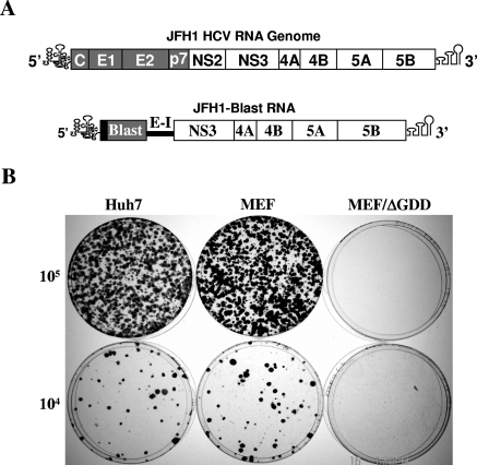
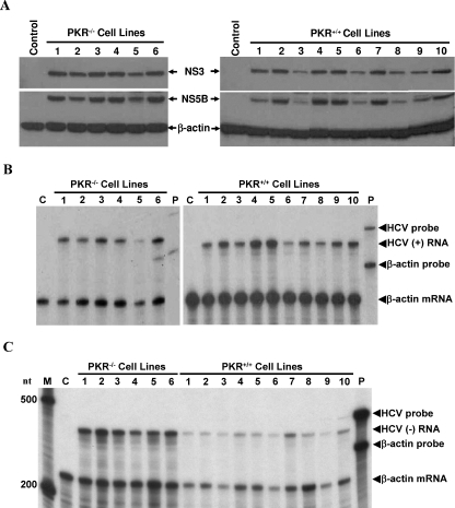
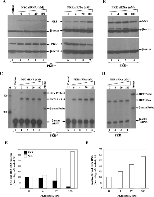
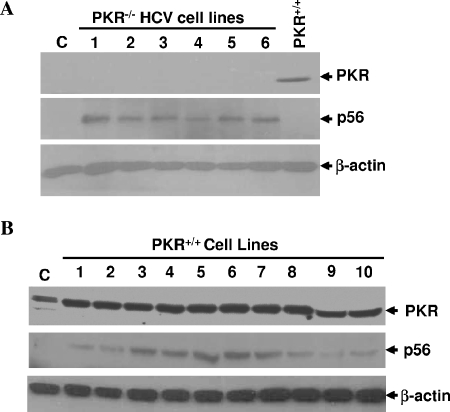
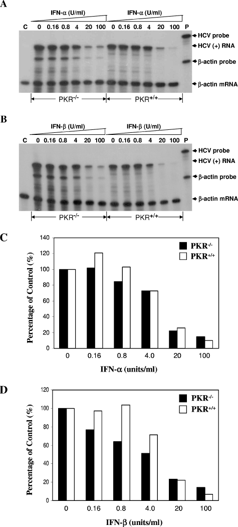
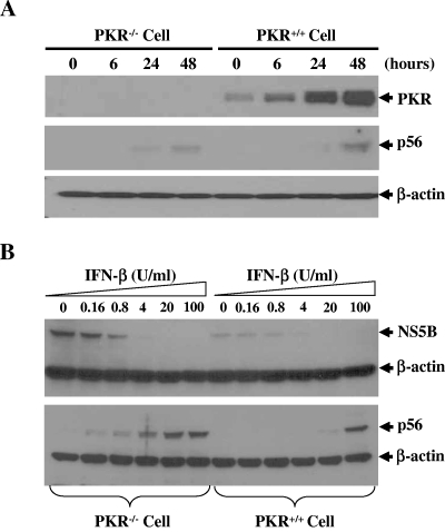
Hepatitis C virus (HCV) infection causes chronic hepatitis and is currently treated with alpha interferon (IFN-alpha)-based therapies. The underlying mechanisms of chronic HCV infection and IFN-based therapies, however, have not been defined. Protein kinase R (PKR) was implicated in the control of HCV replication and mediation of IFN-induced antiviral response. In this report, we demonstrate that a subgenomic RNA replicon of genotype 2a HCV replicated efficiently in mouse embryonic fibroblasts (MEFs), as determined by cell colony formation efficiency and the detection of HCV proteins and both positive- and negative-strand RNAs. Additionally, the subgenomic HCV RNA was found to replicate more efficiently in the PKR knockout (PKR(-/-)) MEF than in the wild-type (PKR(+/+)) MEF. The knockdown expression of PKR by specific small interfering RNAs significantly enhanced the level of HCV RNA replication, suggesting that PKR is involved in the control of HCV RNA replication. The level of ISG56 (p56) was induced by HCV RNA replication, indicating the activation of PKR-independent antiviral pathways. Furthermore, IFN-alpha/beta inhibited HCV RNA replication in PKR(-/-) MEFs as efficiently as in PKR(+/+) MEFs. These findings demonstrate that PKR-independent antiviral pathways play important roles in controlling HCV replication and mediating IFN-induced antiviral effect. Our findings also provide a foundation for the development of transgenic mouse models of HCV replication and set a stage to further define the roles of cellular genes in the establishment of chronic HCV infection and the mediation of intracellular innate antiviral response by using MEFs derived from diverse gene knockout animals.


Figures

FIG. 1.
FIG. 1.
A. Schematic of HCV RNA genome organization and the subgenomic JFH1-BLAST RNA. The JFH1-BLAST RNA consists of the 5′UTR, the core N-terminal 12-amino-acid coding sequence fused in frame with the blasticidin resistance gene (BLAST), the encephalomyocarditis virus internal ribosomal entry site (E-I), the NS3-NS5B coding region, and the 3′UTR. B. Cell colony formation induced by JFH1-BLAST RNA replication in Huh7 and MEF cells. Two micrograms of JFH1-BLAST or JFH1-BLAST/ΔGDD was transfected into 8 × 10⁶ Huh7 cells or MEFs by electroporation (7). Cells were seeded onto 100-mm culture dishes at different densities (as indicated). At 24 h posttransfection, cell colony formation was determined by selection with the addition of 5 to 10 μg/ml blasticidin for about 2 to 3 weeks. The blasticidin-containing medium was changed every 3 to 4 days. Cell clones were visualized by staining with a crystal violet solution and photographed (7).
FIG. 2.
FIG. 2.
A. Determination of HCV NS3 and NS5B proteins by Western blot analysis. Upon selection with blasticidin, the PKR+/+ and PKR−/− MEF cell lines (numbered on the top) resistant to blasticidin were picked up and expanded. Cells were lysed in a RIPA buffer as described previously (8). Twenty-five micrograms of each cell lysate was electrophoresed in a sodium dodecyl sulfate-10% polyacrylamide gel and then transferred onto a nitrocellulose membrane. The HCV NS3 and NS5B proteins were detected by Western blotting using monoclonal antibodies specific to NS3 and NS5B, respectively (8). β-Actin was used as an internal control and was detected by using a monoclonal antibody against β-actin (Sigma). Proteins were visualized by chemiluminescent staining (Roche). Parental cells without HCV RNA were used as controls, as indicated on the top. B. Determination of positive-stranded HCV RNA by RPA. Total cellular RNAs were extracted with Trizol reagent (Invitrogen) from PKR−/− and PKR+/+ cell lines that contain the JFH1-BLAST RNA. A total of 10 μg RNA was used to hybridize with 3 × 10⁴ cpm of [32P]UTP-labeled mouse β-actin RNA probe (Ambion) and 105 cpm of the negative-stranded HCV 3′UTR RNA-containing probe. The RPA was performed by following the manufacturer's procedures for RPA III kits (Ambion). Upon digestion with RNase A/T1, the RNA products protected from RNase digestion were analyzed in 6% polyacrylamide-7.7 M urea gels. C. Quantification of negative-stranded (−) HCV RNA by RPA. The radiolabeled positive-stranded HCV 5′UTR RNA was used as a probe. Cell lines are the same as those numbered on the tops of panels A and B. The levels of HCV RNAs were quantified with a PhosphorImager using the levels of mouse β-actin mRNA as controls to normalize amounts of total RNAs used in the assay. Both (−)3′UTR and (+)5′UTR RNA probes contains about 40 nucleotides (nt) of unpaired region so that the protected HCV RNA products migrated faster than undigested HCV RNA probes. Sizes of RNA markers are indicated on the left by numbers (nucleotides), and arrows on the right highlight the probes and protected RNA products. HCV (−) RNA, negative-stranded HCV RNA products; and HCV (+) RNA, positive-stranded HCV RNA products. Total RNAs from parental PKR−/− and PKR+/+ MEFs without HCV RNA replication were used as negative controls, respectively (indicated by the letter C). P, probe only.
FIG. 3.
FIG. 3.
Effects of PKR knockdown expression by siRNAs on HCV RNA replication. JFH1-BLAST RNA-harboring PKR+/+ and PKR−/− MEFs were transfected with increasing concentrations of PKR SMARTpool siRNAs or a nonspecific control siRNA, as indicated on the top. A concentration of 100 nM of siRNAs was mixed with DharmaFECT lipid reagent and then diluted with medium to 20 and 4 nM. The mixtures of siRNAs and lipid were transferred onto cells in a 6-well culture plate, with incubation for 3 days. The levels of PKR and HCV NS3 proteins were determined by Western blot analysis (A and B), and positive-stranded HCV RNAs were determined by RPA, as described in the legend to Fig. 2 and in Materials and Methods (C and D). The JFH1-BLAST RNA-harboring PKR−/− MEFs were used as controls for nonspecific effects of PKR siRNAs on HCV RNA replication (B and D). Parental cells without HCV RNA replication were used as negative controls as indicated on the top. Numbers on the top indicate the concentrations (in nanomolars) of siRNAs. PKR+/+ and PKR−/− MEFs are shown at the bottom. Arrows highlight corresponding proteins, RNA probes, and RNA products. E. Relative levels of PKR and HCV NS3 proteins in PKR+/+ MEFs upon PKR siRNA treatment. The levels of PKR and HCV NS3 proteins in panel A were determined by densitometry. The levels of PKR and NS3 proteins in cells without PKR siRNA treatment were used as controls and were considered 100%. The relative levels of PKR and NS3 proteins in PKR siRNA-treated PKR+/+ MEFs (A) were calculated as percentages of control (y axis) and plotted against the concentrations (in nanomolars) of PKR-specific siRNA (x axis). F. Correlation of the relative levels of positive-stranded HCV RNA and PKR siRNA concentrations. The percentage of control of positive-stranded HCV RNA was calculated in the same way as that described for panel E. The relative levels of HCV RNA are plotted against PKR siRNA concentration (in nanomolars). NSC, nonspecific control.
FIG. 4.
FIG. 4.
Detection of cellular proteins PKR and p56 by Western blot analysis. Western blot analysis is described in Materials and Methods. The JFH1-BLAST RNA-harboring PKR−/− (A) and PKR+/+ (B) MEF cell lines are the same as those shown in Fig. 2 (numbered on the top). Arrows on the right highlight the cellular proteins PKR and p56. β-Actin was used as an internal control to normalize amounts of proteins used between samples. C, negative control.
FIG. 5.
FIG. 5.
Inhibition of HCV RNA replication by IFN-α/β in PKR−/− and PKR+/+ MEFs. The JFH1-BLAST RNA-containing PKR−/− and PKR+/+ MEFs were treated with increasing concentrations of either mouse IFN-α (A) or mouse IFN-β (B). At 48 h after the addition of IFN, total cellular RNAs were extracted with Trizol reagent (Invitrogen). The levels of positive-stranded HCV RNA were determined by RPA, as described in the legend to Fig. 2. IFN concentrations (in units/milliliter) are shown by numbers on the top. The HCV RNA-harboring PKR−/− and PKR+/+ MEFs are indicated at the bottom. Total RNAs extracted from parental PKR−/− and PKR+/+ MEFs were used as negative controls, as indicated by C on the top. The RNA probes and protected positive-stranded HCV RNA products are highlighted by arrows on the right. C. Comparison of the relative levels of positive-stranded HCV RNA in response to mouse IFN-α treatment between PKR−/− and PKR+/+ MEFs. The data shown in panel A were quantified by PhosphorImager analysis. The relative level of positive-stranded HCV RNA was calculated as a percentage of control, considering the level of HCV RNA without IFN treatment (control) as 100%. The relative levels of HCV RNA (in percentages) are plotted against IFN-α concentrations (in units/milliliter). D. Comparison of mouse IFN-β inhibitory activity against HCV RNA replication between PKR−/− and PKR+/+ MEFs. The data in panel B were quantified with a PhosphorImager and converted to percentages of control in the same way as that described for panel C. The relative levels (in percentages) of HCV RNA (y axis) are plotted against IFN-β concentrations (in units/milliliter; x axis).
FIG. 6.
FIG. 6.
Induction of cellular gene products by IFN-α/β treatment in PKR−/− and PKR+/+ MEFs. (A) Induction of PKR and p56 by mouse IFN-α. The JFH1-BLAST RNA-harboring PKR−/− and PKR+/+ MEF cell lines were treated with 100 U/ml of mouse IFN-α. At different time points (shown on the top), IFN-α-treated cells were lysed in a RIPA buffer. The levels of cellular proteins PKR, p56, and β-actin were determined by Western blot analysis, as described in Materials and Methods. (B) Correlation of p56 expression and suppression of HCV NS5B protein expression. The JFH1-BLAST RNA-harboring PKR−/− and PKR+/+ MEFs were treated with increasing concentrations of mouse IFN-β for 48 h. The levels of NS5B and p56 were determined by Western blot analysis.

Similar articles

Cited by

References

    1. Ali, S., and G. Kukolj. 2005. Interferon regulatory factor 3-independent double-stranded RNA-induced inhibition of hepatitis C virus replicons in human embryonic kidney 293 cells. J. Virol. 79:3174-3178. - PMC - PubMed
    1. Ali, S., C. Pellerin, D. Lamarre, and G. Kukolj. 2004. Hepatitis C virus subgenomic replicons in the human embryonic kidney 293 cell line. J. Virol. 78:491-501. - PMC - PubMed
    1. Bigger, C. B., K. M. Brasky, and R. E. Lanford. 2001. DNA microarray analysis of chimpanzee liver during acute resolving hepatitis C virus infection. J. Virol. 75:7059-7066. - PMC - PubMed
    1. Blight, K. J., A. A. Kolykhalov, and C. M. Rice. 2000. Efficient initiation of HCV RNA replication in cell culture. Science 290:1972-1975. - PubMed
    1. Blight, K. J., J. A. McKeating, J. Marcotrigiano, and C. M. Rice. 2003. Efficient replication of hepatitis C virus genotype 1a RNAs in cell culture. J. Virol. 77:3181-3190. - PMC - PubMed

Publication types

MeSH terms