. 2006 Aug;80(15):7416-26.
doi: 10.1128/JVI.00196-06.

Role for Wee1 in inhibition of G2-to-M transition through the cooperation of distinct human papillomavirus type 1 E4 proteins

Affiliations

Role for Wee1 in inhibition of G2-to-M transition through the cooperation of distinct human papillomavirus type 1 E4 proteins

Gillian L Knight et al. J Virol. 2006 Aug.

Abstract

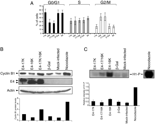
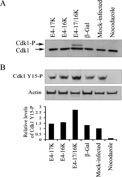
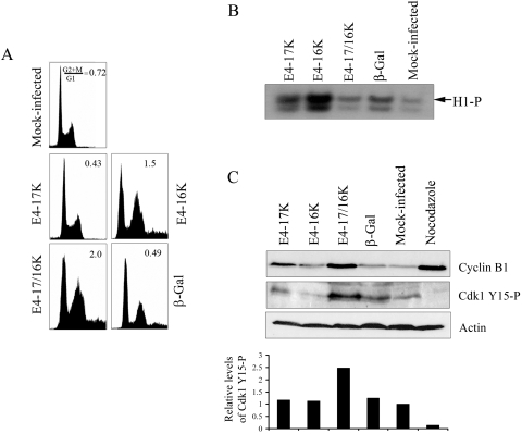
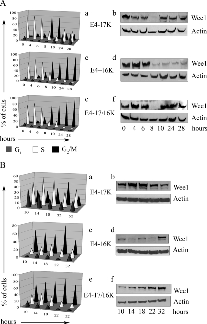
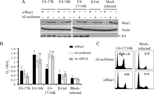
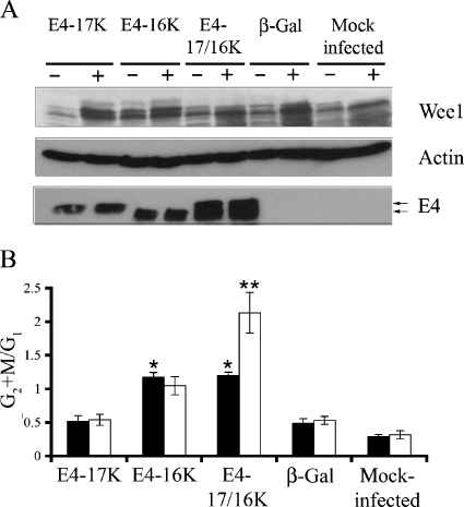
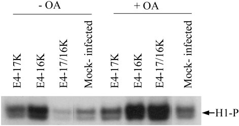
The infectious cycle of human papillomavirus type 1 (HPV1) is accompanied by abundant expression of the full-length E1;E4 protein (17-kDa) and smaller E4 polypeptides (16-, 11-, and 10-kDa) that arise by sequential loss of N-terminal E1;E4 sequences. HPV1 E4 inhibits G(2)-to-M transition of the cell cycle. Here, we show that HPV1 E4 proteins mediate inhibition of cell division by more than one mechanism. Cells arrested by coexpression of E1;E4 (E4-17K) and a truncated protein equivalent to the 16-kDa species (E4-16K) contain inactive cyclin B1-cdk1 complexes. Inactivation of cdk1 is through inhibitory Tyr(15) phosphorylation, with cells containing elevated levels of Wee1, the kinase responsible for inhibitory cdk1 phosphorylation. Consistent with these findings, overexpression of Wee1 enhanced the extent to which E4-17K/16K-expressing cells arrest in G(2), indicating that maintenance of Wee1 activity is necessary for inhibition of cell division induced by coexpression of the two E4 proteins. Moreover, we have determined that depletion of Wee1 by small interfering RNA (siRNA) alleviates the G(2) block imposed by E4-17K/16K. In contrast however, maintenance of Wee1 activity is not necessary for G(2)-to-M inhibition mediated by E4-16K alone, as overexpression or depletion of Wee1 does not influence the G(2) arrest function of E4-16K. Cells arrested by E4-16K expression contain low levels of active cyclin B1-cdk1 complexes. We hypothesize that differential expression of HPV1 E4 proteins during the viral life cycle determines the host cell cycle status. Different mechanisms of inhibition of G(2)-to-M transition reinforce the supposition that distinct E4 functions are important for HPV replication.


Figures

FIG. 1.
FIG. 1.
Differences in the level of cyclin B1 protein and cdk1 activity between HeLa cells arrested in G2 by E4-16K expression or combined expression of E4-17K and E4-16K. (A) Cell cycle analysis of HeLa cells either mock infected (MI) or infected with rAds expressing HPV1 E4 proteins (17K, 16K, or both [17/16K]) or the control protein β-galactosidase (β-Gal). Data from seven independent experiments are shown as the means ± standard deviations. The proportion of cells in the G0/G1, S, and G2/M phases were deconvoluted from the frequency histogram by using Multicycle dedicated cell cycle analysis software. The mean G2+M:G1 ratios are 0.33 (E4-17K), 1.53 (E4-16K), 1.65 (E4-17/16K), 0.35 (β-Gal), and 0.34 (MI). (B) Western blot analysis of cyclin B1 and E4 expression in HeLa cell lysates. HeLa cells treated with nocodazole were used as a control population of cells arrested in mitosis. β-Actin represents a protein loading control. The histogram shows the relative levels of cyclin B1, following normalization to actin levels. (C) Level of histone H1 phosphorylation (H1-P) following in vitro phosphorylation ([γ-32P]ATP) using cyclin B1-cdk1 complexes immunoprecipitated from HeLa cell lysates using a cyclin B1 antibody. The histogram shows the relative intensities of H1-P from four independent experiments. Equivalent levels of cyclin B1 and cdk1 were immunoprecipitated for all samples (data not shown).
FIG. 2.
FIG. 2.
Inhibition of G2-to-M transition following coexpression of E4-17/16K proteins correlates with increased levels of phosphorylated cdk1. (A) cdk1 protein expression profiles in HeLa cells either mock infected or infected with rAds for 72 h. HeLa cells treated with nocodazole were used as a control population of cells arrested in mitosis. (B) Western blot analysis of cell lysates with an antibody that detects inhibitory phosphorylation of cdk1 on Tyr15 (Cdk1 Y15-P). As a protein loading control, the membrane was probed with an antibody against β-actin. The histogram shows cdk1 Y15-P levels relative to total cdk1 protein.
FIG. 3.
FIG. 3.
Activity of cdk1 in SCC-12F keratinocytes arrested by HPV1 E4 proteins. (A) Cell cycle profiles of SCC-12F cells either mock infected or infected with rAds for 72 h. (B) Level of histone H1 phosphorylation (H1-P) following in vitro phosphorylation ([γ-32P]ATP) using cyclin B1-cdk1 complexes immunoprecipitated from SCC-12F lysates with a cyclin B1 antibody. Autoradiograph shown is representative of two experiments. Equivalent levels of cyclin B1 and cdk1 were immunoprecipitated for all samples (data not shown). (C) Western blot analysis of expression of cyclin B1 and cdk1 phosphorylated on Tyr15 (Cdk1 Y15-P) in SCC-12F lysates. As a protein loading control, the membrane was probed with an antibody against β-actin. The histogram shows cdk1 Y15-P levels relative to total cdk1 protein.
FIG. 4.
FIG. 4.
Level of Wee1 kinase is stabilized in HeLa cells arrested in G2 following coexpression of the E4-17K and E4-16K proteins. Wee1 protein expression in synchronous HeLa cells infected with rAds expressing E4-17K or E4-16K or coinfected with both viruses 0 to 28 h (A) or 10 to 32 h (B) after release from a double thymidine block. Graphical representation of cell cycle progression is shown in a, c, and e. Western blot analysis of Wee1 levels, with β-actin acting as a protein loading control, is shown in b, d, and f.
FIG. 5.
FIG. 5.
Depletion of Wee1 by siRNA inhibits G2 arrest induced by E4-17/16K protein expression. (A) Western blot analysis of the Wee1 protein in HeLa cells either mock transfected or transfected with siRNAs specific for Wee1 (siWee1) or Luciferase (siLuciferase), followed by mock infection or infection with rAds expressing HPV1 E4 proteins or β-galactosidase. Expression of E4 was confirmed in the appropriate infections, and β-actin acted as a protein loading control. (B) The histogram shows G2+M:G1 ratios of data from four independent experiments, shown as means ± standard deviations. The single asterisk indicates a significant (99.9%) decrease in the G2+M:G1 ratio of siWee1-transfected E4-17/16K cells compared to that of mock-transfected (no siRNA) or siLuciferase-transfected E4-17/16K cells. The double asterisk indicates a significant (99.9%) G2 arrest in cells expressing the E4-16K or E4-17/16K protein compared to cells expressing β-galactosidase (no siRNA) or mock-infected cells (no siRNA). (C) Cell cycle profiles of E4-17/16K-expressing cells and control cells treated with siLuciferase or siWee1.
FIG. 6.
FIG. 6.
Extent of G2 arrest is enhanced in E4-17/16K-expressing cells by overexpression of Wee1. (A) Western blot analysis of Wee1 protein levels in HeLa cells either mock transfected (−) or transfected (+) with Wee1 expression plasmid. Fluorescence microscopy of cells cotransfected with a green fluorescent protein expression plasmid indicated efficient transfection of 66.7% ± 13.1% (of GFP-positive cells). Expression of E4 was confirmed in the appropriate infections, and β-actin acted as a protein loading control. (B) Histogram of G2+M:G1 ratios in mock (closed bars) and Wee1 (open bars) transfectants. Data from four independent experiments are shown as means ± standard deviations. The single asterisk indicates a significant (99.9%) G2 arrest of cells expressing E4-16K or E4-17/16K compared to cells expressing β-galactosidase. The double asterisk indicates a significant (99.99%) increase in the G2+M:G1 ratio of Wee1-transfected E4-17/16K cells compared to that of mock-transfected E4-17/16K cells.
FIG. 7.
FIG. 7.
Inhibition of PP2A abrogates inactivation of cdk1 activity in cells coexpressing E4-17/16K proteins. HeLa cells either mock infected or infected with rAds expressing the different E4 proteins were treated with 10 μM okadaic acid (+OA) or DMSO control (−OA). The level of cdk1 activity was determined by histone H1 phosphorylation (H1-P) using cyclin B1-cdk1 complexes immunoprecipitated by a cyclin B1 antibody. Equivalent levels of cyclin B1 and cdk1 were immunoprecipitated for all samples (data not shown). The autoradiograph shown is representative of three experiments.
FIG. 8.
FIG. 8.
E4-17K and E4-16K form a heteromeric complex. Western blot analysis (MAb 4.37) of HA tag immune complexes isolated from HeLa cells transfected with a 17K-HA or 16K expression plasmid or cells cotransfected with both plasmids (17K-HA plus 16K). Control cells were transfected with empty vector (pcDNA3.0). Note that the MAb 4.37 detects both the 17K and 16K forms of E4. The asterisk in the HA immunoprecipitation from 17K-HA-transfected cells indicates the presence of a low level of processed 16-kDa E4 polypeptide.

Similar articles

Cited by

References

    1. Ashmole, I., P. H. Gallimore, and S. Roberts. 1998. Identification of conserved hydrophobic C-terminal residues of the human papillomavirus type 1 E1E4 protein necessary for E4 oligomerisation in vivo. Virology 240:221-231. - PubMed
    1. Boudreau, R. T., and D. W. Hoskin. 2005. The use of okadaic acid to elucidate the intracellular role(s) of protein phosphatase 2A: lessons from the mast cell model system. Int. Immunopharmacol. 5:1507-1518. - PubMed
    1. Breitburd, F., O. Croissant, and G. Orth. 1987. Expression of human papillomavirus type-1 E4 gene products in warts, p. 115-122. In B. M. Steinberg, J. Brandsma, and L. B. Taichman (ed.), Papillomaviruses: cancer cells, vol. 5. Cold Spring Harbor Laboratory Press, Cold Spring Harbor, N.Y.
    1. Brown, D. R., C. R. Brown, and E. E. Lehr. 2004. Intracellular expression patterns of the human papillomavirus type 59 E1/E4 protein in COS cells, keratinocytes, and genital epithelium. Intervirology 47:321-327. - PubMed
    1. Davy, C. E., D. J. Jackson, K. Raj, W. L. Peh, S. A. Southern, P. Das, R. Sorathia, P. Laskey, K. Middleton, T. Nakahara, Q. Wang, P. J. Masterson, P. F. Lambert, S. Cuthill, J. B. Millar, and J. Doorbar. 2005. Human papillomavirus type 16 E1^E4-induced G2 arrest is associated with cytoplasmic retention of active Cdk1/cyclin B1 complexes. J. Virol. 79:3998-4011. - PMC - PubMed

Publication types

MeSH terms

LinkOut - more resources