. 2006 Aug;80(15):7469-80.
doi: 10.1128/JVI.02677-05.

Influenza virus receptor specificity and cell tropism in mouse and human airway epithelial cells

Affiliations

Influenza virus receptor specificity and cell tropism in mouse and human airway epithelial cells

Aida Ibricevic et al. J Virol. 2006 Aug.

Abstract

Recent human infections caused by the highly pathogenic avian influenza virus H5N1 strains emphasize an urgent need for assessment of factors that allow viral transmission, replication, and intra-airway spread. Important determinants for virus infection are epithelial cell receptors identified as glycans terminated by an alpha2,3-linked sialic acid (SA) that preferentially bind avian strains and glycans terminated by an alpha2,6-linked SA that bind human strains. The mouse is often used as a model for study of influenza viruses, including recent avian strains; however, the selectivity for infection of specific respiratory cell populations is not well described, and any relationship between receptors in the mouse and human lungs is incompletely understood. Here, using in vitro human and mouse airway epithelial cell models and in vivo mouse infection, we found that the alpha2,3-linked SA receptor was expressed in ciliated airway and type II alveolar epithelial cells and was targeted for cell-specific infection in both species. The alpha2,6-linked SA receptor was not expressed in the mouse, a factor that may contribute to the inability of some human strains to efficiently infect the mouse lung. In human airway epithelial cells, alpha2,6-linked SA was expressed and functional in both ciliated and goblet cells, providing expanded cellular tropism. Differences in receptor and cell-specific expression in these species suggest that differentiated human airway epithelial cell cultures may be superior for evaluation of some human strains, while the mouse can provide a model for studying avian strains that preferentially bind only the alpha2,3-linked SA receptor.


Figures
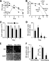
FIG. 1.
FIG. 1.
Influenza virus production and cell survival of MDCK cells and mTEC. (A) Virus production in MDCK cells compared to mTEC cultures. Cells were infected with influenza virus rWSN using the virus titer indicated, and media were collected daily for plaque assay. (B) Cell number following viral inoculation of MDCK cells and mTEC as in panel A. Data are means of duplicate samples from a representative experiment (n = 3). (C) mTEC number (means ± standard deviations) determined by DAPI staining following incubation with medium or influenza virus (1 × 10⁶ PFU/insert, 24 h) as in panel A. There were significant differences (P < 0.05) in cell numbers at the indicated days between medium- and virus-treated samples (*) and between virus-treated samples at day 1 and at day 7 (**). (D) Quantification of influenza virus M2 expression in mTEC (means ± standard deviations). M2 was detected by immunostaining following virus inoculation as for panel C. M2 expression at day 1 and day 4 was significantly different (P < 0.05) from that on day 7 and day 11 (*). (E) Reinfection of mTEC cultures. Naive (uninfected) or previously infected (10 days earlier) mTEC were incubated with virus (rWSN, 1 × 10⁶ PFU/sample). One day later, cells were immunostained for M2 expression. (F) M2 expression and virus production (means ± standard deviations) from cells in panel E were significantly different between samples (*, P < 0.05). Data in panels C to F are representative of three independent experiments.
FIG. 2.
FIG. 2.
Influenza virus infects ciliated mouse tracheal epithelial cells. (A) Photomicrograph of representative fields of mTEC cultures inoculated with virus (H1N1, rWSN, 1 × 10⁶ PFU/insert, 24 h), then immunostained with the ciliated cell marker β-tubulin-IV (β-tub), detected with FITC (green), and labeled with DAPI (blue) at the indicated day. Bar, 200 μm. (B) Quantification of ciliated and nonciliated cells under conditions as in panel A. Cells (identified by DAPI staining) were quantified for the presence or absence of β-tubulin-IV. Shown are means ± standard deviations from two independent experiments. A significant difference (P < 0.05) between ciliated cells incubated with medium versus virus is indicated (*). (C) M2 (red) expression in ciliated cells (β-tub, green) 1 day after infection as in panel A. (Left) Photomicrograph at low magnification (bar, 100 μm); (right) photomicrograph at high magnification (bar, 10 μm). (D) Apoptosis in ciliated cells 2 days after infection as in panel A. Shown are photomicrographs of mTEC stained for activated caspase 3 (red) with γ-tubulin (γ-tub, green) and for annexin V (red) with foxj1 (green). Bars, 10 μm. (E) Effect of virus concentration on mTEC infection. mTEC cultures were inoculated for 1 h with rWSN using the indicated concentration. Twenty-four hours later, cultures were evaluated for virus production by a TCID50 assay, and the percentage of ciliated cells infected (means ± standard deviations of the percentage of ciliated cells expressing M2) was determined using immunostaining as for panel C. There was no significant difference (P > 0.05) in the percentage of infected ciliated cells when virus concentrations equal to or greater than 10 × 106 PFU were used (*).
FIG. 3.
FIG. 3.
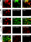
Selective expression of α2,3-linked sialic acid in ciliated mTEC. (A) Localization of α2,3- or α2,6-linked SA by binding of MAA and SNA lectins, respectively, in MDCK cells. Cells were incubated with biotinylated MAA (red) or SNA (green) and detected by streptavidin-labeled fluorescent dyes. (B) mTEC bind MAA but not SNA. mTEC were either incubated with biotinylated MAA (left) (red), pretreated with unconjugated MAA followed by biotinylated MAA (center) (red), or incubated with SNA only (right) (green). (C) MAA binds ciliated mTEC. mTEC were incubated with biotinylated MAA (red) and immunostained for β-tubulin-IV (β-tub) (green). (D) Effect of lectin pretreatment on influenza virus (rWSN, 1 × 10⁶ PFU/insert) inoculation (low-magnification images). Shown is M2 expression in mTEC after virus inoculation of cells pretreated with medium (left), MAA (middle), or SNA (right). Bar, 100 μm. (E) Expression of SeV protein in ciliated cells. mTEC were either inoculated with SeV (1 × 10⁵ PFU, 24 h) and then immunostained for SeV protein (red) and β-tub (green) (left) (high magnification) or pretreated with either medium (center) (low magnification [bar, 100 μm]) or MAA (right) (low magnification [bar, 100 μm]) and then immunostained for SeV protein. All bars represent 10 μm except as noted.
FIG. 4.
FIG. 4.
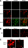
Expression of α2,3-linked sialic acid in preciliated mTEC. (A) MAA binding during differentiation of mTEC cultures. mTEC were cultured until confluent, then differentiated using ALI conditions. At the indicated ALI day (d), mTEC were incubated with biotinylated MAA (red) and immunostained with γ-tubulin (green), a marker of ciliated cell basal bodies. MAA binding was detected at ALI day 1, prior to the presence of organized apical basal bodies shown at ALI day 7. Bar, 10 μm. (B) Effect of mTEC differentiation on influenza virus infection. Shown is M2 expression from the indicated ALI day in mTEC infected with rWSN (1 × 10⁶ PFU/insert, 24 h). Bar, 100 μm. (C) Virus infection of mTEC cultures from cilium-deficient Foxj1−/− mice. (Left) MAA binding was detected as for panel A. Bar, 10 μm. (Right) Foxj1−/− mTEC were infected with virus and M2 detected as for panel B. Bar, 100 μm.
FIG. 5.
FIG. 5.
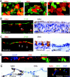
Selective expression of the α2,3-linked SA receptor in mouse lungs and influenza virus infection in vivo. (A) Expression of the α2,3-linked SA receptor (red) in ciliated airway epithelial cells (green) in fixed, paraffin-embedded sections (5 μm) from the mouse large airway. (B) The α2,3-linked SA receptor (red) is expressed in type II (SPC, pro-surfactant protein C, green) (top) but not in type I (T1α, green) (bottom) alveolar epithelial cells. Arrows indicate cells with MAA staining. (C) M2 expression in ciliated cells from lungs of mice inoculated with influenza virus A/WS/33 intranasally using 104 PFU analyzed at the indicated day for M2 expression by the immunoperoxidase method (brown) and counterstained with hematoxylin. Two different examples of infected ciliated cells at day 3 are shown. (D) Low-magnification image of virus infection (as in panel C) in large airways and type II alveolar epithelial cells detected by immunoperoxidase (M2) (top) and immunofluorescence (HA, green; SPC, red) (bottom). (Inset) SPC expression in type II cells. Bars for panels A to C, 10 μm; bar for panel D, 100 μm.
FIG. 6.
FIG. 6.
Cell-selective localization of influenza virus receptors in human airway epithelial cells. (A) hTEC cultures were either incubated with biotinylated MAA (red) (left) or SNA (red) (center) and colocalized with the ciliated cell marker β-tubulin (β-tub, green) or incubated with both MAA (red) and SNA (green) (right). (B) Photomicrographs of MAA binding in sections of human lung detected by immunofluorescence (left) and immunoperoxidase (right). Expression of MAA in ciliated cells and a subpopulation of basal epithelial cells (bc) is shown. (C) Photomicrographs of SNA binding in ciliated and goblet cells (arrows, left) using methods as in panel B. (D) Representative reconstructed (x,z axis) confocal microscopy image of hTEC cultures showing localization of SNA binding (red) with goblet cells (MUC5AC, green) but not with Clara cells (CCSP, blue). (E) Detection of receptors in alveoli of human lung sections with MAA (left) and SNA (center) using immunoperoxidase and colocalization of MAA (red) with the type II alveolar epithelial cell marker SPC (green) using immunofluorescence (DAPI, blue) (right). Panels A to E are representative of expression in at least five different donors. Bars, 10 μm.
FIG. 7.
FIG. 7.
Human airway epithelial cell-specific tropism of influenza virus strains that preferentially bind to α2,3- or α2,6-linked SA. hTEC cultures were incubated with the indicated virus strain and analyzed by immunostaining. (A) rWSN (H1N1; 1 × 10⁶ PFU/insert; 22 h) virus (M2, red) infects ciliated cells (β-tub, green) only. (B) A/California/7/2004 (H3N2; 2 × 10⁶ PFU/insert, 22 h) virus infects ciliated cells and nonciliated (nc) cells identified as goblet cells (arrows). Cells expressing M2 (red) were colocalized with β-tubulin (green) (left) or MUC5AC (green) (right). (C) Quantification of M2 expression in ciliated (c) and nonciliated (nc) mTEC or hTEC infected with A/California/7/2004 as in panel B and A/rUdorn/72 (2 × 10⁴ PFU/insert, 20 h). Percentages of infected cells are means ± standard deviations.

Similar articles

Cited by

References

    1. Angata, T., and A. Varki. 2002. Chemical diversity in the sialic acids and related alpha-keto acids: an evolutionary perspective. Chem. Rev. 102:439-469. - PubMed
    1. Baum, L. G., and J. C. Paulson. 1990. Sialyloligosaccharides of the respiratory epithelium in the selection of human influenza virus receptor specificity. Acta Histochem. Suppl. 40:35-38. - PubMed
    1. Bean, W. J., M. Schell, J. Katz, Y. Kawaoka, C. Naeve, O. Gorman, and R. G. Webster. 1992. Evolution of the H3 influenza virus hemagglutinin from human and nonhuman hosts. J. Virol. 66:1129-1138. - PMC - PubMed
    1. Beigel, J. H., J. Farrar, A. M. Han, F. G. Hayden, R. Hyer, M. D. de Jong, S. Lochindarat, T. K. Nguyen, T. H. Nguyen, T. H. Tran, A. Nicoll, S. Touch, and K. Y. Yuen. 2005. Avian influenza A (H5N1) infection in humans. N. Engl. J. Med. 353:1374-1385. - PubMed
    1. Bender, C., H. Hall, J. Huang, A. Klimov, N. Cox, A. Hay, V. Gregory, K. Cameron, W. Lim, and K. Subbarao. 1999. Characterization of the surface proteins of influenza A (H5N1) viruses isolated from humans in 1997-1998. Virology 254:115-123. - PubMed

Publication types

MeSH terms