. 2006 Aug;80(15):7636-44.
doi: 10.1128/JVI.00567-06.

Structure of A197 from Sulfolobus turreted icosahedral virus: a crenarchaeal viral glycosyltransferase exhibiting the GT-A fold

Affiliations

Structure of A197 from Sulfolobus turreted icosahedral virus: a crenarchaeal viral glycosyltransferase exhibiting the GT-A fold

Eric T Larson et al. J Virol. 2006 Aug.

Abstract

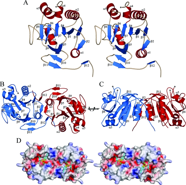
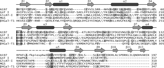
Sulfolobus turreted icosahedral virus (STIV) was the first icosahedral virus characterized from an archaeal host. It infects Sulfolobus species that thrive in the acidic hot springs (pH 2.9 to 3.9 and 72 to 92 degrees C) of Yellowstone National Park. The overall capsid architecture and the structure of its major capsid protein are very similar to those of the bacteriophage PRD1 and eukaryotic viruses Paramecium bursaria Chlorella virus 1 and adenovirus, suggesting a viral lineage that predates the three domains of life. The 17,663-base-pair, circular, double-stranded DNA genome contains 36 potential open reading frames, whose sequences generally show little similarity to other genes in the sequence databases. However, functional and evolutionary information may be suggested by a protein's three-dimensional structure. To this end, we have undertaken structural studies of the STIV proteome. Here we report our work on A197, the product of an STIV open reading frame. The structure of A197 reveals a GT-A fold that is common to many members of the glycosyltransferase superfamily. A197 possesses a canonical DXD motif and a putative catalytic base that are hallmarks of this family of enzymes, strongly suggesting a glycosyltransferase activity for A197. Potential roles for the putative glycosyltransferase activity of A197 and their evolutionary implications are discussed.


Figures

FIG. 1.
FIG. 1.
(A) Stereo image of the A197 monomer. The ribbon diagram depicts secondary structural elements of A197 (β-strands are blue, α-helices are red, and loops are wheat), which are labeled in ascending order from the N terminus to the C terminus. *, N- and C-terminal ends of the disordered region within the α4-α5 loop (residues 139 to 147). (B) Ribbon diagram of the A197 homodimer looking into the putative active site. One monomer is blue, and the second is red. The orientation of the blue monomer is rotated slightly counterclockwise with respect to that depicted in Fig. 1A. Secondary structural elements and the termini of the disordered region within the α4-α5 loops are labeled as in panel A. (C) Relative to panel B, the A197 homodimer has been rotated 90° about the depicted axis. The putative active site now runs along the top of the figure. (D) Stereo image of the electrostatic surface of the A197 homodimer. The orientation is identical to that in panel B, serving to highlight the putative active site. *, termini of the disordered region within the α4-α5 loop. The putative donor substrate-binding sites are marked with stick representations of the superpositioned donor substrate of GnT I. The electrostatic potential was mapped to the surface of A197 with SPOCK (13), using a probe radius of 1.4 Å, a temperature of 353°K, an ionic strength of 0.15 M, and protein and solvent dielectric constants of 4 and 80, respectively. The color ramp of the surface is from −15 kT/e (red, acidic) to 15 kT/e (blue, basic). All images were prepared and rendered using PyMOL (17).
FIG. 2.
FIG. 2.
Ribbon diagrams of A197 and the equivalent domains of two structural neighbors. The relative orientations are the same in all panels and are identical to that in Fig. 1A. (A) Residues 1 to 197 of the A197 monomer. (B) Residues 106 to 317 of rabbit GnT I (PDB identifier [ID] 1FOA) (62). Secondary structural elements that are shared with A197 are labeled accordingly. The donor substrate (UDP-GlcNAc) is depicted with sticks, and the coordinated divalent manganese cation (Mn2+) is shown as a pink sphere in the active site. The C-terminal end of the structure is marked with dots to indicate the continuation of the polypeptide leading to an additional domain that is not present in A197. (C) Residues 75 to 310 of human GlcAT-I (PDB ID 1FGG) (48). Labels are consistent with those in panels A and B. UDP in the donor-binding site and Galβ1-3Gal in the acceptor-binding site are shown as sticks. The coordinated manganese cation is shown as a pink sphere. (D) Superposition of A197 (blue), GnT I (orange), and GlcAT-I (yellow) with detail of key GT-A glycosyltransferase active-site features. Residues constituting the DXD motif (Asp61, Glu62, and Asp63) and catalytic base (Asp151) of A197 are shown in sticks, as are the equivalent residues in GnT I (Glu211, Asp212, Asp213, and Asp291, respectively) and GlcAT-I (Asp194, Asp195, Asp196, and Glu281, respectively). For clarity, only the ribbon diagram of A197 is shown. The green mesh depicts difference electron density (contoured at 6σ) for the manganese binding site; it is found adjacent to the DXD motif and coincides nicely with the expected metal binding site. In GnT I and GlcAT-I, the DXD motif is involved in coordination of the diphosphate moiety of the donor substrate through the intervening Mn2+ ion, while the catalytic base is appropriately positioned for proton extraction from the hydroxyl moiety of the acceptor sugar substrate. Superpositions were prepared using LSQKAB (28). All images were prepared and rendered using PyMOL (17).
FIG. 3.
FIG. 3.
Structure-based sequence alignment of A197 (PDB identifier [ID] 2C0N) with GT-A domains of close structural neighbors. GnT I, residues 106 to 317 of rabbit N-acetylglucosaminyltransferase I (PDB ID 1FOA; family GT13; inverting mechanism) (62); GlcAT-I, residues 75 to 310 of human β1,3-glucuronyltransferase I (PDB ID 1FGG; family GT43; inverting mechanism) (48); MGS, residues 2 to 99 of Rhodobacter marinus mannosylglycerate synthase (PDB ID 2BO4; family GT78; retaining mechanism) (19); β4Gal-T1, residues 180 to 347 of bovine β1,4-galactosyltransferase T1 (PDB ID 1FGX; family GT7; inverting mechanism) (21). The secondary structural elements of A197 are mapped above the alignment (arrows denote β-strands, and rounded rectangles denote α-helices). Long stretches of sequence that are not structurally equivalent to A197 have been removed from the alignment for conciseness and are marked by double shills. The missing residues of the A197 α4-α5 loop are in lowercase in the alignment and are marked with dashes in the secondary structure map. Identical residues are highlighted in gray, and the DXD motif and catalytic base are in boldface and are boxed. The structure-based sequence alignment was created with the 3DCoffee@igs web server (http://www.igs.cnrs-mrs.fr/Tcoffee/) (51), using Msap_pair to compute the library and slight manual adjustment around the gaps with consideration of the pairwise structural alignments created by LSQMAN (30).

Similar articles

Cited by

References

    1. Albers, S.-V., S. M. Koning, W. N. Konings, and A. J. M. Driessen. 2004. Insights into ABC transport in Archaea. J. Bioenerg. Biomembr. 36:5-15. - PubMed
    1. Allen, M., D. Willits, M. Young, and T. Douglas. 2003. Constrained synthesis of cobalt oxide nanomaterials in the 12-subunit protein cage from Listeria innocua. Inorg. Chem. 42:6300-6305. - PubMed
    1. Altschul, S. F., T. L. Madden, A. A. Schäffer, J. Zhang, Z. Zhang, W. Miller, and D. J. Lipman. 1997. Gapped BLAST and PSI-BLAST: a new generation of protein database search programs. Nucleic Acids Res. 25:3389-3402. - PMC - PubMed
    1. Arnold, H. P., W. Zillig, U. Ziese, I. Holz, M. Crosby, T. Utterback, J. F. Weidmann, J. K. Kristjanson, H. P. Klenk, K. E. Nelson, and C. M. Fraser. 2000. A novel lipothrixvirus, SIFV, of the extremely thermophilic crenarchaeon Sulfolobus. Virology 267:252-266. - PubMed
    1. Bailey, S. 1994. The CCP4 suite—programs for protein crystallography. Acta Crystallogr. Sect. D Biol. Crystallogr. 50:760-763. - PubMed

Publication types

MeSH terms

Associated data

LinkOut - more resources