. 2006 Aug;80(15):7645-57.
doi: 10.1128/JVI.00206-06.

Alpha interferon potently enhances the anti-human immunodeficiency virus type 1 activity of APOBEC3G in resting primary CD4 T cells

Affiliations

Alpha interferon potently enhances the anti-human immunodeficiency virus type 1 activity of APOBEC3G in resting primary CD4 T cells

Keyang Chen et al. J Virol. 2006 Aug.

Abstract

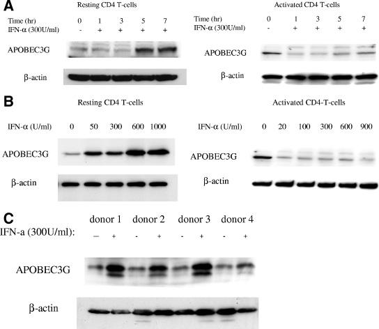
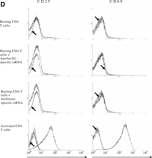
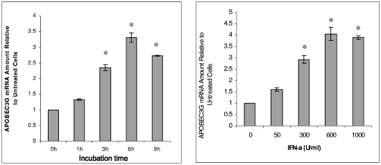
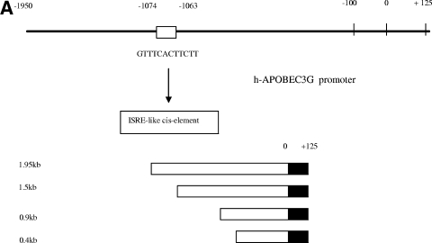
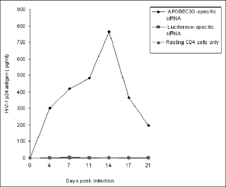
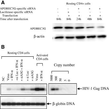
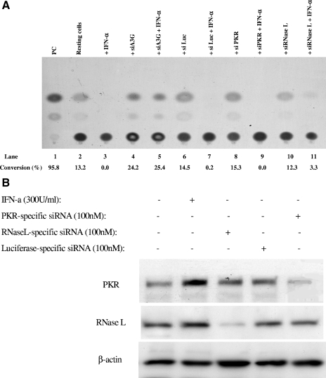
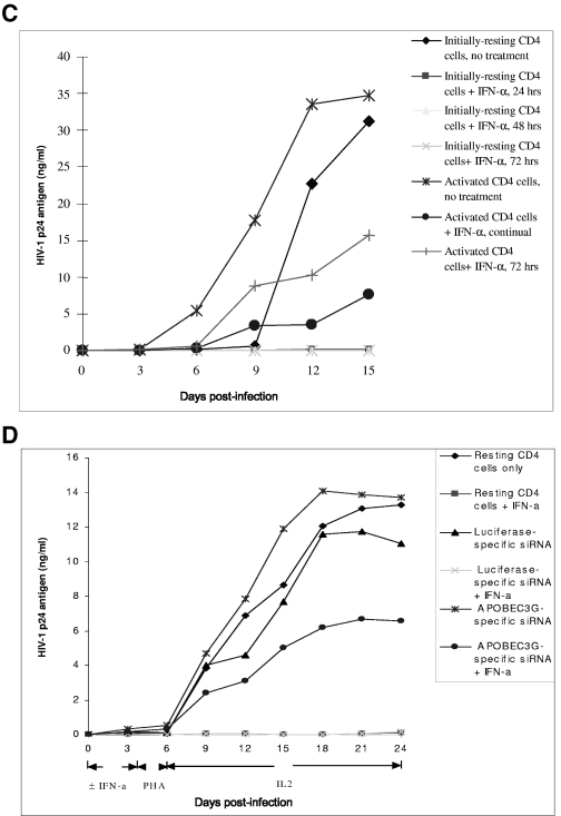
The interferon (IFN) system, including various IFNs and IFN-inducible gene products, is well known for its potent innate immunity against wide-range viruses. Recently, a family of cytidine deaminases, functioning as another innate immunity against retroviral infection, has been identified. However, its regulation remains largely unknown. In this report, we demonstrate that through a regular IFN-alpha/beta signal transduction pathway, IFN-alpha can significantly enhance the expression of apolipoprotein B mRNA-editing enzyme-catalytic polypeptide-like 3G (APOBEC3G) in human primary resting but not activated CD4 T cells and the amounts of APOBEC3G associated with a low molecular mass. Interestingly, short-time treatments of newly infected resting CD4 T cells with IFN-alpha will significantly inactivate human immunodeficiency virus type 1 (HIV-1) at its early stage. This inhibition can be counteracted by APOBEC3G-specific short interfering RNA, indicating that IFN-alpha-induced APOBEC3G plays a key role in mediating this anti-HIV-1 process. Our data suggest that APOBEC3G is also a member of the IFN system, at least in resting CD4 T cells. Given that the IFN-alpha/APOBEC3G pathway has potent anti-HIV-1 capability in resting CD4 T cells, augmentation of this innate immunity barrier could prevent residual HIV-1 replication in its native reservoir in the post-highly active antiretroviral therapy era.


Figures

FIG. 1.
FIG. 1.
Expression of APOBEC3G is up-regulated by IFN-α in human resting CD4 T cells. (A) Time course study. Recombinant IFN-α (300 U/ml; Sigma) was added to the cultures of resting (left) or activated (right) primary CD4 T cells. The expression of APOBEC3G protein was detected at various time points. (B) Dose-dependent effect of IFN-α upon APOBEC3G expression in resting (left) or activated (right) CD4 T cells. Resting or activated CD4 T cells were treated with IFN-α at different concentrations. The cell lysates were prepared at 7 h posttreatment and subjected to Western blotting. (C) The resting CD4 T cells were prepared from four independent healthy blood donors and were treated with IFN-α for 7 h. The cell lysates were then prepared and subjected to Western blot analysis. Loading controls were carried out by detecting β-actin expression with an immunoblotting assay using mouse monoclonal antibody to human β-actin. The data (A to C) represent at least three independent experiments. (D) Flow cytometric analysis of activation status of resting CD4 T cells after nucleofection or activated CD4 T cells. The cells were labeled with anti-CD25 fluorescein isothiocyanate antibody, anti-CD69 allophycocyanin antibody, or an isotype control (arrows) and subjected to fluorescence-activated cell sorter analysis. Similar results were obtained using cells from four different donors.
FIG. 1.
FIG. 1.
Expression of APOBEC3G is up-regulated by IFN-α in human resting CD4 T cells. (A) Time course study. Recombinant IFN-α (300 U/ml; Sigma) was added to the cultures of resting (left) or activated (right) primary CD4 T cells. The expression of APOBEC3G protein was detected at various time points. (B) Dose-dependent effect of IFN-α upon APOBEC3G expression in resting (left) or activated (right) CD4 T cells. Resting or activated CD4 T cells were treated with IFN-α at different concentrations. The cell lysates were prepared at 7 h posttreatment and subjected to Western blotting. (C) The resting CD4 T cells were prepared from four independent healthy blood donors and were treated with IFN-α for 7 h. The cell lysates were then prepared and subjected to Western blot analysis. Loading controls were carried out by detecting β-actin expression with an immunoblotting assay using mouse monoclonal antibody to human β-actin. The data (A to C) represent at least three independent experiments. (D) Flow cytometric analysis of activation status of resting CD4 T cells after nucleofection or activated CD4 T cells. The cells were labeled with anti-CD25 fluorescein isothiocyanate antibody, anti-CD69 allophycocyanin antibody, or an isotype control (arrows) and subjected to fluorescence-activated cell sorter analysis. Similar results were obtained using cells from four different donors.
FIG. 2.
FIG. 2.
IFN-α enhances APOBEC3G mRNA expression in resting CD4 T cells. Total cellular RNA was extracted from resting CD4 T cells at 7 h after the addition of IFN-α (300 U/ml) into cell culture. The mRNA level of APOBEC3G was analyzed by real-time RT-PCR. (Left) Time course study. The amounts of APOBEC3G mRNA at 3 h, 6 h, and 9 h are significantly higher than that at time zero (asterisk, P < 0.001, t test). (Right) Dose-dependent experiment. The amounts of APOBEC3G mRNA induced by 300 U/ml, 600 U/ml, and 1,000 U/ml of IFN-α are significantly higher than that without IFN-α treatment (*, P < 0.001, t test).
FIG. 3.
FIG. 3.
Transcriptional regulation of APOBEC3G expression by IFN-α. (A) (Top) The APOBEC3G promoter at a 1.95-kb length was amplified using genomic DNA from H9 cells as a template and subjected to sequential deletion analysis. An ISRE-like sequence was identified in 1.95-kb DNA. (Bottom) The APOBEC3G promoters at various lengths (0.4 kb, 0.9 kb, 1.5 kb, and 1.95 kb) were constructed and placed upstream of the luciferase reporter gene. (B) Resting (left) or activated (right) CD4 T cells were transfected with various chimeric plasmids, followed by treatment with or without IFN-α (300 U/ml). Cell lysates were prepared at 24 h posttransfection, and luciferase activity was examined. The addition of IFN-α into resting CD4 T cells significantly enhances the activities of the 1.5-kb and 1.9-kb promoters (*, P < 0.001, t test) (right). (C) The resting (left) or activated (right) CD4 T cells were transfected, respectively, with a 1.5-kb promoter plasmid or plasmid containing a 1.5-kb promoter with a mutant ISRE-like sequence and treated with or without various siRNAs or treated with or without IFN-α (300 U/ml). The cell lysates were harvested at 24 h posttransfection, followed by detection of luciferase activity. In resting CD4 T cells, compared with the activity of the 1.5-kb promoter, the 1.5-kb M promoter, the 1.5-kb M promoter treated with IFN-α, or the 1.5-kb promoter treated with IFN-α and IRF9-specific siRNA, the activity of the 1.5-kb promoter treated with IFN-α or the 1.5-kb promoter plus green fluorescent protein (GFP)-specific siRNA treated with IFN-α was significantly increased (*, P < 0.001, t test). (D) The resting CD4+ T cells were transfected with or without IRF9-specific siRNA. At certain time points, the cell lysates were subjected to immunoblotting using anti-IRF9 antibody (left). The resting CD4 T cells transfected with or without various siRNAs were treated with or without IFN-α (300 U/ml). Cells were collected at 7 h post-IFN-α treatment, and the APOBEC3G level (right) was analyzed using immunoblotting. NC, negative control plasmid without any promoter; PC, positive control plasmid containing cytomegalovirus promoter; 1.5kb-M, 1.5-kb chimeric plasmid containing mutations in the ISRE-like sequence; siIRF9, IRF9-specific siRNA; siGFP, GFP-specific siRNA.
FIG. 3.
FIG. 3.
Transcriptional regulation of APOBEC3G expression by IFN-α. (A) (Top) The APOBEC3G promoter at a 1.95-kb length was amplified using genomic DNA from H9 cells as a template and subjected to sequential deletion analysis. An ISRE-like sequence was identified in 1.95-kb DNA. (Bottom) The APOBEC3G promoters at various lengths (0.4 kb, 0.9 kb, 1.5 kb, and 1.95 kb) were constructed and placed upstream of the luciferase reporter gene. (B) Resting (left) or activated (right) CD4 T cells were transfected with various chimeric plasmids, followed by treatment with or without IFN-α (300 U/ml). Cell lysates were prepared at 24 h posttransfection, and luciferase activity was examined. The addition of IFN-α into resting CD4 T cells significantly enhances the activities of the 1.5-kb and 1.9-kb promoters (*, P < 0.001, t test) (right). (C) The resting (left) or activated (right) CD4 T cells were transfected, respectively, with a 1.5-kb promoter plasmid or plasmid containing a 1.5-kb promoter with a mutant ISRE-like sequence and treated with or without various siRNAs or treated with or without IFN-α (300 U/ml). The cell lysates were harvested at 24 h posttransfection, followed by detection of luciferase activity. In resting CD4 T cells, compared with the activity of the 1.5-kb promoter, the 1.5-kb M promoter, the 1.5-kb M promoter treated with IFN-α, or the 1.5-kb promoter treated with IFN-α and IRF9-specific siRNA, the activity of the 1.5-kb promoter treated with IFN-α or the 1.5-kb promoter plus green fluorescent protein (GFP)-specific siRNA treated with IFN-α was significantly increased (*, P < 0.001, t test). (D) The resting CD4+ T cells were transfected with or without IRF9-specific siRNA. At certain time points, the cell lysates were subjected to immunoblotting using anti-IRF9 antibody (left). The resting CD4 T cells transfected with or without various siRNAs were treated with or without IFN-α (300 U/ml). Cells were collected at 7 h post-IFN-α treatment, and the APOBEC3G level (right) was analyzed using immunoblotting. NC, negative control plasmid without any promoter; PC, positive control plasmid containing cytomegalovirus promoter; 1.5kb-M, 1.5-kb chimeric plasmid containing mutations in the ISRE-like sequence; siIRF9, IRF9-specific siRNA; siGFP, GFP-specific siRNA.
FIG. 4.
FIG. 4.
IFN-α-induced ISGF3 complex binds to ISRE-like sequence. (A) The ISRE-like sequence in the h-APOBEC3G promoter specifically binds to nuclear proteins from resting CD4 T cells treated with IFN-α. Resting CD4 cells were treated with or without IFN-α (300 U/ml) for 7 h, and nuclear proteins were extracted. 32P-labeled nucleotides containing the ISRE-like sequence or its mutant were incubated with or without nuclear proteins. As a control, anti-STAT2 antibody was added for supershifting. The mixtures were resolved in a 6% native PAGE gel. The gel was dried and autoradiographed overnight. (B) Chromatin immunoprecipitation assay. The resting CD4 T cells were treated with or without IFN-α and fixed with formaldehyde. After sonication to break down long chromatin filament, the samples were subjected to immunoprecipitation with anti-STAT2 or anti-β-actin antibody. DNA was then extracted from the precipitated complex and subjected to PCR with two primer pairs. The PCR products were analyzed by electrophoresis.
FIG. 5.
FIG. 5.
Effect of IFN-α upon APOBEC3G in an LMM complex. The resting primary CD4 T cells were treated with or without IFN-α (300 U/ml) and harvested at 7 h after stimulation. The supernatants of cell lysates were concentrated and loaded into an FPLC column with 0.05 M sodium phosphate at a flow rate of 1.0 ml/min. Eluted fractions were subjected to SDS-PAGE, followed by an immunoblotting assay with anti-APOBEC3G antibody. Cell lysates from the activated CD4 T cells with or without IFN-α treatment were also analyzed as controls.
FIG. 6.
FIG. 6.
APOBEC3G-specific siRNA makes the resting CD4 T cells purified from fresh human PBMC permissive to wild-type HIV-1 replication. The resting CD4 T cells (3 × 10⁶) were transfected with or without APOBEC3G-specific siRNA (100 nmol/ml) or luciferase-specific siRNA (100 nmol/ml). After 72 h, the transfected cells were infected with HIV-1NL4-3 viruses (multiplicity of infection, 0.1). After being washed, the infected cells were cultured in RPMI 1640-conditioned medium without any mitogen or cytokine stimulation. The p24 antigen of HIV-1 viruses in the supernatant was detected via ELISA every 3 or 4 days.
FIG. 7.
FIG. 7.
APOBEC3G-mediated inhibitory effect of IFN-α upon intracellular reverse transcription. (A) Resting CD4 T cells were transfected with APOBEC3G-specific siRNA (100 nmol/ml) or luciferase-specific siRNA (100 nmol/ml). Cells were collected at different time points after transfection, and APOBEC3G levels were detected by an immunoblotting assay. (B) Resting or activated CD4 T cells, transfected with or without various siRNAs, were infected by DNase-treated HIV-1 viruses. The infected cells were treated with or without IFN-α. At 48 h postinfection, the cells were harvested and viral gag DNA was detected with PCR, using SK38/SK39 as the primer pair and SK19 as the probe. As a control, β-globin DNA was also detected.
FIG. 8.
FIG. 8.
IFN-α potently inhibits HIV-1 replication in initially resting CD4 T cells through APOBEC3G. (A) APOBEC3G-specific siRNA mediates potent inhibition of IFN-α upon HIV-1 infectivity in a single-round viral infection. Three plasmids, pMD.G, pCMVΔR8.2, and HIV-CAT (derived from pHR′ by replacing the LacZ gene with the CAT gene), were transfected into 293T cells (18). The supernatants were harvested after 72 h, and the recombinant viruses were further concentrated by ultracentrifugation. Conversely, the resting CD4 T cells purified from human PBMC were transfected with various siRNAs. After 72 h, the cells were infected with the concentrated recombinant viruses (20 ng p24 equivalent per cell sample [2 × 10⁶]) with or without IFN-α treatment. After 4 h, the unbound viruses were washed off and the infected cells were maintained in a conditioned medium with or without IFN-α (300 U/ml). At 96 h postinfection, IFN-α was removed and PHA and IL-2 were added to activate the cells. After 48 h, the cells were harvested and a CAT assay was performed. (B) Effect of IFN-α and siRNA upon the expression of PKR and RNase L in resting CD4 T cells. The resting CD4 T cells purified from human PBMC were treated with IFN-α (300 U/ml) or transfected with PKR-specific siRNA or RNase L-specific siRNA. At 7 h posttreatment or 72 h posttransfection, the cells were harvested and subjected to Western blot analysis, using anti-PKR antibody (BD), anti-RNase L antibody (Abcam), or anti-β-actin antibody. (C) HIV-1NL4-3 viruses were allowed to infect resting or PHA-activated CD4 T cells (3 × 10⁶) (multiplicity of infection, 0.1). Simultaneously, the infected cells were treated with or without IFN-α (300 U/ml) for various periods. For the infected activated CD4 T cells, the culture was maintained in RPMI 1640-conditioned medium containing IL-2 (25 U/ml). The HIV-1 p24 antigen in the supernatant was harvested every 3 days and detected by ELISA. For the infected resting CD4 T cells, however, PHA (5 μg/ml) was added into the cultures at 72 h postinfection. After 48 h, PHA was removed and the activated cells were cultured in RPMI 1640-conditioned medium containing IL-2 (25 U/ml). The HIV-1 p24 antigen in the supernatant was harvested every 3 days and detected by ELISA. (D) The resting CD4 T cells (3 × 10⁶) were transfected with or without APOBEC3G-specific siRNA or luciferase-specific siRNA. After 72 h, the transfected or untransfected resting CD4 T cells were infected with HIV-1NL4-3 viruses (multiplicity of infection, 0.1) for 4 h. Simultaneously, the infected cells were treated with or without IFN-α (300 U/ml) for 96 h. After IFN-α was washed off, PHA (5 μg/ml) was added to stimulate the cells for 48 h. After PHA was removed, the activated cells were cultured in RPMI 1640-conditioned medium containing IL-2 (25 U/ml). The HIV-1 p24 antigen in the supernatant was harvested every 3 days and detected by ELISA. These data represent at least three independent experiments.
FIG. 8.
FIG. 8.
IFN-α potently inhibits HIV-1 replication in initially resting CD4 T cells through APOBEC3G. (A) APOBEC3G-specific siRNA mediates potent inhibition of IFN-α upon HIV-1 infectivity in a single-round viral infection. Three plasmids, pMD.G, pCMVΔR8.2, and HIV-CAT (derived from pHR′ by replacing the LacZ gene with the CAT gene), were transfected into 293T cells (18). The supernatants were harvested after 72 h, and the recombinant viruses were further concentrated by ultracentrifugation. Conversely, the resting CD4 T cells purified from human PBMC were transfected with various siRNAs. After 72 h, the cells were infected with the concentrated recombinant viruses (20 ng p24 equivalent per cell sample [2 × 10⁶]) with or without IFN-α treatment. After 4 h, the unbound viruses were washed off and the infected cells were maintained in a conditioned medium with or without IFN-α (300 U/ml). At 96 h postinfection, IFN-α was removed and PHA and IL-2 were added to activate the cells. After 48 h, the cells were harvested and a CAT assay was performed. (B) Effect of IFN-α and siRNA upon the expression of PKR and RNase L in resting CD4 T cells. The resting CD4 T cells purified from human PBMC were treated with IFN-α (300 U/ml) or transfected with PKR-specific siRNA or RNase L-specific siRNA. At 7 h posttreatment or 72 h posttransfection, the cells were harvested and subjected to Western blot analysis, using anti-PKR antibody (BD), anti-RNase L antibody (Abcam), or anti-β-actin antibody. (C) HIV-1NL4-3 viruses were allowed to infect resting or PHA-activated CD4 T cells (3 × 10⁶) (multiplicity of infection, 0.1). Simultaneously, the infected cells were treated with or without IFN-α (300 U/ml) for various periods. For the infected activated CD4 T cells, the culture was maintained in RPMI 1640-conditioned medium containing IL-2 (25 U/ml). The HIV-1 p24 antigen in the supernatant was harvested every 3 days and detected by ELISA. For the infected resting CD4 T cells, however, PHA (5 μg/ml) was added into the cultures at 72 h postinfection. After 48 h, PHA was removed and the activated cells were cultured in RPMI 1640-conditioned medium containing IL-2 (25 U/ml). The HIV-1 p24 antigen in the supernatant was harvested every 3 days and detected by ELISA. (D) The resting CD4 T cells (3 × 10⁶) were transfected with or without APOBEC3G-specific siRNA or luciferase-specific siRNA. After 72 h, the transfected or untransfected resting CD4 T cells were infected with HIV-1NL4-3 viruses (multiplicity of infection, 0.1) for 4 h. Simultaneously, the infected cells were treated with or without IFN-α (300 U/ml) for 96 h. After IFN-α was washed off, PHA (5 μg/ml) was added to stimulate the cells for 48 h. After PHA was removed, the activated cells were cultured in RPMI 1640-conditioned medium containing IL-2 (25 U/ml). The HIV-1 p24 antigen in the supernatant was harvested every 3 days and detected by ELISA. These data represent at least three independent experiments.

Similar articles

Cited by

References

    1. Berglund, O., K. Engman, A. Ehrnst, J. Andersson, K. Lidman, B. Akerlund, A. Sonnerborg, and O. Strannegard. 1991. Combined treatment of symptomatic human immunodeficiency virus type 1 infection with native interferon-alpha and zidovudine. J. Infect. Dis. 163:710-715. - PubMed
    1. Bishop, K. N., R. K. Holmes, A. M. Sheehy, N. O. Davidson, S. J. Cho, and M. H. Malim. 2004. Cytidine deamination of retroviral DNA by diverse APOBEC proteins. Curr. Biol. 14:1392-1396. - PubMed
    1. Bogerd, H. P., H. L. Wiegand, B. P. Doehle, K. K. Lueders, and B. R. Cullen. 2006. APOBEC3A and APOBEC3B are potent inhibitors of LTR-retrotransposon function in human cells. Nucleic Acids Res. 34:89-95. - PMC - PubMed
    1. Bukrinsky, M. I., T. L. Stanwick, M. P. Dempsey, and M. Stevenson. 1991. Quiescent T lymphocytes as an inducible virus reservoir in HIV-1 infection. Science 254:423-427. - PMC - PubMed
    1. Chiu, Y. L., V. B. Soros, J. F. Kreisberg, K. Stopak, W. Yonemoto, and W. C. Greene. 2005. Cellular APOBEC3G restricts HIV-1 infection in resting CD4+ T cells. Nature 435:108-114. - PubMed

Publication types

MeSH terms