. 2006 Aug;80(15):7714-28.
doi: 10.1128/JVI.00207-06.

A poxvirus host range protein, CP77, binds to a cellular protein, HMG20A, and regulates its dissociation from the vaccinia virus genome in CHO-K1 cells

Affiliations

A poxvirus host range protein, CP77, binds to a cellular protein, HMG20A, and regulates its dissociation from the vaccinia virus genome in CHO-K1 cells

Jye-Chian Hsiao et al. J Virol. 2006 Aug.

Abstract

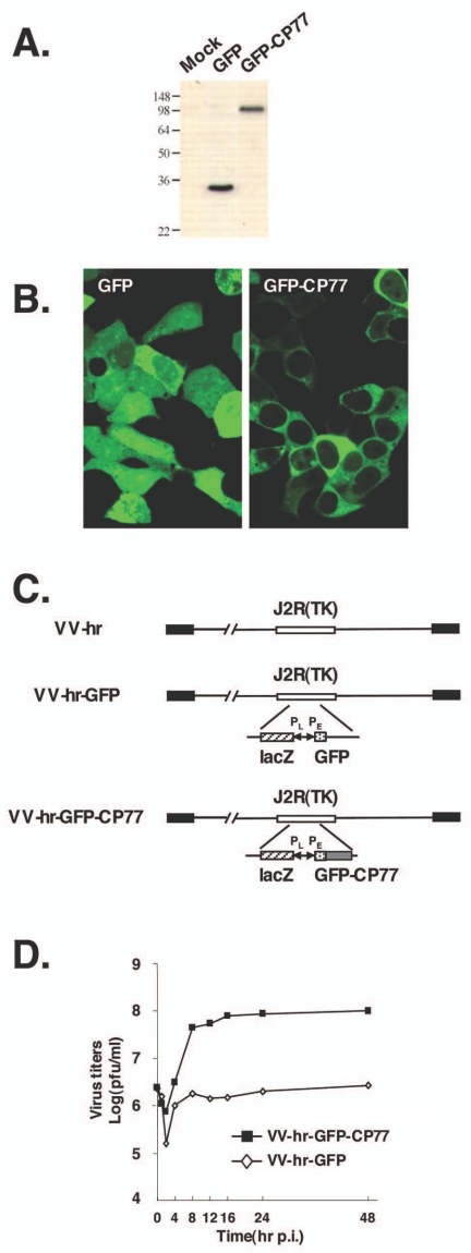
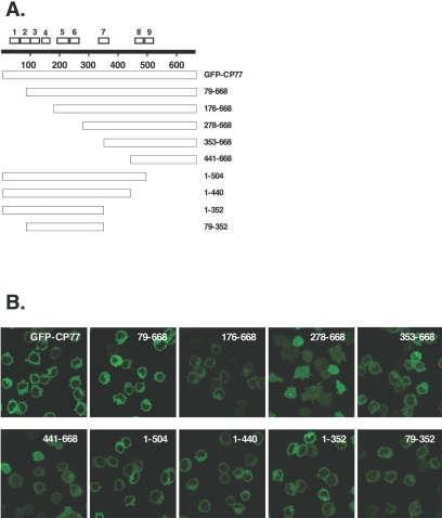
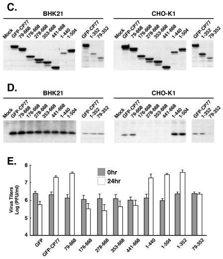
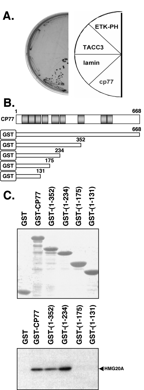
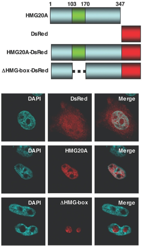
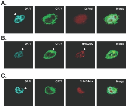
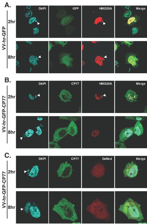
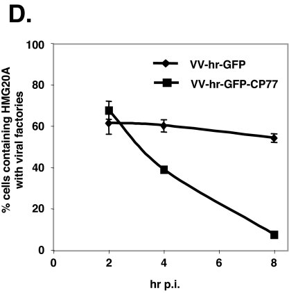
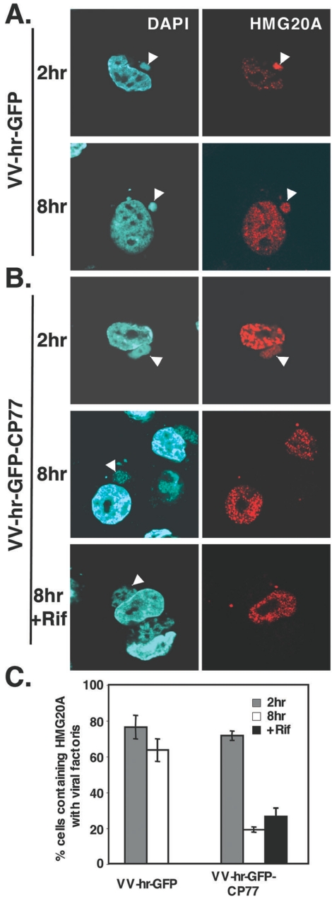
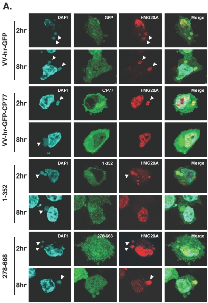
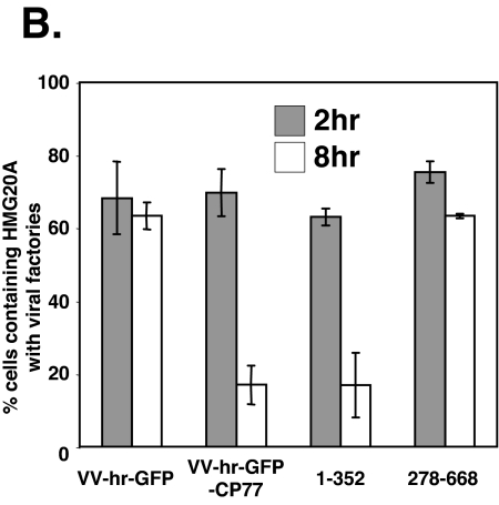
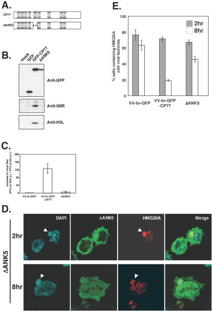
Vaccinia virus does not grow in Chinese hamster ovary (CHO-K1) cells in the absence of a viral host range factor, cowpox protein CP77. In this study, CP77 was fused to the C terminus of green fluorescence protein (GFP-CP77) and a series of nested deletion mutants of GFP-CP77 was constructed for insertion into a vaccinia virus host range mutant, VV-hr, and expressed from a viral early promoter. Deletion mapping analyses demonstrated that the N-terminal 352 amino acids of CP77 were sufficient to support vaccinia virus growth in CHO-K1 cells, whereas the C-terminal residues 353 to 668 were dispensable. In yeast two-hybrid analyses, CP77 bound to a cellular protein, HMG20A, and GST pulldown analyses showed that residues 1 to 234 of CP77 were sufficient for this interaction. After VV-hr virus infection of CHO-K1 cells, HMG20A was translocated from the nucleus to viral factories and bound to the viral genome via the HMG box region. In control VV-hr-infected CHO-K1 cells, binding of HMG20A to the viral genome persisted from 2 to 8 h postinfection (h p.i.); in contrast, when CP77 was expressed, the association of HMG20A with viral genome was transient, with little HMG20A remaining bound at 8 h p.i. This indicates that dissociation of HMG20A from viral factories correlates well with CP77 host range activity in CHO-K1 cells. Finally, in cells expressing a CP77 deletion protein (amino acids 277 to 668) or a DeltaANK5 mutant that did not support vaccinia virus growth and did not contain the HMG20A binding site, HMG20A remained bound to viral DNA, demonstrating that the binding of CP77 to HMG20A is essential for its host range function. In summary, our data revealed that a novel cellular protein, HMG20A, the dissociation of which from viral DNA is regulated by CP77, providing the first cellular target regulated by viral host range CP77 protein.


Figures

FIG. 1.
FIG. 1.
Construction and expression of GFP-CP77. (A) Immunoblot analysis of GFP and GFP-CP77, expressed from the cytomegalovirus promoter, in transfected 293T cells with anti-GFP Ab (1:4,000) (B) Confocal microscopy of GFP and GFP-CP77 in transfected 293T cells. (C) Construction of recombinant VV-hr expressing GFP or GFP-CP77. VV-hr is a vaccinia virus mutant that contains an 18-kb deletion at the left end of the viral genome (13). VV-hr-GFP and VV-hr-GFP-CP77 were generated by inserting an expression cassette containing GFP or GFP-CP77 ORF driven by an early promoter (PE) and the lacZ gene driven by a viral late promoter (PL) into the tk (J2R) locus, as described previously (29). (D) One-step growth curve analysis of recombinant VV-hr viruses in CHO-K1 cells. CHO-K1 cells were infected with VV-hr-GFP or VV-hr-GFP-CP77 at an MOI of 5 PFU per cell, and cell lysates were harvested at 0, 2, 4, 8, 12, 16, 24, or 48 h p.i. for virus titer determination on BHK-21 cells.
FIG. 2.
FIG. 2.
GFP-CP77 deletion mutant viruses in infected CHO-K1 cells. (A) CP77 deletion constructs used. The white boxes at the top represent the nine ankyrin repeats in CP77. (B) Immunofluorescence analysis of CP77 deletion proteins. CHO-K1 cells were infected with each of the recombinant viruses expressing CP77 deletion proteins at an MOI of 5 PFU per cell, fixed with paraformaldehyde at 2 h p.i., and photographed using confocal microscopy. (C) Immunoblots of CP77 deletion proteins in infected BHK-21 and CHO-K1 cells. Cells were infected with the CP77 deletion mutant viruses shown in panel A and harvested at 2 h p.i. for immunoblot analysis using anti-GFP Ab (1:4,000). (D) Immunoblots of viral G8R protein in infected BHK-21 and CHO-K1 cells. Cells were infected with the CP77 deletion mutant viruses shown in panel A and harvested at 6 h p.i. for immunoblot analysis using anti-G8R Ab (1:1,000). (E) Growth of CP77 deletion viruses in CHO-K1 cells. CHO-K1 cells were infected as described for panel B and harvested at 0 or 24 h p.i., and virus titers in cell lysates were determined by plaque assays on BHK-21 cells.
FIG. 2.
FIG. 2.
GFP-CP77 deletion mutant viruses in infected CHO-K1 cells. (A) CP77 deletion constructs used. The white boxes at the top represent the nine ankyrin repeats in CP77. (B) Immunofluorescence analysis of CP77 deletion proteins. CHO-K1 cells were infected with each of the recombinant viruses expressing CP77 deletion proteins at an MOI of 5 PFU per cell, fixed with paraformaldehyde at 2 h p.i., and photographed using confocal microscopy. (C) Immunoblots of CP77 deletion proteins in infected BHK-21 and CHO-K1 cells. Cells were infected with the CP77 deletion mutant viruses shown in panel A and harvested at 2 h p.i. for immunoblot analysis using anti-GFP Ab (1:4,000). (D) Immunoblots of viral G8R protein in infected BHK-21 and CHO-K1 cells. Cells were infected with the CP77 deletion mutant viruses shown in panel A and harvested at 6 h p.i. for immunoblot analysis using anti-G8R Ab (1:1,000). (E) Growth of CP77 deletion viruses in CHO-K1 cells. CHO-K1 cells were infected as described for panel B and harvested at 0 or 24 h p.i., and virus titers in cell lysates were determined by plaque assays on BHK-21 cells.
FIG. 3.
FIG. 3.
The N-terminal region from aa 1 to 234 of CP77 binds to HMG20A in vitro. (A) Yeast two-hybrid analyses identified that HMG20A specifically interacts with CP77 bait and not with baits such as lamin, TACC3, and ETK-PH. (B) The diagram shows CP77 protein with the nine ankyrin repeats (gray boxes). The GST-CP77 deletion constructs used are shown below; the numbers represent the C-terminal amino acids in each constructs. (C). GST pulldown with purified recombinant GST-CP77 deletion proteins. Each purified proteins were separated on SDS-PAGE, stained with Coomassie blue, and photographed. For the pulldown assays, 10 μg of each purified recombinant protein bound to glutathione-agarose beads was incubated with virus-infected cell lysates overexpressing V5-tagged HMG20A. The beads were then washed, and the pulled-down HMG20A protein was analyzed in immunoblot with anti-V5 Ab (1:5,000).
FIG. 4.
FIG. 4.
HMG20A is a nuclear protein. A schematic representation of the HMG20A constructs is shown at the top of the figure. The green box represents the HMG box region that binds to DNA. CHO-K1 cells were transfected with pDsRed, pHMG20A-DsRed, or pΔHMG-box-DsRed plasmids that express DsRed fusion protein from a cytomegalovirus promoter and cell images collected at 24 h posttransfection by confocal laser scanning microscopy (Carl Zeiss, Gottingen, Germany) using a 100 × objective lens with excitation/emission wavelengths of 563 nm/582 nm for DsRed, 359 nm/461 nm for DAPI, and 484 nm/510 nm for enhanced GFP.
FIG. 5.
FIG. 5.
Colocalization of CP77 with HMG20A at viral factories in infected CHO-K1 cells. CHO-K1 cells on coverslips were transfected with pDsRed (A), pHMG20A-DsRed (B), and pΔHMG-box-DsRed (C) plasmids and then infected with recombinant VV-hr-GFP-CP77 at an MOI of 10 PFU per cell. At 4 h p.i., the cells were fixed, permeabilized, and stained with DAPI (0.5 mg/ml) for 5 min. Cell images were collected with confocal laser scanning microscopy (Carl Zeiss, Gottingen, Germany) using a 100 × objective lens. The white arrows show viral factories where CP77 colocalized with HMG20A.
FIG. 6.
FIG. 6.
HMG20A dissociates from viral factories in CHO-K1 cells expressing CP77. (A to C) CHO-K1 cells were transfected with HMG20A-DsRed (A and B) or DsRed (C), infected with recombinant VV-hr-GFP (A) or VV-hr-GFP-CP77 (B and C) at an MOI of 10 PFU per cell, and then collected at 2 and 8 h p.i., fixed, permeabilized, and stained with DAPI (0.5 mg/ml). Cell images were collected by confocal laser scanning microscopy (Carl Zeiss, Gottingen, Germany) using a 100 × objective lens. The white arrowheads show the locations of viral factories in the cells. (D) Quantification of cells containing HMG20A associated with viral factories. For each experiment performed at 2, 4, and 8 h p.i., ∼150 cells were individually counted to obtain the numbers of cells containing HMG20A at viral factories and cells not containing HMG20A at viral factories, and the percentage of cells with HMG20A at viral factories was calculated.
FIG. 6.
FIG. 6.
HMG20A dissociates from viral factories in CHO-K1 cells expressing CP77. (A to C) CHO-K1 cells were transfected with HMG20A-DsRed (A and B) or DsRed (C), infected with recombinant VV-hr-GFP (A) or VV-hr-GFP-CP77 (B and C) at an MOI of 10 PFU per cell, and then collected at 2 and 8 h p.i., fixed, permeabilized, and stained with DAPI (0.5 mg/ml). Cell images were collected by confocal laser scanning microscopy (Carl Zeiss, Gottingen, Germany) using a 100 × objective lens. The white arrowheads show the locations of viral factories in the cells. (D) Quantification of cells containing HMG20A associated with viral factories. For each experiment performed at 2, 4, and 8 h p.i., ∼150 cells were individually counted to obtain the numbers of cells containing HMG20A at viral factories and cells not containing HMG20A at viral factories, and the percentage of cells with HMG20A at viral factories was calculated.
FIG. 7.
FIG. 7.
HMG20A dissociates from viral factories in CHO-K1 cells does not require virion assembly. CHO-K1 cells were transfected with HMG20A-DsRed, infected with recombinant VV-hr-GFP (A) or VV-hr-GFP-CP77 (B) at an MOI of 10 PFU per cell, and then collected at 2 and 8 h p.i., fixed, permeabilized, and stained with DAPI (0.5 mg/ml). When rifampin was used, it was added to the infected cells after virus infection at a final concentration of 100 μg/ml and remained in medium until cell harvesting. Cell images were collected by confocal laser scanning microscopy (Carl Zeiss, Gottingen, Germany) using a 100 × objective lens. The white arrowheads show the locations of viral factories in the cells. (C) Quantification of cells containing HMG20A associated with viral factories. For each experiment performed at 2 and 8 h p.i., ∼150 cells were individually counted to obtain the numbers of cells containing HMG20A at viral factories and cells not containing HMG20A at viral factories, and the percentage of cells with HMG20A at viral factories was calculated.
FIG. 8.
FIG. 8.
Binding of CP77 to HMG20A is required for HMG20A dissociation from viral factories. (A) CHO-K1 cells were transfected with plasmid expressing HMG20A-DsRed, then infected with VV-hr-GFP, VV-hr-GFP-CP77, VV-hr-GFP-CP77(1-352), or VV-hr-GFP-CP77(278-668) as described above, and fixed at 2 and 8 h p.i. for confocal immunofluorescence analyses. The white arrowheads point to viral factories. (B) Quantification of cells containing HMG20A associated with viral factories. For each experiment, ∼150 cells were individually counted to obtain the numbers of cells containing HMG20A at viral factories or not containing HMG20A at viral factories, and the percentage of cells showing HMG20A at viral factories was calculated.
FIG. 8.
FIG. 8.
Binding of CP77 to HMG20A is required for HMG20A dissociation from viral factories. (A) CHO-K1 cells were transfected with plasmid expressing HMG20A-DsRed, then infected with VV-hr-GFP, VV-hr-GFP-CP77, VV-hr-GFP-CP77(1-352), or VV-hr-GFP-CP77(278-668) as described above, and fixed at 2 and 8 h p.i. for confocal immunofluorescence analyses. The white arrowheads point to viral factories. (B) Quantification of cells containing HMG20A associated with viral factories. For each experiment, ∼150 cells were individually counted to obtain the numbers of cells containing HMG20A at viral factories or not containing HMG20A at viral factories, and the percentage of cells showing HMG20A at viral factories was calculated.
FIG. 9.
FIG. 9.
The ANK5 domain is required for CP77 h activity and for HMG20A dissociation from viral factories. (A) Schematic representation of the CP77 ΔANK5 mutant construct. The dotted line represents the deletion of aa 191 to 231. The gray boxes represent ankyrin repeats. (B) Immunoblot analyses of viral proteins in cells infected with ΔANK5 mutant virus. CHO-K1 cells were mock infected or infected with VV-hr-GFP, VV-hr-GFP-CP77, or ΔANK5 as described in the text and harvested at 2 and 6 h p.i. for immunoblotting using anti-GFP Ab (1:4,000), anti-G8R Ab (1:1,000), or anti-H3L Ab (1:2,000), respectively. (C) The CP77 ΔANK5 mutant has lost the hr activity. CHO-K1 cells were mock infected or infected with VV-hr-GFP, VV-hr-GFP-CP77, or ΔANK5 virus as described in the legend to Fig. 2E; cells were harvested at 0 and 24 h p.i., and virus titers in lysates were determined on BHK-21 cells. (D) The ΔANK5 mutant protein did not dissociate HMG20A from viral factories at 8 h p.i. CHO-K1 cells were transfected with a plasmid expressing HMG20A-DsRed, then infected with ΔANK5 mutant virus as described above, and fixed at 2 and 8 h p.i. for confocal immunofluorescence analyses. The white arrowheads point to viral factories. (E) Quantification of cells containing HMG20A associated with viral factories. Cells were individually counted as described in the text to obtain the numbers of cells containing HMG20A at viral factories or not containing HMG20A at viral factories, and the percentage of cells showing HMG20A at viral factories was calculated.

Similar articles

Cited by

References

    1. Ali, A. N., P. C. Turner, M. A. Brooks, and R. W. Moyer. 1994. The SP-1 gene of rabbitpox virus determines host range and is required for hemorrhagic pock formation. Virology 202:305-314. - PubMed
    1. Bair, C.-H., C.-S. Chung, I. A. Vasilevskaya, and W. Chang. 1996. Isolation and characterization of a Chinese hamster ovary mutant cell line with altered sensitivity to vaccinia virus killing. J. Virol. 70:4655-4666. - PMC - PubMed
    1. Baroudy, B. M., S. Venkatesan, and B. Moss. 1983. Structure and replication of vaccinia virus telomeres. Cold Spring Harbor Symp. Quant. Biol. 47:723-729. - PubMed
    1. Barry, M., S. Hnatiuk, K. Mossman, S. F. Lee, L. Boshkov, and G. McFadden. 1997. The myxoma virus M-T4 gene encodes a novel RDEL-containing protein that is retained within the endoplasmic reticulum and is important for the productive infection of lymphocytes. Virology 239:360-377. - PubMed
    1. Beattie, E., K. L. Denzler, J. Tartaglia, M. E. Perkus, E. Paoletti, and B. L. Jacobs. 1995. Reversal of the interferon-sensitive phenotype of a vaccinia virus lacking E3L by expression of the reovirus S4 gene. J. Virol. 69:499-505. - PMC - PubMed

Publication types