. 2006 Oct 1;576(Pt 1):215-34.
doi: 10.1113/jphysiol.2006.113035. Epub 2006 Jul 13.

Distinct ASIC currents are expressed in rat putative nociceptors and are modulated by nerve injury

Affiliations

Distinct ASIC currents are expressed in rat putative nociceptors and are modulated by nerve injury

Olivier Poirot et al. J Physiol. .

Abstract

The H(+)-gated acid-sensing ion channels (ASICs) are expressed in dorsal root ganglion (DRG) neurones. Studies with ASIC knockout mice indicated either a pro-nociceptive or a modulatory role of ASICs in pain sensation. We have investigated in freshly isolated rat DRG neurones whether neurones with different ASIC current properties exist, which may explain distinct cellular roles, and we have investigated ASIC regulation in an experimental model of neuropathic pain. Small-diameter DRG neurones expressed three different ASIC current types which were all preferentially expressed in putative nociceptors. Type 1 currents were mediated by ASIC1a homomultimers and characterized by steep pH dependence of current activation in the pH range 6.8-6.0. Type 3 currents were activated in a similar pH range as type 1, while type 2 currents were activated at pH < 6. When activated by acidification to pH 6.8 or 6.5, the probability of inducing action potentials correlated with the ASIC current density. Nerve injury induced differential regulation of ASIC subunit expression and selective changes in ASIC function in DRG neurones, suggesting a complex reorganization of ASICs during the development of neuropathic pain. In summary, we describe a basis for distinct cellular functions of different ASIC types in small-diameter DRG neurones.


Figures

Figure 1
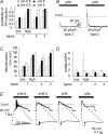
Figure 1. Three distinct ASIC current types are present in small-diameter rat DRG neurones
A, size distribution of ASIC current-responding neurones. Neurones were stimulated by extracellular acidification from the conditioning pH 7.4 to the stimulating pH 5. Plotted is the frequency of detecting an ASIC current as a function of neurone diameter (n = 7–64 per diameter range, except for the 12.5–15 μm range, where n = 2). B, principle of the distinction in three different neurone types based on their ASIC currents. Indicated are the limiting values of the time constant of open-channel inactivation τ and IpH4/IpH6 ratio (for current types 2 and 3). Representative traces of each ASIC current type are shown, evoked by a 5 s acidification to pH 4 (grey) or pH 6 (black). Horizontal scale bar is 2 s, vertical scale bar is 500 pA (type 1, left trace), 2 nA (types 1 right trace and type 2) and 4 nA (type 3).
Figure 2
Figure 2. Correlation with nociceptor markers
A, capsaicin (10 μm) was perfused in a pH 7.4 solution for 5 s. Left panel, a typical current trace; right panel, bar graph showing capsaicin-induced current density for neurones expressing current types 1–3 (n = 14–38). B, action potential shape. Action potentials were induced by short depolarizing current injection in current-clamp mode. The figure shows a representative action potential with (left panel) and without (right panel) inflection in the falling phase and illustrates the different parameters that were measured and are presented in Table 1 of Supplemental data. C, the top panel is a phase contrast image; the bottom panel is a fluorescence image of the same cells after labelling by FITC–IB4. The arrow points to the IB4-positive cell. Scaling bar represents 25 μm. D, bar graph showing the proportion of small IB4-negative (left) and IB4-positive neurones (right) without ASIC current (white), and type 1 (hatched), type 2 (black) and type 3 (cross-hatched).
Figure 3
Figure 3. Biophysical properties of ASIC currents
The left panels illustrate the protocols, the middle panels show representative traces of type 3 and the right panels represent the analysis. Data of types 1–3 are represented and compared with recombinant ASIC1a and ASIC3. A, pH dependence of ASIC current activation. Right panel, pH dependence of ASIC activation. Peak currents normalized to the response to pH 4 are plotted as a function of the stimulating pH and fitted to the Hill equation. Fit parameters are listed in Table 1. B, pH dependence of steady-state-inactivation (SSIN). Right panel, the IpH6 current response, normalized to the IpH6 at conditioning pH 8, is plotted against the conditioning pH. Lines are fits to the Hill equation, whose parameters are listed in Table 1. The symbols are the same as in A. C, time course of ASIC current recovery from inactivation in DRG neurones. Right panel, IpH6 of the second stimulation, normalized to the IpH6 of the first stimulation, plotted against the duration of the interval at pH 7.4 between the two stimulations. The time constants for recovery, obtained from an exponential fit to the data (lines), are indicated in Table 1. The symbols are the same as in A.
Figure 4
Figure 4. Action potential induction by extracellular acidification in DRG neurones
Experiments were done in the current-clamp mode. For type 1 currents, data have been subdivided for neurones expressing type 1 current at IpH6 density < 50 pA pF−1, indicated ‘low’ or > 50 pA pF−1, ‘high’. A, dependence of the probability of AP induction on stimulating pH for neurone types 1–3. B, traces from two type 2 neurones. The IpH6 density is indicated for each neurone under the trace. C, dependence of the depolarization ΔVm on stimulating pH for neurone types 1–3. The filling pattern of the bars has the same meaning as in A. Two-way ANOVA showed dependence on stimulating pH and ASIC type, P < 0.05. D, number of APs per stimulation as a function of the stimulating pH for neurone types 1–3. The filling pattern of the bars has the same meaning as in A. This analysis includes only experiments with successful AP induction. Two-way ANOVA showed dependence on stimulating pH and ASIC type, P < 0.05. E, current-clamp traces from a type 1 neurone (IpH6 density = 199 pA pF−1) in response to 5 s extracellular acidification to increasingly acidic pH. The insets show a part of the trace on an extended time scale.
Figure 5
Figure 5. Relative contribution of ASICs and TRPV1 to the H+-gated current in DRG neurones
A, representative traces of pH 6-induced current in presence or absence of the TRPV1 inhibitor capsazepine (10 μm). Left, current with a type 2 transient component and a large sustained phase TRPV1-dependent component, which is blocked by capsazepine. Right, a type 3 transient current without TRPV1 component, mediated by ASICs and not inhibited by capsazepine. B, comparison of TRPV1-dependent IpH6 density and ASIC-dependent IpH6 density in neurones expressing current types 1–3 (n = 6–24). *TRPV1-dependent and ASIC-dependent IpH6 density are different (P < 0.01).
Figure 6
Figure 6. Comparison with ASIC currents in large-diameter DRG neurones
A, principle of the distinction of ASIC current types 4–7. Indicated are the limiting values of the time constant of open-channel inactivation τ, the IpH4/IpH5 ratio (types 4 and 5) and the sustained/peak current ratio (Is/Ip, types 6 and 7). Representative traces of each ASIC current type are shown, evoked by a 5 s acidification to pH 4 (grey) or pH 5 (black). The horizontal bar is 2 s except for type 5 where it is 4 s. The vertical bar is 200 pA for types 4 and 6, 2 nA for type 5 and 8 nA for type 7. B, pH dependence of ASIC activation. Peak currents normalized to the response to pH 4 are plotted as a function of the stimulating pH and fitted to the Hill equation (see Fig. 3 and Table 1) (n = 2–12). Note that type 4 and 5 currents did not saturate at pH values as acidic as pH 4 and were therefore not fitted. Fit parameters for type 6 were 6.58 ± 0.04 (pH0.5,1), 5.00 ± 0.05 (pH0.5,2), 2.2 ± 0.4 (Hn1) and 6.8 (Hn2) and the fraction of component 1 was 0.63 ± 0.03. Fit parameters for type 7 were 6.66 ± 0.08 (pH0.5,1), 5.13 ± 0.20 (pH0.5,2), 2.4 ± 0.7 (Hn1) and 3.1 ± 3.6 (Hn2) and the fraction of component 1 was 0.68 ± 0.07. C, pH dependence of open-channel-inactivation kinetics. The time constant of channel inactivation (τ, ms) is indicated for responses to pH 6, 5 and 4. *τ values are pH dependent (P < 0.05, n = 5–14). D, sustained/peak current ratio (Is/Ip) (n = 5–25).
Figure 7
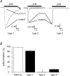
Figure 7. Inhibition of ASIC currents by the venom of P. cambridgei
The venom was used at a dilution of 1: 20 000 in all experiments and applied in the conditioning solution. A, representative traces showing venom effects on ASIC currents of type 1, 2 and 3. B, summary of P. cambridgei venom inhibition of recombinant ASIC1a IpH6 (open bar) and IpH6 of neurone types 1–3 (filled bars) (n = 7–32).
Figure 8
Figure 8. Modulation of ASIC currents by Zn2+
A, representative current traces in response to extracellular acidification to pH 6.4 for the time indicated by the bar, in the absence (black) or the presence (grey) of 300 μm Zn2+. B, relative changes in IpH6.4 in the presence of 300 μm Zn2+. Data from cloned ASICs are represented as open bars and data from neuronal ASICs as filled bars.
Figure 9
Figure 9. ASIC regulation in small diameter DRG neurones in neuropathic pain
A–C, electrophysiological characterization. A, mean IpH6 density in (tibial) injured and (sural) non-injured neurones under control conditions (sham) and after SNI. B, as in A, however, IpH6 density is shown for each of the current types 1–3 (type 1, hatched bars; type 2, filled bars; type 3, cross-hatched bars). ASIC current types were identified based on their open-channel inactivation kinetics, IpH6/IpH4 ratio and the fraction of the sustained current (Table 1). C, proportion of neurone types 1–3, as well as of neurones expressing no ASIC currents before and after SNI. Note the appearance of a ‘new’ current type 8 in injured neurones after SNI (n = 29–41 neurones per condition). D–F, results of the quantitative real-time RT-PCR analysis. D, relative mRNA expression levels of five ASIC subunits in DRGs under control conditions (n = 4 animals). E, relative changes of mRNA levels after SNI. F, relative changes of mRNA levels after SNL. In SNL, non-injured (grey) and injured (black) neurones are present in different DRGs and can therefore be separated. *P < 0.05.

Similar articles

Cited by

References

    1. Akopian AN, Chen CC, Ding YN, Cesare P, Wood JN. A new member of the acid-sensing ion channel family. Neuroreport. 2000;11:2217–2222. - PubMed
    1. Amir R, Devor M. Chemically mediated cross-excitation in rat dorsal root ganglia. J Neurosci. 1996;16:4733–4741. - PMC - PubMed
    1. Amir R, Kocsis JD, Devor M. Multiple interacting sites of ectopic spike electrogenesis in primary sensory neurons. J Neurosci. 2005;25:2576–2585. - PMC - PubMed
    1. Baron A, Schaefer L, Lingueglia E, Champigny G, Lazdunski M. Zn2+ and H+ are coactivators of acid-sensing ion channels. J Biol Chem. 2001;276:35361–35367. - PubMed
    1. Baron A, Waldmann R, Lazdunski M. ASIC-like, proton-activated currents in rat hippocampal neurons. J Physiol. 2002;539:485–494. - PMC - PubMed

Publication types

MeSH terms

LinkOut - more resources