

Effects of single-lung inflation on inspiratory muscle function in dogs
- PMID: 16840517
- PMCID: PMC1995645
- DOI: 10.1113/jphysiol.2006.112797
Effects of single-lung inflation on inspiratory muscle function in dogs
Abstract
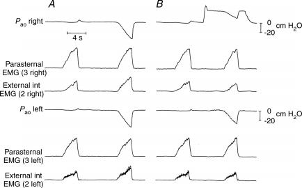
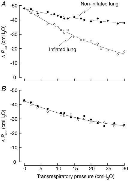
After single-lung transplantation (SLT) for emphysema, a hyperinflated (native) lung operates in parallel with a normal (transplanted) lung. The interpulmonary distribution of the changes in pleural pressure (DeltaP(pl)) during breathing, however, is unknown. To approach the problem, two endotracheal tubes were inserted in the right and left main stem bronchi of anaesthetized dogs, one lung was passively inflated, and the values of inspiratory DeltaP(pl) over the two lungs were assessed by measuring the changes in airway opening pressure (DeltaP(ao)) in the two tubes during occluded breaths. With single-lung inflation, DeltaP(ao) decreased in both lungs, but the decrease in the inflated lung was invariably larger than in the non-inflated lung; when transrespiratory pressure in the inflated lung was set at 30 cmH(2)O, DeltaP(ao) in this lung was 27.7 +/- 2.0% of the value of functional residual capacity (FRC), whereas DeltaP(ao) in the non-inflated lung was 74.4 +/- 4.5% (P < 0.001). This difference was abolished after the ventral mediastinal pleura was severed. The ribs in both hemithoraces were displaced cranially with inflation, such that the displacement in the contralateral hemithorax was 75% of that in the ipsilateral hemithorax, and parasternal intercostal activity remained unchanged. These observations suggest that in patients with SLT for emphysema (1) the inspiratory DeltaP(pl) over the transplanted lung are greater than those over the native lung and (2) this difference results primarily from the greater pressure-generating ability of the inspiratory muscles, in particular the diaphragm, on the transplanted side.
Figures


 ).
).
Similar articles
-
Effects of inflation on the coupling between the ribs and the lung in dogs.J Physiol. 2004 Mar 1;555(Pt 2):481-8. doi: 10.1113/jphysiol.2003.057026. Epub 2003 Dec 23. J Physiol. 2004. PMID: 14694150 Free PMC article.
-
The effect of lung inflation on the inspiratory action of the canine parasternal intercostals.J Appl Physiol (1985). 2006 Mar;100(3):858-63. doi: 10.1152/japplphysiol.00739.2005. Epub 2005 Nov 17. J Appl Physiol (1985). 2006. PMID: 16293705
-
Bilateral impact on the lung of hemidiaphragmatic paralysis in the dog.Respir Physiol Neurobiol. 2009 Mar 31;166(1):68-72. doi: 10.1016/j.resp.2009.02.004. Epub 2009 Feb 21. Respir Physiol Neurobiol. 2009. PMID: 19429521
-
[The respiratory muscles in emphysema. The effects of thoracic distension].Rev Mal Respir. 2000 Apr;17(2):449-57. Rev Mal Respir. 2000. PMID: 10859763 Review. French.
-
Effect of acute inflation on the mechanics of the inspiratory muscles.J Appl Physiol (1985). 2009 Jul;107(1):315-23. doi: 10.1152/japplphysiol.91472.2008. Epub 2009 Mar 5. J Appl Physiol (1985). 2009. PMID: 19265064 Review.
Cited by
-
Regional diaphragm volume displacement is heterogeneous in dogs.Am J Physiol Regul Integr Comp Physiol. 2017 Mar 1;312(3):R443-R450. doi: 10.1152/ajpregu.00270.2016. Epub 2017 Jan 18. Am J Physiol Regul Integr Comp Physiol. 2017. PMID: 28100474 Free PMC article.
References
-
- Boriek AM, Rodarte JR, Wilson TA. Kinematics and mechanics of midcostal diaphragm of dog. J Appl Physiol. 1997;83:1068–1075. - PubMed
-
- Cappello M, De Troyer A. Interaction between the left and right intercostal muscles in airway pressure generation. J Appl Physiol. 2000;88:817–820. - PubMed
-
- Cassart M, Verbandt Y, de Francquen P, Gevenois PA, Estenne M. Diaphragm dimensions after single-lung transplantation for emphysema. Am J Respir Crit Care Med. 1999;159:1992–1997. - PubMed
-
- Danon J, Druz WS, Goldberg NB, Sharp JT. Function of the isolated paced diaphragm and the cervical accessory muscles in C1 quadriplegics. Am Rev Respir Dis. 1979;119:909–919. - PubMed
Publication types
MeSH terms
LinkOut - more resources
Full Text Sources
Medical