. 2006 Oct 1;576(Pt 1):235-56.
doi: 10.1113/jphysiol.2006.111336. Epub 2006 Jul 13.

Kv7/KCNQ/M-channels in rat glutamatergic hippocampal axons and their role in regulation of excitability and transmitter release

Affiliations

Kv7/KCNQ/M-channels in rat glutamatergic hippocampal axons and their role in regulation of excitability and transmitter release

K Vervaeke et al. J Physiol. .

Abstract

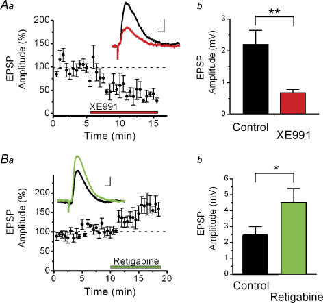
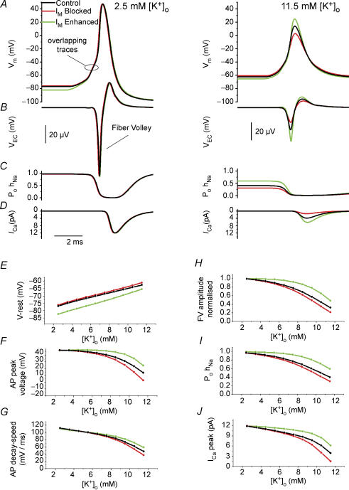
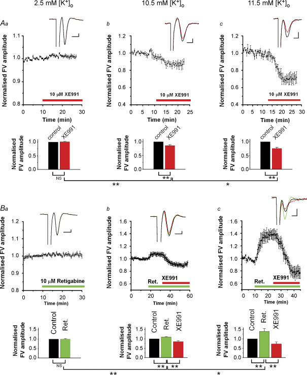
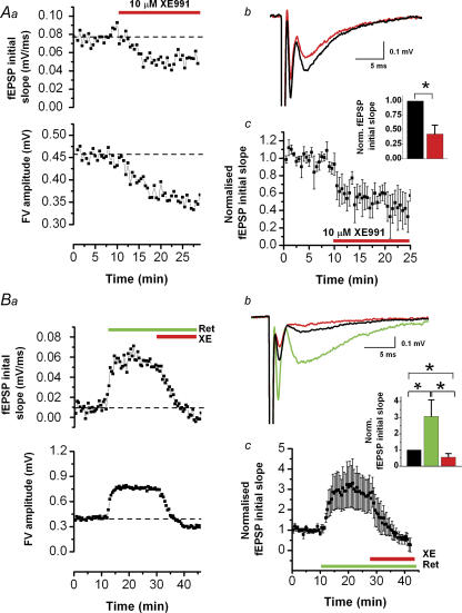
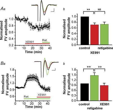
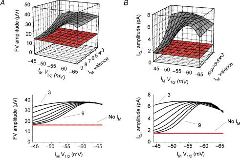
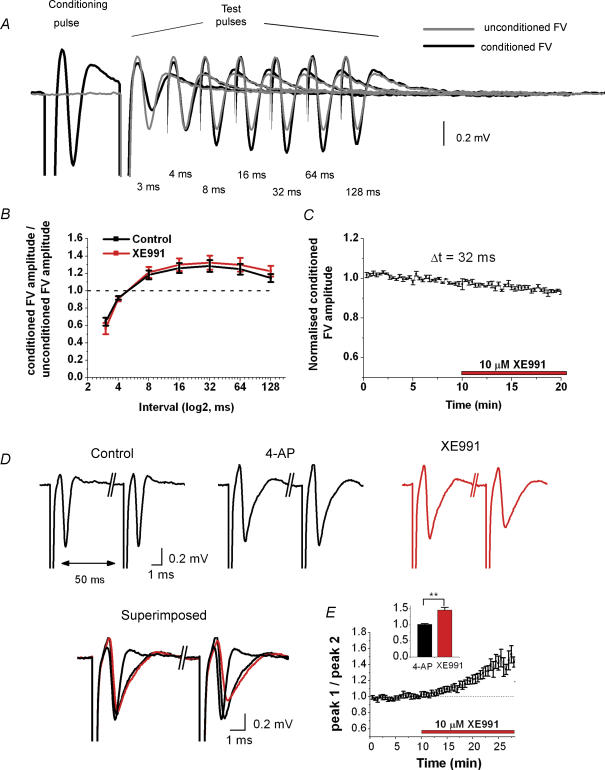
M-current (I(M)) plays a key role in regulating neuronal excitability. Mutations in Kv7/KCNQ subunits, the molecular correlates of I(M), are associated with a familial human epilepsy syndrome. Kv7/KCNQ subunits are widely expressed, and I(M) has been recorded in somata of several types of neurons, but the subcellular distribution of M-channels remains elusive. By combining field-potential, whole-cell and intracellular recordings from area CA1 in rat hippocampal slices, and computational modelling, we provide evidence for functional M-channels in unmyelinated axons in the brain. Our data indicate that presynaptic M-channels can regulate axonal excitability and synaptic transmission, provided the axons are depolarized into the I(M) activation range (beyond approximately -65 mV). Here, such depolarization was achieved by increasing the extracellular K(+) concentration ([K(+)](o)). Extracellular recordings in the presence of moderately elevated [K(+)](o) (7-11 mm), showed that the specific M-channel blocker XE991 reduced the amplitude of the presynaptic fibre volley and the field EPSP in a [K(+)](o)-dependent manner, both in stratum radiatum and in stratum lacknosum moleculare. The M-channel opener, retigabine, had opposite effects. The higher the [K(+)](o), the greater the effects of XE991 and retigabine. Similar pharmacological modulation of EPSPs recorded intracellularly from CA1 pyramidal neurons, while blocking postsynaptic K(+) channels with intracellular Cs(+), confirmed that active M-channels are located presynaptically. Computational analysis with an axon model showed that presynaptic I(M) can control Na(+) channel inactivation and thereby affect the presynaptic action potential amplitude and Ca(2+) influx, provided the axonal membrane potential is sufficiently depolarized. Finally, we compared the effects of blocking I(M) on the spike after-depolarization and bursting in CA3 pyramidal neuron somata versus their axons. In standard [K(+)](o) (2.5 mm), XE991 increased the ADP and promoted burst firing at the soma, but not in the axons. However, I(M) contributed to the refractory period in the axons when spikes were broadened by a low dose 4-aminopyridine (200 microm). Our results indicate that functional Kv7/KCNQ/M-channels are present in unmyelinated axons in the brain, and that these channels may have contrasting effects on excitability depending on their subcellular localization.


Figures

Figure 3
Figure 3. XE991 reduced and retigabine increased EPSPs recorded from CA1 pyramidal neurons in elevated [K+]o (11.5 mM)
Whole-cell recordings of EPSPs in CA1 pyramidal cells, in response to stimulation in stratum radiatum. In the patch pipette, all K+ ions were replaced by 140 mm Cs+ to block postsynaptic K+ channels. Aa, summary time course (mean ± s.e.m.) of the effect of 10 μm XE991 on the EPSP amplitude in the presence of 50 μmdl-APV and 5 μm bicuculline (n = 5). The membrane potential was maintained at −78 mV during each experiment. Inset shows averages of five consecutive traces before (black) and after (red) application of XE991. Ab, XE991 reduced the EPSP amplitude from 2.2 ± 0.4 to 0.68 ± 0.10 mV (n = 4, P = 0.002). Ba and b, in similar conditions as in Aa and b, 20 μm retigabine increased the EPSP amplitude from 2.4 ± 0.6 to 4.5 ± 0.9 mV (n = 6, P = 0.045). Scale bars for insets in Aa and Bb: 5 ms, 1 mV.
Figure 7
Figure 7. Blocking IM increased the ADP and promoted burst firing in CA3 pyramidal neuron somata in standard [K+]o (2.5 mM)
CA3 pyramidal neurons were recorded with sharp electrodes in the presence of DNQX (50 μm), dl-APV (50 μm) and bicuculline (5 μm). The prestimulus membrane potential was maintained at −60 mV by steady current injection. A brief current pulse (10 ms, 0.5 nA) was injected every 10 s to evoke a single action potential (Aa). A single spike is followed by an after-depolarization (ADP), and medium and slow after-hyperpolarizations (mAHP and sAHP). Inset, the spike and AHPs on a compressed time scale. Ab–d, application of 10 μm XE991 increased the ADP and promoted burst firing followed by a prominent sAHP. B, time course of the XE991-induced changes in ADP amplitude and firing (same experiment as in Aa–d). When the cell fired only a single spike, the area of the ADP is indicated by ○. Bursts are indicated by |, in different rows for bursts consisting of 2, 3 or 4 spikes. Note that the number of spikes during a burst progressively increased. C, summary plot of the effect of XE991 on the ADP area, for all cells tested (n = 7; control, 254 ± 38 mV ms−1; XE991, 433 ± 87 mV ms−1; P = 0.036).
Figure 5
Figure 5. Computational modelling of the presynaptic intracellular response to IM modulation, based on extracellular data
In the axon model, an action potential was evoked by an intracellular short current pulse (1 ms duration), 600 μm from the soma, and recorded 200 μm orthodromically from the initiation site. A–D, various responses in either 2.5 mm (left panels) or 11.5 mm[K+]o (right panels) in control conditions (black) and when IM was either removed (red) or enhanced by a negative shift in its activation curve (green). From top to bottom: A, intracellular potential (Vm); B, extracellular potential (VEC); C, open probability of INa inactivation particle (PohNa); and D, the Ca2+ current during the action potential (ICa). E–J, same simulation as in A–D, but repeated for various [K+]o values, ranging from 2.5 to 11.5 mm.E, resting membrane potential V-rest. F, action potential peak voltage. G, rate of action potential repolarization (decay speed). H, normalized FV amplitude. I, open probability of the INa inactivation particle (PohNa). J, peak Ca2+ current, ICa, in response to the action potential.
Figure 1
Figure 1. In stratum radiatum, XE991 reduced, and retigabine increased, the presynaptic compound action potential (FV) in an [K+]o-dependent manner
Aa–c, summary time course of the effect of the M-channel blocker XE991 on the FV amplitude (mean ± s.e.m.) recorded in stratum radiatum in response to stimulation of presynaptic fibres in stratum radiatum. The effects of XE991 at three different [K+]o levels are shown: 2.5 mm (a, n = 5), 10.5 mm (b, n = 5) and 11.5 mm (c, n = 5). Insets show the average of 5 consecutive sweeps of the FV before (black) and after (red) application of 10 μm XE991. Bars show the summary of the XE991 effects on the FV amplitude (mean ± s.e.m.) as shown in Aa–c, respectively. Ba–c, similar to Aa–c, but the M-channel opener retigabine was added before XE991 (a, n = 5; b, n = 5; c, n = 5). NS, P > 0.05; *0.01 < P < 0.05; **P < 0.01. Scale bars for insets in Aa–c and Ba–c: 1 ms, 0.1 mV. All experiments were performed in the presence of blockers of synaptic transmission: 50 μm DNQX, 50 μmdl-APV and 5 μm bicuculline free base.
Figure 2
Figure 2. In stratum radiatum, XE991 reduced and retigabine increased the field postsynaptic responses in elevated [K+]o (11.5 mM)
All responses were evoked by stimulation of presynaptic fibres in stratum radiatum and were recorded in stratum radiatum in the presence of 11.5 mm[K+]o, 50 μm dl-APV and 5 μm bicuculline. Aa, effect of 10 μm XE991 on fEPSP initial slope (upper panel) and fibre volley (FV) amplitude (lower panel). Ab, average of 5 consecutive traces before (black) and after (red) application of XE991 (same experiment as in Aa). Ac, summary time course (mean ± s.e.m.) of experiments performed as in Aa–c (n = 5). XE991 reduced the fEPSP initial slope by a factor 0.43 ± 0.15. Ba, effect of 10 μm retigabine and subsequent application of 10 μm XE991 on fEPSP initial slope and FV amplitude. Bb, average of 5 consecutive traces before (black) and after application of retigabine (green), and XE991 (red) (same experiment as in Ba). Bc, summary time course (mean ± s.e.m.) of experiments obtained as in Ba and b (n = 5). Retigabine increased the fEPSP initial slope by a factor 3.1 ± 1.0 and subsequent application of XE991 reduced the fEPSP initial slope by a factor 0.56 ± 0.22 compared with the control (*0.01 < P < 0.05).
Figure 4
Figure 4. In stratum lacunosum moleculare, XE991 reduced and retigabine increased the presynaptic compound action potentials (FV) in elevated [K+]o (11.5 mM)
Aa, summary time course (mean ± s.e.m.) of the FV amplitude recorded in the stratum lacunosum moleculare in response to stimulation of presynaptic fibres in stratum lacunosum moleculare. Inset shows average of 5 consecutive traces of the FV in control conditions (black), after application of 10 μm XE991 (red) and followed by application of 10 μm retigabine (green). Ab, XE991 decreased the FV amplitude by a factor 0.71 ± 0.03. Subsequent application of retigabine did not further affect the FV (0.73 ± 0.02). Ba, as in Aa, but retigabine was applied before XE991. Bb, retigabine increased the FV by a factor 1.47 ± 0.16 while subsequent application of XE991 decreased the FV by a factor 0.89 ± 0.13 compared with the control (**P < 0.01; NS, not significant). DNQX (50 μm), dl-APV (50 μm) and bicuculline (5 μm) were added more than10 min before the onset of each recordings. Scale bars for insets in Aa and Ba: 1 ms, 0.05 mV.
Figure 6
Figure 6. In the model, the effects of IM on the presynaptic compound action potential (FV) and spike-evoked ICa in elevated [K+]o (11.5 mM) is robust for a wide range of IM parameter values
An action potential was evoked by an intracellular short current pulse (1 ms duration) in the axon model, 600 μm from the soma, and recorded 200 μm from the initiation site (in the orthodromic direction). FV amplitude (A) and spike-evoked ICa peak amplitude (B) are shown as functions of the IM steady state activation curve parameters V1/2 and valence (steepness). Grey, control; red, no IM
Figure 8
Figure 8. The M-channel blocker XE991 affects paired-pulse modulation of the presynaptic compound action potential (FV) in stratum radiatum after spike broadening (2.5 mM [K+]o)
A, fibre volleys (FVs) recorded under control conditions in response to test pulses (80 μs duration), preceded by a stronger conditioning pulse (120 μs duration) at various time intervals (black traces). The FV was facilitated maximally for a pulse interval of ∼32 ms, compared with unconditioned FVs (grey traces). For clarity, the stimulus artifacts are deleted for stimulation intervals 4–128 ms. DNQX (50 μm), dl-APV (50 μm) and bicuculline (5 μm) were present in the bath throughout the experiment. B, the ratio between conditioned and unconditioned FV amplitude is plotted against interstimulus interval (logarithmic axis). The values obtained under control conditions (black, n = 5) or in the presence of 10 μm XE991 (red, n = 5) were not statistically different for any of the tested intervals. C, summary time course of the conditioned FV amplitude measurements during application of 10 μm XE991, for an interstimulus interval of 32 ms (mean ± s.e.m., n = 5). D, fibre volleys evoked by paired-pulse stimulation in stratum radiatum (interval, 50 ms). Bath application of 200 μm 4-aminopyridine (4-AP) broadened the FV, and subsequent application of 10 μm XE991 caused depression of the second FV (red). E, average time course of the FV1/FV2 amplitude ratio. This ratio increased from 1.00 ± 0.03 to 1.50 ± 0.10 (n = 6, P = 0.0008).

Similar articles

Cited by

References

    1. Alle H, Geiger JR. Combined analog and action potential coding in hippocampal mossy fibers. Science. 2006;311:1290–1293. - PubMed
    1. Allen C, Stevens CF. An evaluation of causes for unreliability of synaptic transmission. Proc Natl Acad Sci U S A. 1994;91:10380–10383. - PMC - PubMed
    1. Amaral DG, Witter MP. The three-dimensional organization of the hippocampal formation: a review of anatomical data. Neuroscience. 1989;31:571–591. - PubMed
    1. Amiry-Moghaddam M, Williamson A, Palomba M, Eid T, de Lanerolle NC, Nagelhus EA, Adams ME, Froehner SC, Agre P, Ottersen OP. Delayed K+ clearance associated with aquaporin-4 mislocalization: phenotypic defects in brains of alpha-syntrophin-null mice. Proc Natl Acad Sci U S A. 2003;100:13615–13620. - PMC - PubMed
    1. Andersen P, Silfvenius H, Sundberg SH, Sveen O, Wigstrom H. Functional characteristics of unmyelinated fibres in the hippocampal cortex. Brain Res. 1978;144:11–18. - PubMed

Publication types

MeSH terms

LinkOut - more resources